


Cochlosomiasis is infection with Cochlosoma anatis, a flagellated protozoon that infects ducks, turkeys, geese, and wild birds. C anatis can cause limited pathological changes in the gut of ducks and turkeys, leading to diarrhea, although the direct pathology is still debated. Microscopic evaluation of intestinal mucosal scrapings can be used to determine infection. There is no treatment or vaccine for cochlosomiasis.
The causative organism of cochlosomiasis is Cochlosoma anatis, the flagellated protozoon found in the intestinal microbiota of birds and mammals. The disease is of economic importance in turkey and duck production.
Etiology of Cochlosomiasis
The causative agent of cochlosomiasis in turkeys and ducks is Cochlosoma anatis (syn. Cochlosoma rostratum), the only species in the genus known to infect birds.
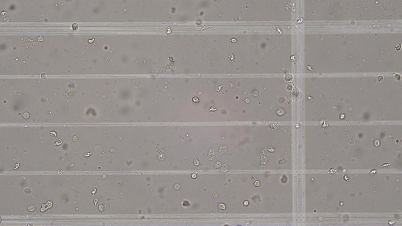
By light microscopy, Cochlosoma anatis is 6–12 mcm long and 4–7 mcm wide, with a characteristic adhesive disc on the anteroventral surface of its pyriform body. It moves in a jerking motion. This protozoon has a single nucleus and uses flagella for movement.
Lateral transmission has been observed with turkeys, but direct contact of the birds was necessary for infection. PCR assay has detected C anatis on house flies; however, no studies have demonstrated that they carry live trophozoites.
Clinical Findings of Cochlosomiasis
There is a debate on the pathogenicity of C anatis in avian species. The exact role of this parasite is unknown because coinfection of viruses, bacteria, and other protozoa with C anatis has led to GI distress, whereas infection with this protozoon alone does not always lead to clinical signs.
Lesions

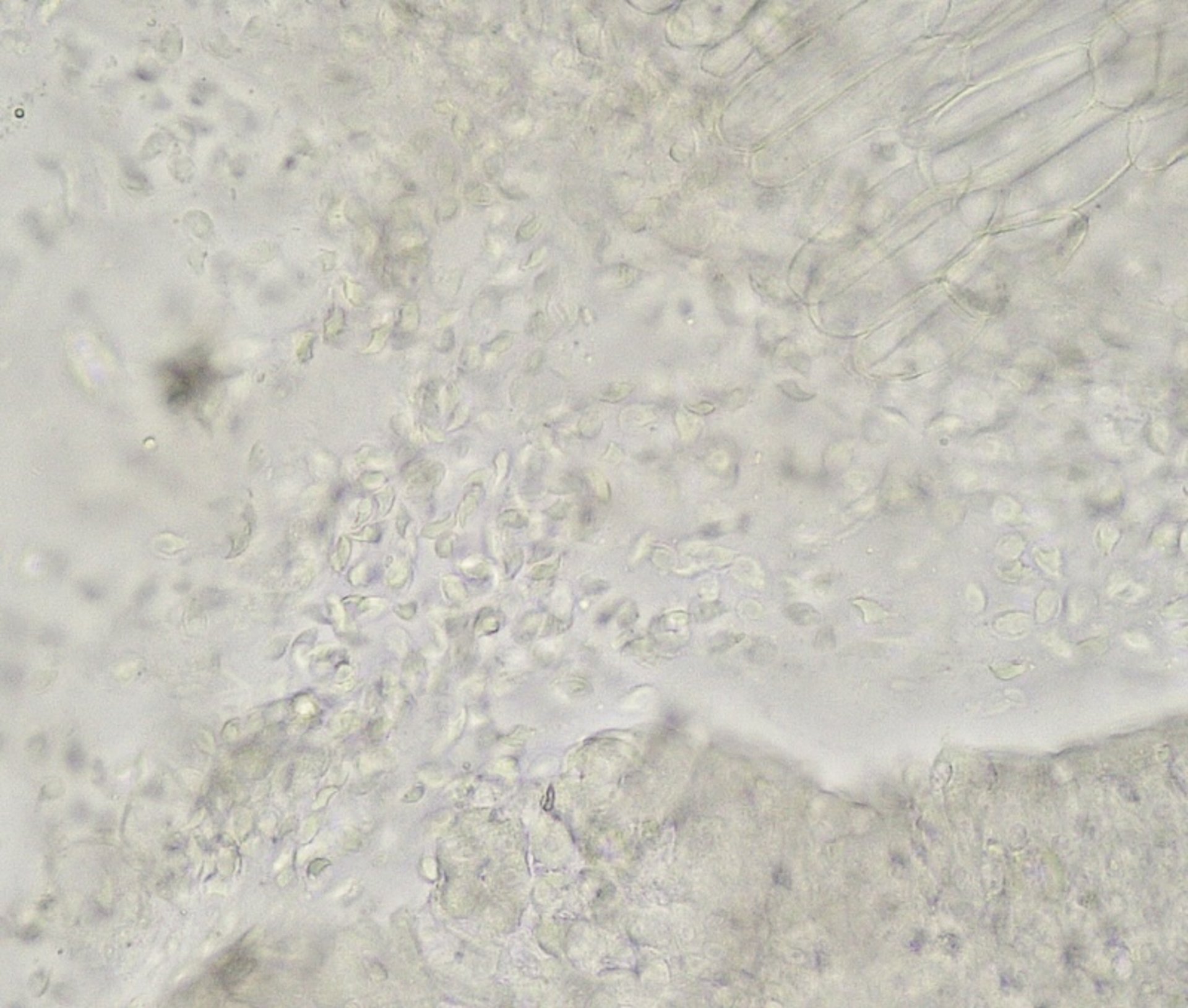
Courtesy of Dr. Robert. B. Beckstead and Kelly Grace Keen.
Scanning electron microscopy has shown C anatis attaching to intestinal mucosa; however, no apparent changes in the intestines have been visualized under light microscopy with the protozoon present (see Cochlosoma parasites, turkey, image and video).
Diagnosis of Cochlosomiasis
Light microscopy and PCR assay are used to identify Cochlosoma anatis in intestinal scrapings and feces. Analysis of samples within two minutes of collection is recommended for proper identification because C anatis becomes immobile when cooled.
Prevention and Treatment of Cochlosomiasis
Wild birds, small rodents, and multiaged flocks have been identified as carriers of C anatis. Proper biosecurity prevents the entry of this parasite to poultry flocks. Disinfection of housing facilities after an outbreak is necessary to prevent contamination of future flocks.
Nitroimidazole and nitarsone have been used as successful treatments for C anatis; however, these products are not approved for use in commercial poultry. Because of concerns of harmful residues, extra-label use of these antimicrobials in poultry is prohibited in the US.
Key Points
Cochlosomiasis is caused by C anatis, a flagellated protozoon that has debatable pathogenicity in avian species.
Intestinal scrapings and fecal material can be used for identification.
No treatments are currently available for cochlosomiasis.


