

Mange is caused by microscopic mites that invade the skin of otherwise healthy animals. The mites cause irritation of the skin, resulting in itching, hair loss, and inflammation. Most types of mange are highly contagious. Both dogs and cats are very susceptible. Horses and other domestic animals can also be infected. There are several types of mange that affect dogs, including canine scabies (sarcoptic mange), ear mites (otodectic mange), walking dandruff (cheyletiellosis), and trombiculosis.
Canine Scabies (Sarcoptic Mange)
This form of mange is caused by the mite Sarcoptes scabiei var canis. This highly contagious parasite is found on dogs worldwide. It is often called canine scabies. Although the mites that cause mange prefer dogs, humans and other animals that come in contact with an infected dog may also become infected. The entire life cycle (17 to 21 days) of these mites is spent on the infested dog. Females burrow tunnels in the skin to lay eggs. Mange is easily spread between animals by contact. Indirect transmission, such as through infested bedding, is less common, but it can occur. The incubation period varies from 10 days to 8 weeks, depending on how severely the dog is infested, part of the body affected, number of mites transmitted, and the individual dog’s health and hygiene.
Canine scabies

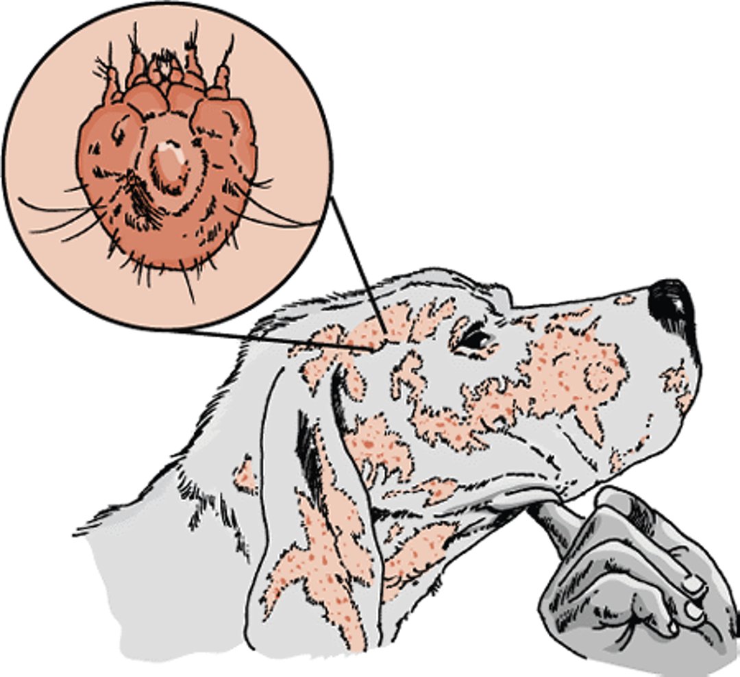
Not all dogs have signs when they are infested with sarcoptic mange mites. Usually, though, the animal will have intense itching that comes on suddenly. The itching is probably caused by sensitivity to the mites’ droppings. Initially, infested skin will erupt with small, solid bumps. Because the dog scratches or bites itself to relieve the itch, these bumps and the surrounding skin are often damaged, causing thick, crusted sores. Secondary yeast or bacterial infections can develop in the damaged skin. Usually, the sores appear first on the abdomen, chest, ears, elbows, and legs. If the mange is not diagnosed and treated, the sores can spread over the entire body. Dogs with longterm, recurring mange develop oily dandruff (seborrhea), severe thickening of the skin with wrinkling and crust build-up, and oozing, weeping sores. Dogs affected this severely can become emaciated and may even die.
“Scabies incognito” is a term used to describe hard-to-diagnose mange. If a dog is regularly bathed and has a well-groomed coat, the mites might be hard to find, even if the dog shows signs of infestation such as itching. The other typical signs of mange—crusts and scales on the skin—are removed by regular bathing.
If mange is suspected, your veterinarian will do a physical examination, including collecting skin scrapings and possibly a stool sample. Some clinics might also use a blood test to diagnose mange. If mites are not found, but the signs are highly suggestive of mange, trial treatment is warranted. Mange is very highly contagious and can spread easily between animals of different species and even to humans. Thus, you should ask your veterinarian for advice on how to avoid contracting mange from your pet.
Treatment should include all dogs and other animals that have been in contact with one another. It may be necessary to clip the hair. The crusts and dirt should be removed by soaking with a medicated (antiseborrheic) shampoo, and an anti-mite dip applied. Lime-sulfur is highly effective and safe for use in young animals. Several dips may be required. Alternatively, internal or topical medicines are also effective. Some internal mange medications are also used for heartworm prevention, so your veterinarian may want to test your dog for heartworms before treatment. Treatment for secondary infections may also be necessary.
Ear Mites (Otodectic Mange)
This form of mange is caused by Otodectes cynotis mites. These mites often infest the external ear, causing inflammation of the ear canal in dogs and especially in cats. Ear mites are usually found deep in the external ear canal, but they are sometimes seen on the body. The infested animal will shake its head and scratch its ear(s). In dogs with normally upright ears, the external ear may droop. The intensity of the itching varies and may be intense. In severe cases, the external ear may be inflamed and produce pus; a torn eardrum is also possible. Dogs with ear mites should be treated with a parasiticide in the ears or on the whole body. Your veterinarian will recommend an appropriate treatment plan that includes medication and ear cleaning instructions. Animals that have contact with infested dogs should also be treated.
Walking Dandruff (Cheyletiellosis)
Cheyletiella yasguri mites cause walking dandruff in dogs. (The dandruff that is seen “walking” is actually the mites moving about on the skin of the dog.) Although these mites often stay on their preferred hosts, infections across species are possible. Walking dandruff is very contagious, especially in kennels, catteries, or multi-pet households. Regular use of certain insecticides to control flea infestations has a side benefit of often controlling the mites that cause walking dandruff. Humans can also be infested with these species of mites. Mites that cause walking dandruff spend their entire 3-week life cycle on their host but can also live up to 10 days in the environment.
Scaling of the skin and infestation along the back are common signs of walking dandruff. Intense itching is frequent, though some animals do not itch at all. Pets that show no signs can carry the mites and transmit them to other pets and humans.
Cheyletiellosis is diagnosed by looking at an animal's skin and examining skin and hair samples with a microscope to identify the presence of mites. The mites and eggs may be hard to find, especially on animals that are bathed often.
In many cases, veterinarians will prescribe topical or body-wide treatments with an insecticide to eliminate the mites. In addition, treating the pet’s environment is necessary to kill mites in bedding, carpets, and other areas. Insecticidal treatment of kennels and other multi-pet communities is required to halt mite infestations.
Owners of pets infested with these mites may want to check with their physicians regarding medication and other steps to control mite infestations in themselves, their family members, and the home environment. It is very easy for these mites to spread from pets to owners.
Canine Demodicosis
The mites that cause canine demodicosis live in small numbers in the hair follicles and sebaceous glands of all dogs. This is normal and causes no signs of disease. However, for reasons not clearly understood, some dogs have large numbers of Demodex canis mites, resulting in inflammation and hair loss. There is evidence of hereditary predisposition for this condition in some dogs. It is strongly suspected that suppression of the immune response to these mites may play a role.
There are 2 clinical forms of canine demodicosis: localized (limited to a small area) and generalized (found on the entire body). Localized demodicosis is usually seen in dogs less than 1 year old. Affected dogs will have 1 to 5 small, isolated areas that are usually hairless, red, and scaly. Itching is mild or absent. A few cases of localized demodicosis progress to the generalized form, though most cases resolve without treatment.
The generalized form of demodicosis can occur in young dogs (juvenile-onset) or in adults (adult-onset). Affected dogs have severe disease with widespread inflammation of the skin. Juvenile-onset generalized demodicosis is the result of an inherited immune system defect. On the other hand, adult-onset generalized demodicosis is often associated with an underlying disease that has suppressed the immune system (such as cancer, Cushing disease, hypothyroidism, or diabetes). Both types of generalized demodicosis can cause hair loss, reddened and swollen skin, increased pigmentation (darkening of the skin), raised lumps that look like acne, and scabs. Secondary bacterial infections (pyodemodicosis) are common. Many dogs with generalized demodicosis also have inflamed foot pads. Other signs can include enlarged lymph nodes, lethargy, fever, and pus-filled inflammation of the deeper layers of skin.
Laboratory analysis of deep skin scrapings is usually used to confirm a diagnosis of demodicosis. In addition, your veterinarian will also want to test your dog for other infections or diseases that may have suppressed the immune system.
Cases of localized demodicosis often resolve without treatment. Generalized demodicosis is a serious disease that requires medical treatment. The outlook for these cases is guarded. Medicated shampoos and dips are often used to treat demodicosis. Prescription medications to kill the mites may be required. In cases where secondary bacterial infections are present, antibiotics may also be prescribed. Skin scrapings are taken at monthly intervals to monitor the number of mites on the dog.
Owners of dogs with demodicosis should understand that treatment of generalized demodicosis can take several months. The prescribed antiparasitic treatment must be continued until at least 2 consecutive negative skin scrapings have been obtained at monthly intervals. Some dogs may need several months of treatment. Recurrence within the first year of treatment is not uncommon.
Because it may be inherited, dogs with demodicosis should not be bred.
Trombiculosis
Trombiculosis is a type of mange caused by the parasitic larval stage of mites of the family Trombiculidae (chiggers). Adults and nymphs look like tiny spiders and live on rotting material. Dogs acquire the larvae by lying on the ground or walking in suitable habitat.
The larvae attach to the host, feed for a few days, and leave when engorged. They are easily identified as tiny, orange-red, oval dots that do not move. These are usually found clustering on the head, ears, feet, or belly. Signs include redness, bumps, hair loss, and crusts. Intense itching can persist even after the larvae have left the animal. Diagnosis is based on history and signs. Your veterinarian will want to exclude other skin disorders that cause itching, such as allergies. Diagnosis is confirmed by careful examination of the affected areas. Skin scrapings might also be examined under the microscope for evidence of 6-legged mite larvae.
Treatment for dogs and other pets with trombiculosis follows the pattern for the general treatment of mange (see above). Medications to kill these mites on your pet may be different from those prescribed for other types of mites. Follow your veterinarian’s treatment program. If the itching has been either severe or extended, antibiotics or other medications may be prescribed to control secondary infections in scratch and bite wounds.
Preventing reinfestation is often difficult. The most useful approach, if feasible, consists of keeping pets away from areas known to harbor mites.
For More Information
Also see professional content regarding mange.


