

Environmental-induced (eg, house dust, house dust mites, pollens of trees, grasses and weeds, molds) or food-induced atopic dermatitis is a common allergic disorder of dogs and cats and frequently causes erythema and pruritus of the pinnae and external ear canals. The allergic condition predisposes to development of secondary bacterial or yeast otitis externa.
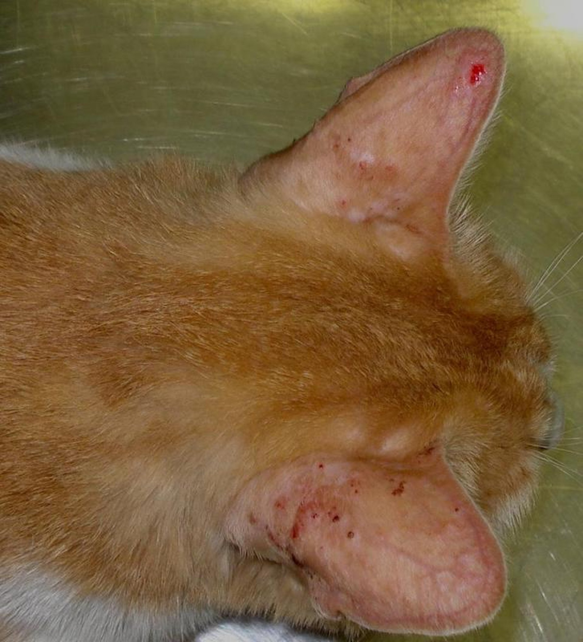
Otitis externa can extend to affect the pinna. Pinnal dermatitis can present with erythema, papules, crusts, and lichenification. In addition, contact hypersensitivity to otic preparations can cause lesions on the inner aspect of the pinna (see contact dermatitis). Other body sites such as the face (ie, periocular region, muzzle, chin), axilla, groin, and feet are also often affected.
For diagnosis, skin cytology is indicated to identify the presence of secondary infections; the most common findings include yeast (Malassezia) and cocci bacteria (Staphylococcus) or a mixed infection.
Topical therapy is in most cases sufficient to control the infection and inflammation. Medicated wipes, creams, lotions, gels, sprays or mousses can be used as delivery based on the ease of application. Active ingredients should be chosen based on the cytological findings. For atopic patients prone to develop pinnal dermatitis, ceramides and topical essential fatty acids can be applied to moisturize the skin and improve the epidermal barrier function. Topical gluccocorticoids can also be used if pruritus is present without a secondary bacterial or yeast infection.

Courtesy of Dr. Michele Corazza.
Feline mosquito hypersensitivity is an allergic reaction to mosquito bites that can cause an ulcerative and crusted dermatitis of the pinnae, nose, and less commonly the footpads, eyelids, chin, and lips of cats. Lesions progress from papules to plaques that can become crusted and ulcerated and coalesce to affect extensive areas. Pruritus is a consistent sign, and regional lymphadenopathy may occur. In severe cases, fever or other systemic signs may develop. Histologically, the lesions are characterized by severe superficial and deep perivascular to interstitial eosinophilic dermatitis, often associated with flame figures, folliculitis, and furunculosis. Clinical differential diagnoses include pemphigus foliaceus, herpesvirus ulcerative dermatitis, other causes of eosinophilic dermatitis (food allergy, atopy, idiopathic), notoedric mange, and dermatophytosis. Treatment includes keeping the cat inside at dusk and down times. Systemic antiinflammatory doses of glucocorticoids may be necessary in severe cases.
For More Information


