

Pheochromocytomas are rare tumors that originate from the adrenal medulla. They have been most commonly reported in dogs, horses, and cattle. Clinical signs associated with pheochromocytomas are nonspecific, and these tumors can be challenging to diagnose. They are sometimes identified as an incidentally discovered adrenal mass on abdominal ultrasonographic evaluation. Surgical removal is the treatment of choice.
A pheochromocytoma is a catecholamine-secreting tumor of chromaffin cells.
Pheochromocytomas occur in domestic species, including dogs and rarely cats. They also occur in horses and cattle.
Incidental masses in the area of the adrenal glands are being discovered with greater frequency because of the increased use of abdominal ultrasonographic evaluation and other imaging techniques. Pheochromocytoma, although rare, should be a differential diagnosis whenever such a mass is identified.
Etiology and Pathogenesis of Pheochromocytomas in Animals
Pheochromocytomas arise from the adrenal medullary chromaffin cells that normally synthesize and secrete the catecholamines epinephrine and norepinephrine.
Epidemiology of Pheochromocytomas in Animals
Pheochromocytomas have been identified more often in dogs (uncommon) than in cats (rare), usually affect only one gland, and tend to occur in older animals. There is no breed predilection. Males may be overrepresented.
Clinical Features of Pheochromocytomas in Animals
Pheochromocytomas are often locally invasive (may result in thrombus formation in the adjacent vena cava) and metastasize in ≥ 25% of cases. Extra-adrenal sites for related tumors (paragangliomas) have been reported in other tissues, including the heart.
Clinical signs of pheochromocytomas are nonspecific and may appear sporadically, possibly related to periodic or intermittent release of catecholamines. In addition, clinical signs may vary depending on which catecholamine predominates.
Pearls & Pitfalls
|
Clinical signs reported in most dogs include the following:
weight loss
anorexia
listlessness
weakness
occasional collapse
Dyspnea and tachycardia may occur, and hypertension is common.
Diagnosis of Pheochromocytomas in Animals
Diagnosis of pheochromocytoma is challenging.

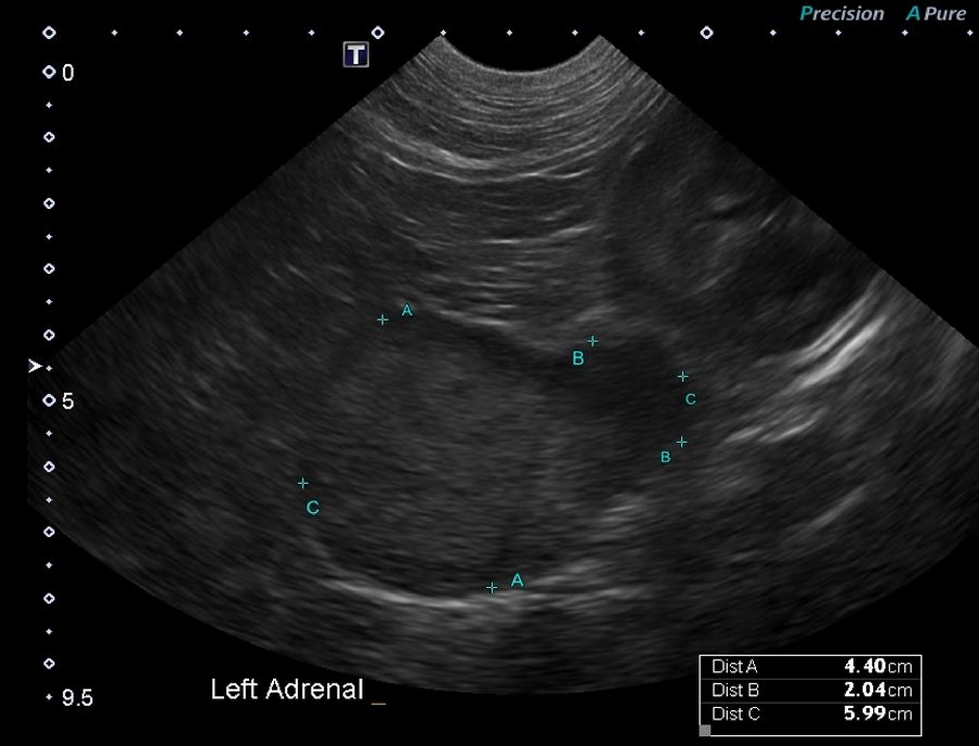
Transverse sonogram of a pheochromocytoma of the left adrenal gland in a dog. The mass is homogeneously echogenic. The ultrasonographic appearance of these lesions is nonspecific.
Courtesy of Dr. Merrilee Holland.
Indirect blood pressure measurement can help establish the diagnosis.
Diagnostic imaging modalities such as ultrasonography and CT are very useful in confirming a suspected adrenal mass. In any patient with a suspected adrenal tumor, thoracic radiography is recommended for detection of pulmonary metastases.
The much more common adrenal disorder hyperadrenocorticism should be included as a differential diagnosis when an adrenal mass is identified. Other differential diagnoses for adrenal nodules and masses include adrenal cortical adenoma or adenocarcinoma, metastasis, and adrenal hyperplasia.
Pearls & Pitfalls
|
Immunohistochemical staining for chromogranin A can distinguish pheochromocytomas from adrenocortical tumors.
Treatment and Prognosis of Pheochromocytomas in Animals
Although surgical removal is the treatment of choice, patients with pheochromocytoma have increased anesthetic risk secondary to the cardiovascular effects of the catecholamines. Surgery is further complicated by the tendency toward local invasion of these tumors and their proximity to large vessels.
The tyrosine kinase inhibitor toceranib phosphate may be useful in the treatment of nonresectable pheochromocytoma.1
Note: toceranib phosphate has also been suggested as a potential treatment for other neuroendocrine tumors, including thyroid carcinoma, insulinoma, and chemodectoma.
Pretreatment with phenoxybenzamine (a noncompetitive alpha adrenergic receptor antagonist) for 1–2 weeks before surgery improves the perioperative mortality rate.
Dogs without metastatic disease have a good longterm prognosis.
Reference
Frezoulis P, Harper A. The role of toceranib phosphate in dogs with non-mast cell neoplasia: a systematic review. Vet Comp Oncol. 2022;2:362-371. doi:10.1111/vco.12799.
Adrenal Medullary Hyperplasia
Diffuse or nodular adrenal medullary hyperplasia appears to precede the development of pheochromocytomas in bulls with C-cell tumors of the thyroid gland. This diffuse proliferation of chromaffin cells is nonencapsulated but compresses the surrounding adrenal cortex. In bulls with prominent diffuse medullary hyperplasia, there are often a few small foci of intense nodular proliferation of medullary cells.
Key Points
Pheochromocytomas are rare tumors that affect the adrenal gland.
Pheochromocytomas have been reported in multiple species, most commonly in dogs.
Clinical signs in affected patients are nonspecific, and the diagnosis can be challenging.
For More Information


