

All of the major groups of animal parasites are found in fish, and apparently healthy wild fish often carry heavy parasite burdens. Parasites with direct life cycles can be important pathogens of cultured fish; parasites with indirect life cycles frequently use fish as intermediate hosts. Knowledge of specific fish hosts greatly facilitates identification of parasites with marked host and tissue specificity, whereas others are recognized because of their common occurrence and lack of host specificity. Examination of fresh smears or biopsies that contain living parasites is often diagnostic.
The most common parasites of fish are protistans ( see Table: Protistan Parasites of Fish). These include species found on external surfaces and species found in specific organs. Most protistans have direct life cycles.
Protistan Parasites of Fish
Parasite group | Parasite species | Tissue | Susceptible Species | Signs | Diagnosis | Treatment | |
|---|---|---|---|---|---|---|---|
External ciliates (motile) | Ichthyophthirius (freshwater), Cryptocaryon (saltwater) | Gills, skin, fins | All | White spots visible on skin or fins; no spots visible if on gills only; sudden high mortality event | Wet mount | Freshwater—formalin, CuSO4, increase salinity to 5 g/L for one week at ≥24°C (75°F) if the fish species will tolerate it Saltwater—formalin, Cu2+, decreased salinity, chloroquine (efficacy not completely demonstrated) | |
Trichodina (freshwater, saltwater), Chilodonella (freshwater), Brooklynella (saltwater) | Gills, skin, fins | All | High respiration rate, piping, excess mucus, flashing, loss of condition | Wet mount | Freshwater—formalin, CuSO4 Saltwater—formalin, Cu2+ | ||
Tetrahymena (freshwater), Uronema, Miamiensis (saltwater) | Skin, eye, muscle | All | Excess mucus, ulcerative skin disease, flashing, intra-ocular lesions, exophthalmos | Wet mount | External infestation: as above, improve sanitation Internal infestation: no treatment | ||
External ciliates (sessile) | Ambiphyra, Apiosoma, Epistylus | Gills, skin, fins | Primarily pond fish | Excess mucus, flashing, piping, loss of condition | Wet mount | Formalin, KMnO4, CuSO4 Management—decrease crowding, correct sanitation | |
External flagellates | Ichthyobodo, Cryptobia (see also Internal flagellates) | Gills, skin, fins | All | Excess mucus, flashing, piping, loss of condition | Wet mount | Formalin, CuSO4, KMnO4, salt | |
External dinoflagellates | Piscinoodinium (freshwater), Amyloodinium (saltwater) | Gills, skin | Tiger barbs, zebrafish, many marine fish, including clownfish and red drum | Mortality, lethargy, piping; small golden spots on skin (not grossly visible if only on gills) | Wet mount | Copper sulfate, chloroquine (nonfood fish only); freshwater dips for marine food fish | |
Internal flagellates | Spironucleus | Lower intestine | All cichlids, bettas, gouramis, many other aquarium fish | Weight loss (anorexia), mortality of fry and juveniles | Wet mount | Metronidazole (non-food fish only) | |
Cryptobia iubilans | Primarily stomach and occasionally upper intestine; can invade spleen, liver, anterior kidney, and other organs | All cichlids | Extreme weight loss, fish may be voracious, but retain a sunken belly | Wet mount, histologic evaluation | None; management—correct sanitation, feeding, stocking density, cull affected fish | ||
Trypanosoma | Blood | Wild-caught loricariids | Anemia, mortality | Wet mount | None | ||
Apicomplexans | Various genera | Intestine | Multiple | Weight loss, mortality | Wet mount, histologic evaluation | Amprolium, toltrazuril, sulfamethazine (efficacy questionable) | |
Protistans Infecting Gills and Skin
Ciliate Parasites of Aquarium Fish
Ciliated protists are among the most common external parasites of fish. Most ciliates have a simple life cycle and divide by binary fission. Ciliates can be motile, attached, or found within the epithelium. The most well-known organism in the latter group is Ichthyophthirius multifiliis, which has a more complex life cycle than the other ciliates.
The infection caused by I multifiliis is referred to as ich or white spot disease. Ichthyophthirius multifiliis is an obligate pathogen that cannot survive without the presence of living fish. All freshwater fish are susceptible, and a similar appearing parasite, Cryptocaryon irritans, is seen in marine species. Ichthyophthirius multifiliis is readily transmitted horizontally via direct exposure to infected fish or via fomites (nets, etc). Fish that survive an outbreak may be refractory to infection in future outbreaks but may also serve as a source of infection to previously unexposed individuals. The parasite invades epithelial tissue of gills, skin, eyes, or fins, leaving a small wound and visible white spot or nodule where each parasite encysts. The organism causes substantial damage because of its unique life cycle, which allows a rapid intensification of infection. Mortality can be rapid and catastrophic.
Clinical signs of ich include lethargy, clamped fins, and dark coloration; white dots are often visible on the skin. Ich is diagnosed by examining small biopsies of skin mucus, fin, and gill where I multifiliis can be easily seen at 40× and 100× magnification. It is large (0.5–1 mm), round, covered with cilia, and has a characteristic horseshoe-shaped macronucleus. Its characteristic movement varies from constantly rotating to ameboid-like. In some infestations, the gills or fins may harbor more organisms than skin, so it is important to examine biopsies from all three locations.
Ich infections require immediate and thorough medical treatment. Formalin or copper are often drugs of choice. Over-the-counter medications for pet fish often contain formalin and malachite green and are effective but, because of regulatory concerns regarding the use of malachite green, should not be dispensed by the veterinarian. Multiple chemical treatments (with intervals determined by water temperature) are required for successful treatment of I multifiliis. At warm temperatures typical of home aquaria (eg, >26°C), infected fish should be treated daily. Three to seven treatments may be required. Constant chemical exposure for at least 3 weeks is generally recommended to control Cryptocaryon in marine systems; lowering salinity to 16–18 g/L is often helpful.
Ichthyophthirius multifiliis has a direct life cycle but has massive reproductive potential from each adult parasite. Adults leave the fish host and encyst in the environment, releasing hundreds of immature parasites (tomites) that must find a host within a specific time frame (days for warmwater fish and weeks for coldwater fish), determined by water temperature. For this reason, leaving a system fallow is one way to prevent reinfection. While encysted, parasitic life stages are refractory to chemical treatment, but cysts can be removed by thorough cleaning and removal of debris from gravel substrates.
Two other important groups of ciliates are motile and move on the surface of skin and gills of fish: Chilodonella spp (which has a marine counterpart, Brooklynella spp) and the trichodinids, which are found on both freshwater and marine fish. Both Chilodonella and Brooklynella are facultative parasites, which must be considered when they are seen on a dead fish. Fish with chilodonelliasis typically lose condition, and copious mucous secretions may be noticed in areas where infestation is most severe. If gills are heavily infested, the fish may show signs of respiratory distress, including rapid breathing and coughing. The gills may be visibly swollen and mucoid. Infected fish may be irritated as evidenced by flashing (scratching) and decreased appetite. Chilodonella can be easily identified from fresh biopsies of infected tissues. They are 0.5–0.7 mm, are somewhat heart-shaped with parallel bands of cilia, and move in a characteristic slow spiral. See table Protistan Parasites of Fishfor treatment.
Several genera of peritrichous ciliates have been grouped together and are collectively referred to as the trichodinids. These include Trichodina, Trichodinella, Tripartiella, and Vauchomia spp. Clinical signs associated with trichodinid infestation are similar to those of chilodonelliasis, although secretion of mucus is not usually as noticeable. Trichodinids are easily identified from biopsies of infected gill or skin mucus. They are readily visible using a light microscope at 40×–100× magnification. Trichodinids move along the surface of infested tissue and appear as little saucers or, from a lateral view, as little bubbles. The body of the organism may be cylindrical, hemispherical, or discoid. Trichodinids are characterized by an attaching disk with a corona of denticles on the adoral sucker surface. For treatment of trichodinids, see Table: Protistan Parasites of Fish. Infestations of Trichodina often indicate poor sanitation or overcrowding, so chemical treatment alone may not be adequate for complete control.
Tetrahymena corlissi, another motile ciliate, is primarily surface dwelling but can invade deeper tissues, including skeletal muscle, the coelom, and ocular fluids. Somewhat similar but unrelated to Tetrahymena are the scuticociliates that affect marine fish. Uronema and Miamiensis, like Tetrahymena, are teardrop-shaped ciliates that, although primarily found on external tissues, can be very tissue invasive. Tetrahymena spp are pear-shaped and 10–20 mcm long, with longitudinal rows of cilia and inconspicuous cytostomes. External infestations of Tetrahymena spp are not uncommon on moribund fish removed from the bottom of a tank or aquarium and are often associated with an environment rich in organic material. As long as Tetrahymena spp are restricted to the external surface of the fish, they are easily eliminated with chemical treatment and sanitation. When they become established internally, they are not treatable and can cause high mortality. Fish with intraocular infections of Tetrahymena spp develop extreme exophthalmos. The parasite is readily identified by examining ocular fluids with a light microscope.
Ambiphyra, Apiosoma, and Epistylis spp are sessile peritrichs that do not feed on the fish host; instead, they attach to the fish, which is often debilitated, and use their cilia to filter and ingest bacteria and small microorganisms in the water column. In low numbers, they cause little harm; however, in high numbers they can cause irritation. Their presence on a fish usually indicates a rich, organic environment. Often, performing large water changes will diminish the population. Salt can also be used to help control the numbers. Ambiphyra and Apiosoma are solitary but can be seen in large groups on a heavily infested fish. Epistylis, Vorticella, and Carchesium are colonial stalked peritrichs.
Flagellate Parasites of Aquarium Fish
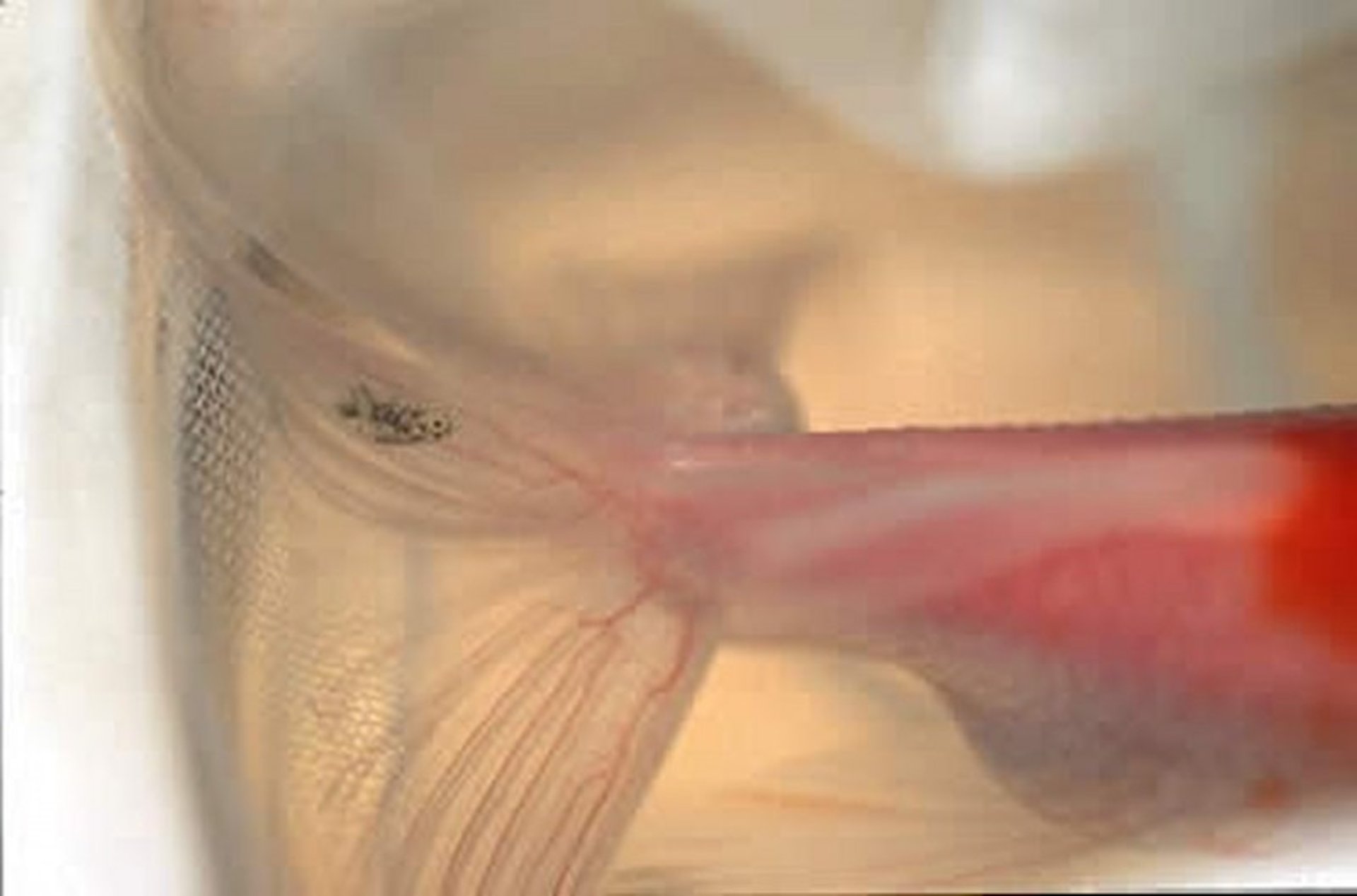
Ichthyobodo spp are some of the most common and smallest (~15 × 5 mcm) flagellated protistan parasites of the skin and gills. A kinetoplastid protist, they are flattened, pear-shaped organisms with two flagella of unequal lengths. These parasites can be found on freshwater or marine fish from a broad geographic range. Ichthyobodo moves in a jerky, spiral pattern, and free-swimming organisms are fairly easy to identify in direct smear preparations. Once attached, the organism can be difficult to see, but movement typical of a flickering flame may be seen under 400× magnification and is characteristic. Affected skin often has a steel-gray discoloration due to copious mucus production (blue slime disease), and gills may appear swollen. Behavioral signs of infestation include lethargy, anorexia, piping, and flashing. Ichthyobodo is readily controlled with formalin, copper sulfate, or potassium permanganate baths. Because the parasite has a direct life cycle, a single treatment should be adequate. If reinfestation occurs, sanitation and quarantine practices should be evaluated.

Courtesy of Dr. Ruth Francis-Floyd.
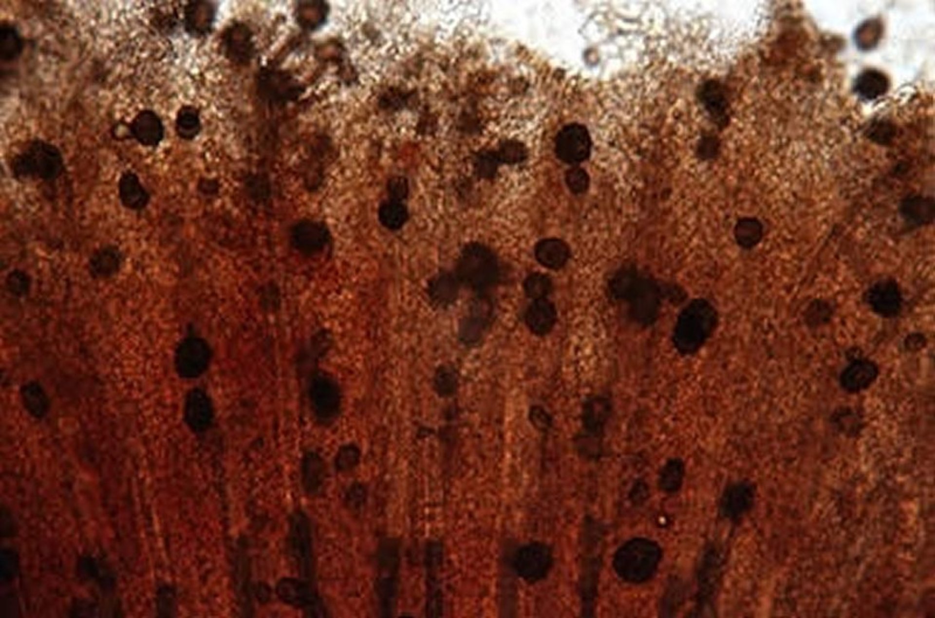
One of the most serious health problems of captive marine fish is the parasitic dinoflagellate Amyloodinium spp. Its freshwater counterpart, Piscinoodinium spp, is frequently seen on zebrafish and some barbs, but can occur on any freshwater fish. Like Amyloodinium it can also result in high mortality. These parasites produce a disease that has been called velvet, rust, gold-dust, and coral disease because of the brownish gold color they impart to infected fish. The pathogenic stages of the organism are pigmented, photosynthetic, nonflagellated, nonmotile algae that attach to and invade the skin and gills during their parasitic existence. When mature, these parasites give rise to cysts that contain numerous flagellated, small, free-swimming stages that can initiate new infections. Control of Amyloodinium is challenging, and the prognosis is guarded. Copper sulfate is the only therapeutic option for food animals in the US, and repeated treatments are necessary to break the life cycle. The disease is particularly problematic in clownfish. The treatments that are most effective are chloroquine, delivered at 10 mg/L as an indefinite bath, and copper sulfate.
Oomycota Parasites of Aquarium Fish
Although the oomycetes (water molds) share some morphological traits with the true fungi, they are more closely related to diatoms, opalinids, and labyrinthulomycetes. Of the many genera of water molds, Saprolegnia, Branchiomyces, and Aphanomyces are the most frequently associated with disease in freshwater fish. These primarily saprophytic organisms are common to freshwater fish around the world. Saprolegnia commonly infects fish eggs and traumatized external tissues of live fish. Gross signs are grayish white, cotton-like growths on the skin, gills, eyes, or fins that may rarely invade deeper tissues of the body. Fish exposed to water temperatures below their optimal range are especially susceptible to Saprolegnia infection.
Microscopically, saprolegniasis can be recognized by making direct smears from the infected tissues and observing the large nonseptate filaments. The sexual stages of the organism can be seen only in cultures of the organism and are required for specific identification. Low nutrient water agar is acceptable for primary isolation of oomycetes, including the genus Saprolegnia. Preventive measures include removal of predisposing causes (eg, improper temperature, inadequate sanitation, excessive chemical treatment, or the presence of dead, infected fish and decaying organic material). If the environment is clean, the temperature is appropriate for the species, and skin pathogens have been eliminated, a single treatment with potassium permanganate, formalin, or hydrogen peroxide is often adequate to control external saprolegniasis. There are some effective over-the-counter products sold through the pet trade that contain malachite green. These dilute solutions should be effective in home aquaria but should not be dispensed by veterinarians and should never be used on food animals. Use in zoological collections is also discouraged.
Branchiomyces is an oomycete that infects gill tissues, and invades the branchial blood vessels. It can cause high mortality. Fish held in water that has high organic content appear to be more susceptible. The multi-branching, nonseptate filaments can be easily seen on a gill biopsy of an infected fish. Copper sulfate, formalin, and potassium permanganate are reported to successfully clear fish of infection.
Epizootic ulcerative syndrome (EUS), caused by Aphanomyces invadans, is a reportable disease and endemic to much of the US and found in low salinity waters around the world. Fish infected with this oomycete usually exhibit ulcerative skin disease. It typically causes an extensive granulomatous response as the filaments invade the skin, skeletal muscles, and adjacent organs. This organism should be suspected when granulomas surrounding large filaments are seen on a scraping of an ulcer. There is no treatment, and fish with clinical signs of disease that is confirmed by histopathologic findings should be euthanized.
Internal Protistan Parasites
Flagellate Parasites of Aquarium Fish

Courtesy of Dr. Sarah Poynton.
Courtesy of Dr. Ruth Francis-Floyd.
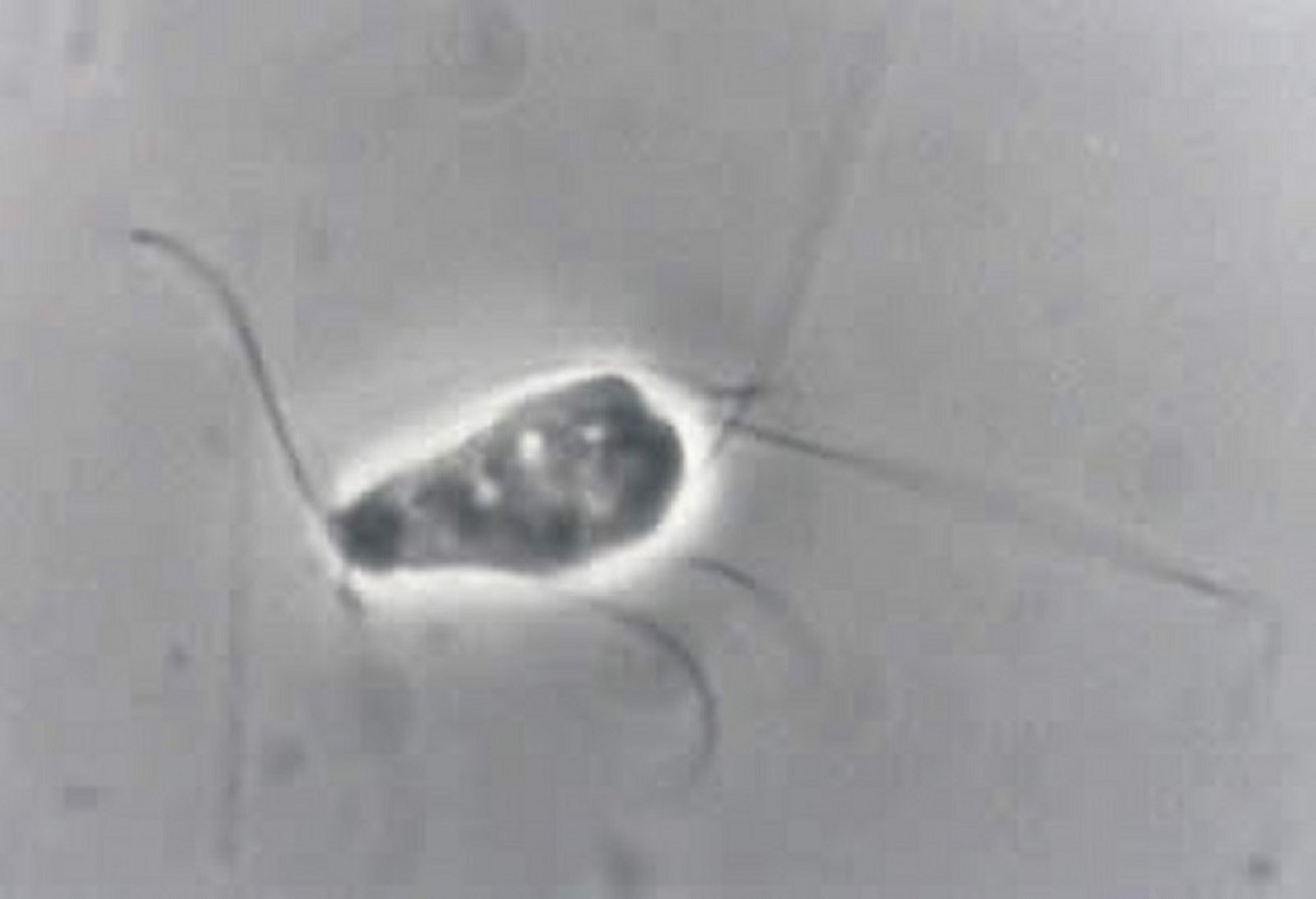
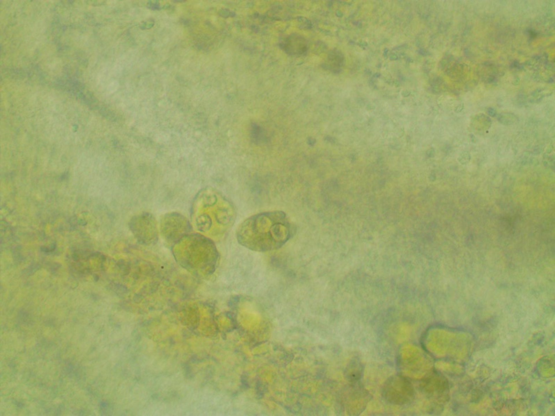
Spironucleus spp are common, small (~9 mcm), bilaterally symmetric, flagellated (four pairs) diplomonad protists most frequently found in the intestinal tract of finfish. Among ornamental fish, the cichlids are highly susceptible. However, they are also frequently seen in the intestinal tract of gourami and some catfish. Pathogenicity of these organisms is variable and correlated with the number present. If there is a loss of condition, or >15 organisms are seen per low-power field on wet mounts of intestinal tissue or contents, then treatment is strongly recommended. The only treatment available for spironucleosis is metronidazole (use only in ornamental species), which should be given orally but can be administered as a bath if fish are anorectic. Chronic infections have been seen in fish maintained in unsanitary or crowded conditions.
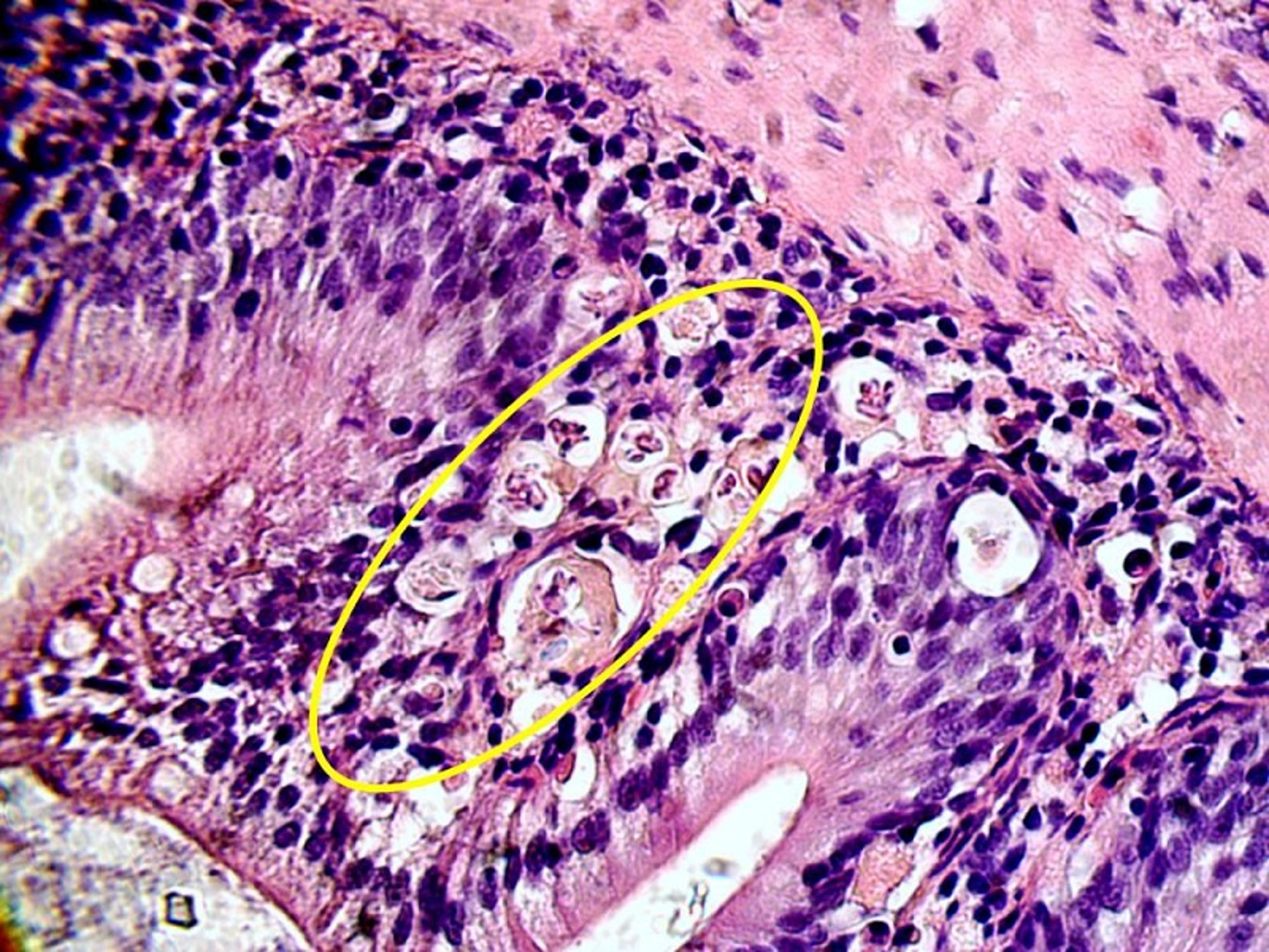
Cryptobia and Trypanosoma spp are slender, elongated (6–20 mcm), actively motile, biflagellated kinetoplastid protistans easily detected in fresh blood and tissue smears of both marine and freshwater finfish. Hematozoic forms are generally described as Trypanosoma and have a well-developed undulating membrane. Trypanosomes can be transmitted by leeches and have been associated with anemia in blue-eyed plecostomus (Panaque suttoni) and other wild-caught loricariids. Cryptobia iubilans has been associated with granulomatous disease in many cichlids including African cichlids, angels, and discus. Clinical disease manifests as severe weight loss and cachexia. Clinically affected fish should be culled. Presumptive diagnosis can be made from microscopic examination of fresh tissue. Typically, granulomas will be found in the stomach, which may be visibly thickened. Fish with severe granulomatous gastritis often have granulomatous disease associated with invasion of adjacent organs. Spleen and anterior kidney are commonly affected, although other organs including cranial nerve may also be damaged. Acid-fast positive material will not be found in granulomas caused by Cryptobia iubilans. Motile flagellates may be visible using magnification of 200× or greater.
Sporozoan Parasites of Aquarium Fish

Courtesy of Dr. Denise Petty.
Courtesy of Dr. Denise Petty.
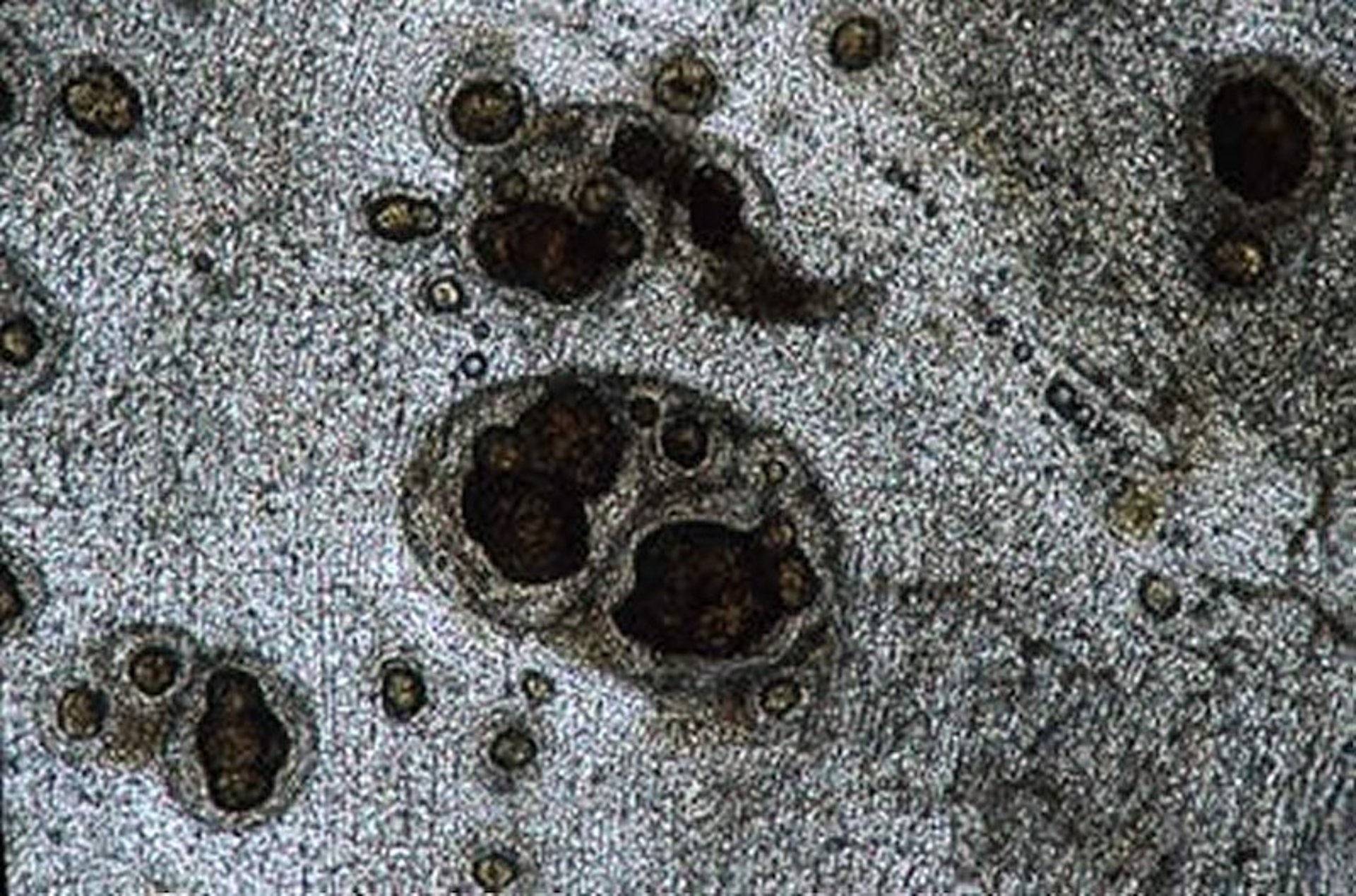
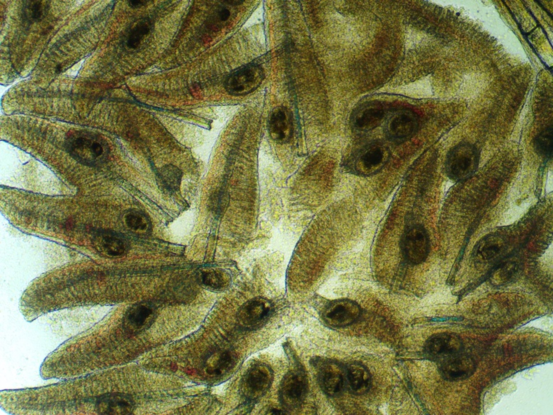
Apicomplexans, although common in freshwater or marine finfish, are more commonly diagnosed on histologic evaluation but can be seen when performing a careful examination of an intestine free of feed. Many species of finfish are affected. Species of the genera Cryptosporidium, Epieimeria, and Goussia have been frequently reported in both freshwater and marine fish. The life cycles of many fish apicomplexans are unknown, and some involve more than one host to complete their development. In addition to intestinal infection, the internal organs may be affected; sporulated Epieimeria-like oocysts and sexual and asexual stages can be found in direct smears and histologic sections of the internal organs.
A number of Eimeria species are found in skates and rays. Of these, Eimeria southwelli has caused high mortality in cownose rays (Rhinoptera bonasus), although it has been reported in other species of rays. Infected cownose rays often present with pale skin and loss of weight despite normal appetite. Fluid obtained by aspiration of the coelomic cavity should be examined for presence of coccidia. Treatment with toltrazuril (10 mg/kg/day, PO, for 5 days) may help control but not eliminate infection. This parasite is highly prevalent in wild-caught cownose rays.
Heavy intestinal infection of Goussia spp has been associated with high mortality in comet goldfish (Carassius auratus). Affected fish are lethargic, frequently lay on their side, and have pale feces. Clumps of cells in the intestine can be seen on a wet mount, but histologic evaluation is often required for diagnosis.
Sulfamethazine (22–24 g/100 kg of fish wt/day in the feed for 50 days at 10°C [50°F]), is used to treat food fish (21-day withdrawal time) in some countries. An FDA-approved form of this drug is not currently available in the US. For aquarium fish, sulfamethazine in the aquarium water (10 mcg/L once a week for 2–3 weeks) has been reported to be preventive, but safety and efficacy data are sparse.
Metazoan Parasites
Common Metazoan Parasites of Fish
Parasites | Tissue | Susceptible Species | Signs | Diagnosis | Treatment | |
|---|---|---|---|---|---|---|
Myxozoans (intermediate host) | Myxosoma cerebralis (whirling disease) | Head, cartilage, backbone | Rainbow trout, salmonids | Blacktail, skeletal deformity | Histologic evaluation, isolation of parasite | None; regulatory concern |
Ceratomyxa shasta | Posterior intestine | Salmonids (Pacific Northwest) | Weight loss, distended and hemorrhagic vent | Wet mount, histologic evaluation | None; regulatory concern | |
Aurantiactinomyxon ictaluri (proliferative gill disease, hamburger gill disease) | Gill | Channel catfish | Mortality, piping | Wet mount, histologic evaluation | None | |
Henneguya | Gill | Channel catfish, other | None, severe hypoxia | Wet mount | None | |
Sphaerospora auratus (renal dropsy) | Kidney | Goldfish (pond-reared) | Mortality, severely enlarged and cystic kidney | Histologic evaluation | None | |
Tetracapsuloides bryosalmonae (proliferative kidney disease) | Kidney | Rainbow trout, all salmonids | Lethargy, darkening, fluid accumulation, exophthalmia | Wet mount, histologic evaluation | None | |
Monogeneans | Gyrodactylus (live bearer, except in Loricaridae) | Skin, fins | Goldfish, koi (predisposed) | Excess mucus, flashing, weight loss | Wet mount | Praziquantel in nonfood fish, formalin |
Family: Ancyrocephalidae Family: Dactylogyridae (egg layers) | Gills | Goldfish, koi, angelfish, discus, channel catfish | Piping, coughing, weight loss, skin lesions | Wet mount | Praziquantel in nonfood fish, formalin | |
Capsalids; Benedenia, Neobenedenia (egg layer) | Epithelium of skin and gills | Marine tropicals, other marine species, marine angelfish | Epithelial lesions, flashing, weight loss, mortality | Visual exam, wet mount | Praziquantel in nonfood fish, formalin | |
Polyopisthocotylea | Gill | Striped bass, hybrid striped bass, Florida pompano | Pale gills, piping, mortality | Visual exam, wet mount | Praziquantel in nonfood fish, formalin | |
Trematodes (intermediate host usually mollusc) | Heterophyidae | Gill | Redtail shark, black shark, rainbow shark, angelfish (freshwater), other pond-reared fish, aquarium fish | Flared gills, hypoxia, piping, do not tolerate shipping and handling | Wet mount | |
Clinostomum | Skeletal muscle | Largemouth bass, centrarchids, hybrid striped bass | Yellow grub | Direct visualization, wet mount | ||
Bolbophorus confusus | Skeletal muscle, viscera | Channel catfish | None | Direct visualization, wet mount | ||
Posthodiplostomum | Viscera, heart, posterior kidney | Largemouth bass, bluegill, centrachids, salmonids, other fish | None | Direct visualization, wet mount | None | |
Diplostomum | Eye (lens) | Fish are intermediate host (unspecified species) | Cataracts, blindness | Direct visualization, histologic evaluation | None | |
Cestodes | Diphyllobothrium latum | Viscera, musculature | Salmonids, other freshwater species | Adhesions, sterility (if gonads affected) | Direct visualization, wet mount | Praziquantel in nonfood fish |
Corallobothrium | Intestine (adult) | Channel catfish | None | Direct visualization, wet mount | Praziquantel in nonfood fish | |
Proteocephalus ambloplitis | Ovary (larval stage) | Largemouth bass | Sterility | Direct visualization, wet mount | Praziquantel in nonfood fish | |
Bothriocephalus acheilognathi | Intestine (adult) | Carp, aquarium fish | Weight loss, enteritis, mortality | Direct visualization | Praziquantel in nonfood fish | |
Acanthocephalids | Acanthocephalus | Intestine | Wild-caught freshwater fish, wild-caught marine fish | Enteritis, mortality | Direct visualization | |
Nematodes (fish as direct host) | Capillaria and Pseudocapillaria | Intestine | Angelfish, discus, zebrafish, tetras, barbs, and other aquarium fish | Weight loss, pot belly | Direct visualization, wet mount | Fenbendazole (25 mg/kg, PO, once a day for 3 days), levamisole, ivermectin (0.05 mg/kg, PO), emamectin (0.35 mg/kg, PO, once a day for 14 days) |
Camallanus | Posterior intestine | Largemouth bass, other centrarchids | Visualize worms protruding from anus | Direct visualization, wet mount | Fenbendazole, levamisole | |
Philometra | Posterior intestine | Aquarium fish | Visualize worms protruding from anus | Direct visualization | Fenbendazole, levamisole | |
Nematodes (fish as intermediate host) | Eustrongylides | Encysted in coelom | Angelfish, other aquarium species | Weight loss, pot belly | Direct visualization | |
Contracaecum | Encysted in viscera | Largemouth bass, centrarchids | Often none | Direct visualization | ||
Leeches | Leech | Skin | Freshwater game fish, aquarium fish | Anemia, weight loss | Direct visualization | None |
Metazoan parasites include the myxozoans, helminths, and crustaceans, and are common in both wild and cultured fish ( see Table: Common Metazoan Parasites of Fish). Fish frequently serve as intermediate or transport hosts for larval parasites of many animals, including humans. Helminths with direct life cycles are most important in dense populations, and heavy parasite burdens are sometimes found. In general, heavy parasite burdens seem to be more common in fish originating from wild sources.
Myxozoan Parasites of Aquarium Fish
Although originally thought to be single-celled organisms, myxozoans are multicellular and closely related to Cnidaria. They are common fish parasites and have life cycles that use invertebrates as definitive hosts and fish for multiplication. Hence, myxozoan infections are often more common in wild fish or fish reared intensively in outdoor fish ponds. The organisms tend to be host- and tissue-specific. Accordingly, expression of the disease is related to the specific pathogen and host and location within the host. For example, coelozoic myxozoans that reside in cavities typically cause little disease, unlike the histozoic forms that reside in tissues. Myxozoan-infected fish in captive display aquaria are not able to transmit the infection unless the necessary intermediate hosts are present.
Myxozoans are divided into two groups, Myxosporea and Malacosporea, and members of both groups are infective to fish. Although a few cases of direct life cycle have been reported, myxosporeans usually have an indirect life cycle, with oligochaete or polychaete worms used as a definitive host. In contrast, bryozoans are used as a definitive host by the malacosporeans.
There are two important myxosporean infections of ornamental fish. Renal dropsy of goldfish is caused by the myxosporean Sphaerospora auratus. The disease is characterized by renal degeneration and ascites and is usually diagnosed by identification of spores in histologic sections of the kidney. Affected fish develop extreme abdominal distention but may have few other clinical signs. Radiographs may reveal a mass in the area of the posterior kidney; definitive diagnosis is made at necropsy and confirmed histologically. No practical treatment is available. Henneguya, a myxosporean occasionally found in ornamental fish, causes white nodular lesions that are usually found in gill tissue and may be grossly visible. Henneguya is easily identified by the forked-tail appendage of the spore, seen microscopically. If ponds are dried and limed heavily, infection can be eliminated, apparently by reduction of the intermediate hosts. Aquarium infection can be self-limiting in the absence of intermediate hosts. Although an occasional cyst may be considered an incidental finding, severe damage has been associated with diffuse distribution of interlamellar cysts.
Myxosporean diseases important in aquaculture include whirling disease and proliferative kidney disease of salmonids and proliferative gill disease (hamburger gill disease) of channel catfish. Whirling disease is caused by Myxobolus cerebralis. Fish are infected as fingerlings when the parasite infects cartilage in the vertebral column and skull, resulting in visible skeletal deformities. Affected fingerlings typically show rapid tail-chasing behavior (whirling) when startled. The disease is also sometimes called blacktail, because the peduncle and tail may darken significantly. Recovered fish remain carriers. Adults do not show behavioral signs, but skeletal deformities associated with infection do not resolve. Whirling disease can be prevented by purchasing uninfected breeding stock and maintaining them in an environment free of the definitive hosts (tubeficid worms). A presumptive diagnosis of whirling disease is made by detection of spores from skulls of infected fish. Diagnosis may be confirmed histologically or serologically. Whirling disease is of regulatory concern in some states.
Monogenean Parasites of Aquarium Fish
The flatworms Monogenea have direct life cycles and are common, highly pathogenic, and obligate parasites most commonly seen on skin and gills. The preferential location of some species is in internal organs such as the esophagus, stomach, posterior kidney, or urinary bladder. Freshwater parasites tend to be ~0.1–0.8 mm long and are best seen microscopically; however, several important species that parasitize marine fish are significantly larger and may be visible grossly. Monogeneans can be identified by their characteristic hold-fast organ, the haptor, which is armed with large and small hooks. Aquarium and cultured fish are subject to a rapid buildup of parasites by continual infection and worm transfer to other fish in the tank or pond. Although many species are host specific, the more common types seen in aquaria are less selective.
The two most common monogeneans in freshwater aquaria are gyrodactylids and ancyrocephalids. Gyrodactylus, a common parasite of many ornamental fish, primarily gives birth to live young and is usually found on skin and eyes; the ancyrocephalids lay eggs and parasitize the gills. Dactylogyrids are egg-laying gill monogeneans commonly seen in cyprinid fish, especially goldfish and koi. High numbers of any of these monogeneans can result in catastrophic mortality of infested fish.
The capsalids are a large group of monogeneans that affect brackish and marine fish and include the genera Neobenedenia and Benedenia, which are important monogeneans in marine fish. They attach and graze on skin including the eye and gill. The capsalids lay sticky eggs easily transmitted via fomites.
Monogenean-infected fish may show behavioral signs of irritation, including flashing and rubbing the sides of their bodies against objects in the aquarium. Fish become pale as colors fade. They breathe rapidly and distend their gill covers, exposing swollen, pale gills. Localized skin lesions appear with scattered hemorrhages and ulcerations. Ulceration of the cornea may become evident if the eyes are involved. Mortality may be high or chronic.
Praziquantel (5 mg/L, prolonged bath) is the treatment of choice for monogenean infection in freshwater and marine ornamental fish. Formalin is the only treatment option for food fish. When using formalin, multiple treatments at weekly intervals are recommended for egg-laying monogeneans because eggs are resistant to chemical treatment. Organophosphates (0.25 mg/L, prolonged bath) have been used successfully in ornamental fish in the past, but treatment with praziquantel is considered more effective. Organophosphates should be avoided in systems containing elasmobranchs and some characins and cichlids. Many monogeneans on marine fish can be partially removed using freshwater dips for 1–5 minutes, depending on the tolerance of the species; however, eggs will not be damaged or removed. To prevent the disease, introduction of infected fish should be avoided.
Trematode Parasites of Aquarium Fish
Courtesy of Dr. Denise Petty.
Trematodes belong to the phylum Platyhelminthes, and this class contains two subclasses, Aspidogastrea and Digenea, both of which contain members that infect fish. Also known as flukes, this group of flatworms has a complex life cycle that includes a variety of host animals, typically beginning with a mollusc. Fish can be a second intermediate or final host, depending on the requirements of the trematode species. Although adults may be found in the intestine of infected fish, it is more common to see the metacercarial stage in fish. Previously this stage was considered to cause little harm to the host, but their invasion into various organs of the fish can result in major damage and even mortality. For example, large numbers of metacercariae encysted in gill tissue can be detrimental to normal function.
Cestode Parasites of Aquarium Fish

Another group of flatworms, the cestodes, are common in fish, which can serve as intermediate, paratenic, and definitive hosts. Fish become parasitized in several ways; when a fish eats an invertebrate that contains a cestode larva, the larva (procercoid) will migrate to a location (usually within the coelom, not inside the intestine) where it develops into a plerocercoid. The migration and encystment of numerous plerocercoids can result in serious harm; however, much depends on the location of the plerocercoids within the fish. If a fish ingests another fish that contains plerocercoids, these will become a mature tapeworm in the intestine. Many of these fish may not show clinical signs of disease, but heavy cestode infestations can inhibit growth, and cause coelomic distension and intestinal impactions.
The anthelmintic of choice is praziquantel (35–125 mg/kg, in feed, every 24 hours for three days). There is no FDA-approved treatment for food fish.
Nematode Parasites of Aquarium Fish
Nematodes are common in wild fish exposed to the intermediate hosts. Fish may be definitive hosts for adult nematodes, or they may act as transport or intermediate hosts for larval nematode forms (anisakids, eustrongylids, and others) that infect higher vertebrate predators, including humans. Encysted or free nematodes can be found in almost any tissue or body cavity of fish. Aquarium and cultured pond fish may be heavily infected if crustacean intermediate hosts are present.
Cyclops and Daphnia spp are common intermediate hosts for Camallanus spp., a nematode that is pathogenic for guppies and other aquarium fish. These blood-red worms can be seen in the intestine and protruding from the anus of affected fish (red worm disease). Capillaria spp are commonly found in aquarium fish, particularly freshwater angelfish, discus, and other cichlids. Heavy infections in juvenile angelfish have been associated with poor growth rates and an inability to withstand shipping and handling. Another capillariid nematode, Pseudocapillaria tometosa, causes severe disease in zebrafish and other freshwater aquarium fish.
Leeches as Parasites of Aquarium Fish
Leeches are parasitic bloodsuckers of fish and also serve as vectors for blood parasites of fish (eg, Trypanosoma, Cryptobia, and haemogregarines). They can produce debilitating anemia due to chronic blood loss and disease. Leech infestations are most common in wild fish, but aquarium and pond infestations can occur by introduction of infested fish and plants. Heavily infested fish have been observed to swim primarily at the water surface, often with their dorsum exposed. Fish exhibiting this behavior also suffer from ulcerative lesions of the exposed skin. Organophosphates (0.25 mg/L, prolonged bath) are somewhat effective but not approved for use in food fish. Further, environmental regulations may restrict use in outdoor ponds. Multiple treatments may be required to control leeches, because eggs are resilient and juveniles may continue to hatch. Preventive measures include avoiding leeches (ie, effective quarantine). Infestations in recreational fishing ponds are often self-limiting.
Crustacean Parasites of Aquarium Fish
Courtesy of Dr. Ruth Francis-Floyd.
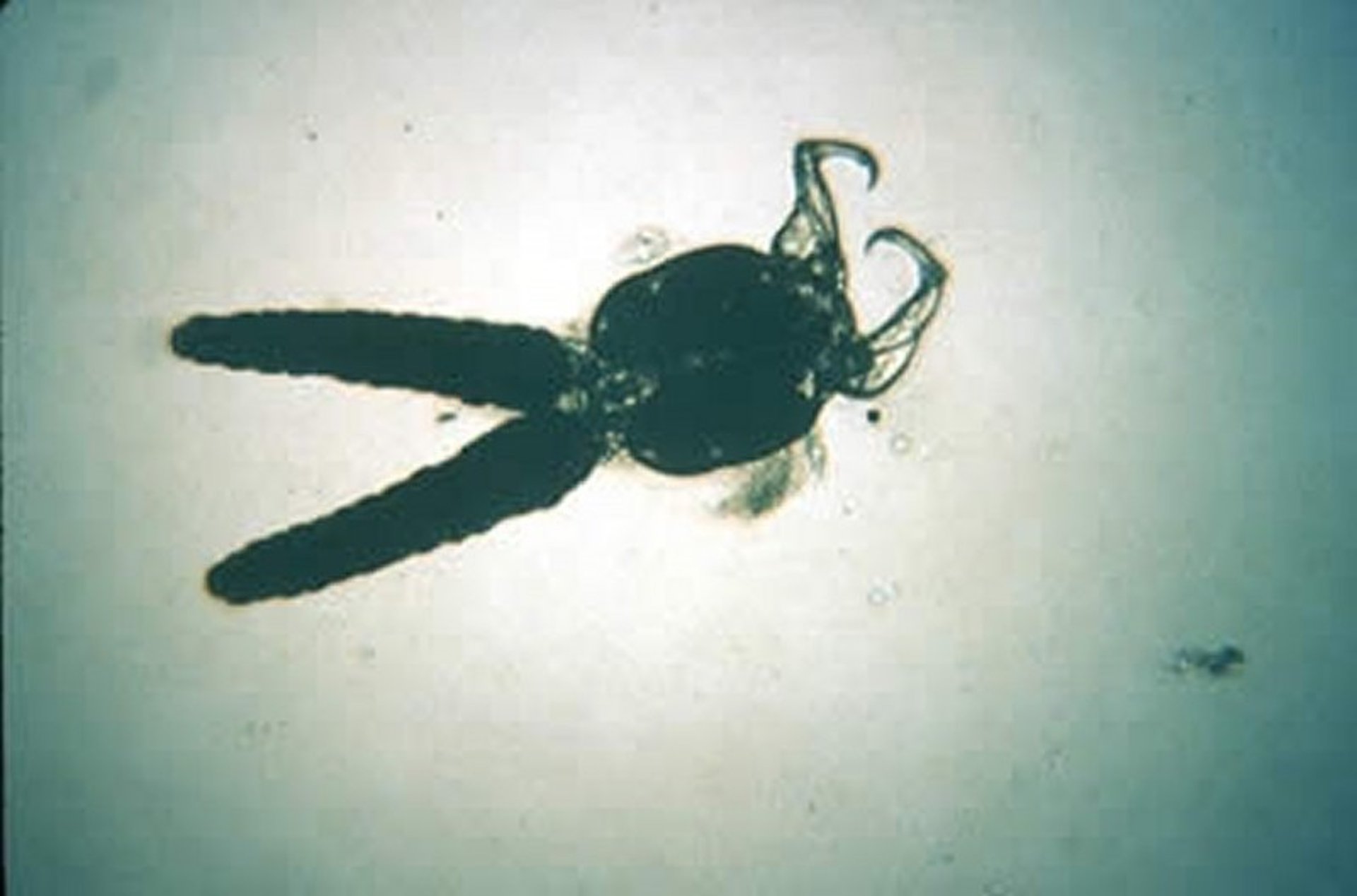
Parasitic crustaceans include copepods and branchiurans. Some copepods, such as the anchor worm, are obligatory parasites of finfish during specific stages of their complicated life cycle. They lose their copepod form, including their appendages, and become rod- or sac-like structures specifically adapted for piercing, holding, feeding, and reproducing. Grossly, they appear as barb-like attachments to the skin or gills, where they feed on blood and tissue fluids. They can cause hemorrhage, anemia, and tissue destruction, as well as provide a portal of entry for other pathogens.
Many different species of these parasites can be found on freshwater and marine fish. The anchor worms, Lernaea spp, are commonly found in a wide variety of aquarium- and pond-reared fish, including goldfish and other cyprinids. Ergasilus spp infest the gills.
Courtesy of Dr. Louise Bauck.

Courtesy of Dr. Louise Bauck.
The branchiurans, often called lice, have flattened bodies adapted for rapid movement over the skin surface. By means of hooks and suckers, they periodically attach for feeding by inserting the piercing mouth part (stylet) into the skin. Sea lice (Lepeophtheirus salmonis) are a major disease problem of pen-reared salmonids. Consultation with a salmonid health specialist is suggested because treatment options are limited and environmental concerns are important. Argulus spp are commonly found on aquarium, pond-reared, and wild freshwater fish.
Diflubenzuron (0.03 mg/L, once) is the most effective treatment for crustacean parasites. It is a chitin synthesis inhibitor and has a long half-life. Because of this and the toxicity of diflubenzuron to all crustaceans, treated water must be retained for 28 days before release.
Organophosphates are somewhat effective in controlling crustacean parasites, but legal restrictions constrain clinical use. Some success has been achieved in treating freshwater fish for parasitic copepods by giving infected fish a 3% (30 g/L) salt dip (<10 minutes, remove fish when it rolls) followed by salt (5 g/L) added to the affected tank for 3 weeks. The increased salinity kills immature forms as they hatch. Diflubenzuron and organophosphates are not approved for use in food fish.
For More Information
Also see pet health content on routine health care for fish and emergencies of fish.


