

Descriptions of viral diseases of fish are rapidly expanding. Viruses are being reported in new species, and interpretation of the significance of findings is also changing. Several viral diseases of ornamental fish are reportable ( see Table: Fish Diseases of Regulatory Concern in the US).
Although viruses of homeothermic animals are cultured at uniform temperatures, fish viruses have wider, but specific, temperature tolerances in fish cell cultures at lower temperatures. Because of this relatively defined temperature range, variation in temperature may enable control, although often it merely induces latency. Because many viral diseases of fish are geographically limited, regulatory agencies and fish farms in disease-free areas consider them exotic diseases and require certification of introduced stocks. Many result in high mortality in young fish and little or no losses in adults, which may become carriers. For these reasons, avoidance of carriers and certification of specific pathogen-free replacement stocks are frequently required.
Specific testing procedures are available. Most vaccines used for control of fish diseases are for bacterial agents; however, use of vaccines to control some viral diseases is being introduced. Drugs are not effective; however, antimicrobials and other drugs may be used to control secondary bacterial infections. Management techniques that minimize stress and crowding, biosecurity measures, and temperature manipulation hold the greatest promise for control of piscine viral diseases.
Herpesviruses (Alloherpesviruses)
Carp Pox
One of the oldest recognized fish diseases, carp pox is caused by Cyprinid herpesvirus-1 (CyHV-1). Common carp and koi Cyprinus carpio is the only susceptible species, and inbred strains appear to be more susceptible to infection. Lesions typically are smooth and raised and may have a milky appearance. They are benign, non-necrotizing areas of epidermal hyperplasia. Severe cases may result in development of papillomatous growths, which may be a site of complicating bacterial infection. Generally, lesions are self-limiting and of minimal clinical significance. Carp pox can be a notable problem in koi because their aesthetic quality, and hence market value, is severely compromised. For the serious koi enthusiast, fish affected with carp pox should be culled during quarantine. If infected fish are introduced to a resident population all susceptible fish, whether exhibiting clinical signs of infection or not, are exposed. Surgical removal of the lesions has not been rewarding.
Koi Herpesvirus
Koi herpesvirus (KHV), caused by Cyprinid herpesvirus-3 (CyHV-3), was first recognized in 1996. It is widespread in the US and considered endemic. Confirmed cases must be reported to the State Veterinarian and the USDA Area Veterinarian in Charge. Because the disease is endemic, regulating bodies report its occurrence to the World Organisation for Animal Health (OIE), but they do not require specific action, such as depopulation, when the disease is reported.
Koi herpesvirus causes clinical disease in koi and common carp Cyprinus carpio. Goldfish (Carassius auratus) and grass carp (Ctenopharyngodon idella) are refractory to clinical disease but may serve as carriers. Koi that are exposed, but survive, may also serve as carriers. Clinical disease is seen at water temperatures of 22°–27°C (72°–81°F), with maximal mortality at temperatures of 22°–25.5°C (72°–78°F). Mortality rates can reach 80%–100%. Fish of any age are susceptible, but mortality rates may be higher in younger fish, especially fry. The most obvious lesions are seen on gill tissues, which are severely affected and develop a mottled red and white appearance with obvious hemorrhage in some cases. Affected fish are lethargic, swim at the surface, and may show behavioral signs of respiratory distress. Severe bacterial or parasitic disease may mask the fact that KHV is the primary cause of gill lesions. The disease is transmitted horizontally by exposure to sick or carrier fish and also by exposure to contaminated water, substrate, or equipment.
When KHV is suspected, affected fish can be shipped to a laboratory for confirmation. Freshly dead specimens shipped on ice and received within 24 hours should be adequate. To confirm the infection in dead animals, PCR assay or virus isolation and identification can be used. Because of the value of koi, there is substantial demand for nonlethal testing protocols. Blood, gill tissue from biopsy, feces, or mucus may be used to assess the status of moribund fish. These tests can result in false-negative results if performed on clinically normal fish.
Indirect tests using blood samples, including ELISA or virus neutralization, are less straightforward. Negative test results could indicate a true negative result or, alternatively, that the infection is in its early stages and the fish has not yet developed a measurable antibody response. A negative test result could potentially be obtained from a previously infected fish that has had a decrease in circulating antibody concentration; however, such a fish could still function as a carrier. There is a misconception among some hobbyists that a negative blood test result is an adequate screening test to exclude carrier status. Practitioners should communicate this clearly to owners because it is easily misunderstood.
If KHV infection is confirmed in a population of koi, depopulation is strongly recommended. Surviving fish are carriers and can serve as a source of infection for naive fish. Although mortality decreases or stops as water temperature approaches 30°C (86°F), survivors will retain carrier status and therefore put other populations at risk.
Prevention of KHV is best accomplished with careful quarantine protocols. A minimum quarantine of 30 days at 24°C (75°F) is recommended to minimize the chance of introducing KHV to an established population of koi. If disease develops during the quarantine period, fish should be evaluated with special consideration for excluding KHV. After quarantine, a new fish may be placed in an isolated area with a few fish from the established population and monitored for signs of disease for at least 2 weeks as an added precaution. If koi are taken to shows, quarantine protocols should be followed every time they return. Koi enthusiasts should be strongly encouraged to attend English-style shows (vs Japanese-style shows), where competing fish are not placed in a common container.
Herpesviral Hematopoietic Necrosis of Fish

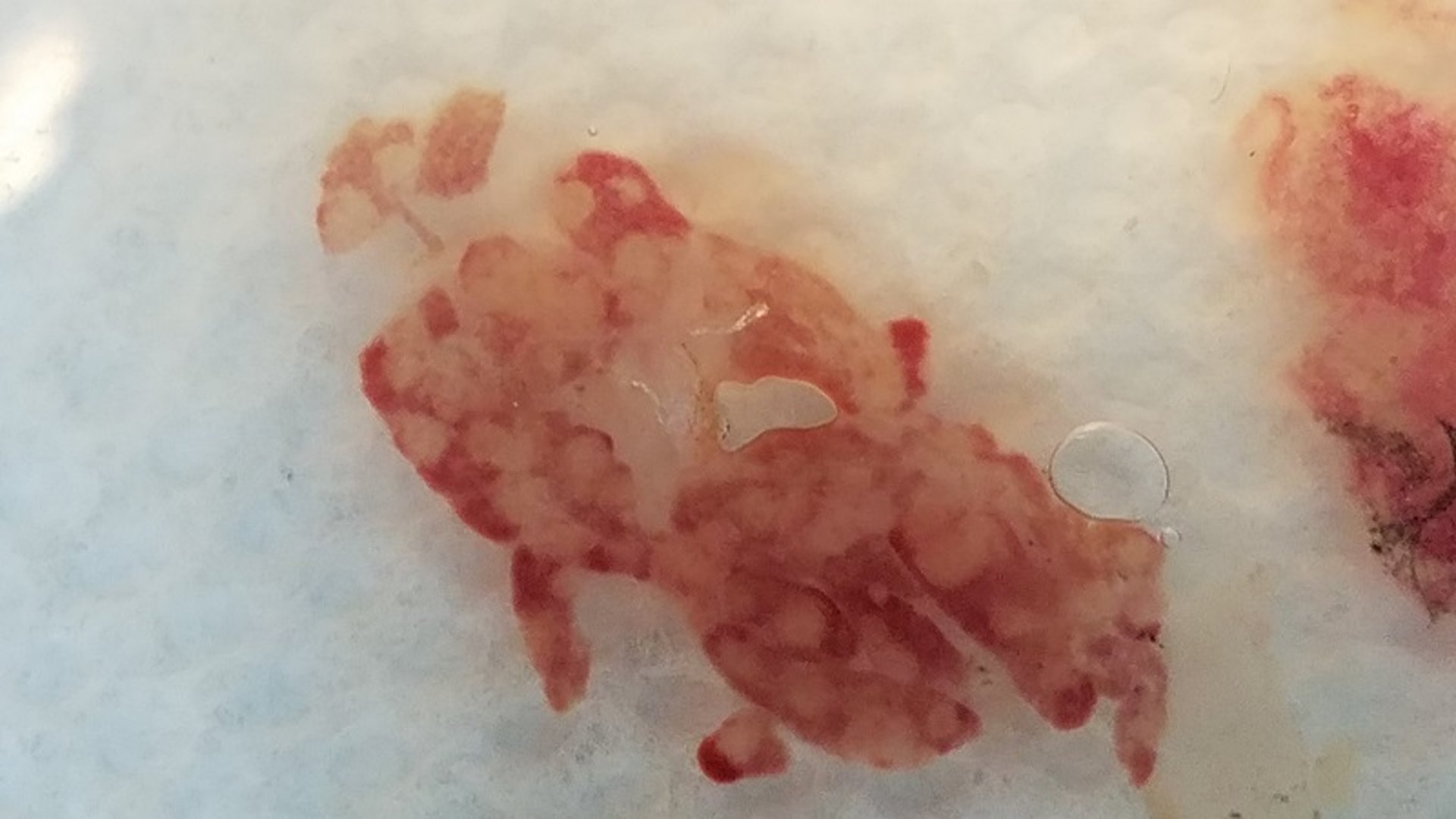
Courtesy of Dr. Denise Petty.
This condition of goldfish is caused by Cyprinid herpesvirus-2 (CyHV-2). It is probably widespread throughout the US, but it is not a reportable disease. Clinically ill goldfish often are anorectic and have pale gills and ascites. At necropsy, the spleen and kidneys (anterior and posterior) are often enlarged and may have grossly visible pale areas. Sections of these organs should be preserved in neutral-buffered 10% formalin for histologic evaluation and frozen for virologic testing. Survivors can be carriers and exhibit clinical signs of disease if subjected to stressors such as transport. Water temperatures between 10° and 22°C will result in replication of the virus that can be detected with quantitative PCR assay. There is no treatment, though elevation of temperature to 28°C (80°F) will result in resolution of clinical signs. However, the fish will continue to be a carrier of this virus.
Herpesvirus of Angelfish
A herpesvirus of angelfish (Pterophyllum spp) has been detected by electron microscopy of skin from moribund angelfish. Affected fish produce copious amounts of skin mucus that gives affected fish a gray sheen. Often, these fish have multiple parasitic infestations and bacterial infections, similar to that of koi herpesvirus in koi. It is suspected that survivors are carriers.
Rhabdoviruses
Viral Hemorrhagic Septicemia of Fish
This reportable disease is caused by a Novirhabdovirus and is a member of the family Rhabdoviridae. This disease is listed by OIE as notifiable. Most of the reported hosts are not ornamental fish, but koi were shown to be susceptible experimentally to genotype IVb. This genotype affects a diverse group of fish in the Great Lakes region of North America.
Spring Viremia of Carp
This acute, virulent, usually hemorrhagic disease of cultured carp is caused by a Vesiculovirus that, like VHS, is a member of the Rhabdoviridae family. The disease is listed as notifiable by the OIE. Historically, it was reported in Europe and the former USSR; however, several outbreaks have been reported in the US between 2002–2007, in both wild fish and cultured ornamental koi. Spring viremia of carp (SVC) is considered a foreign animal disease in the US and must be reported. It causes disease in common carp, including koi, as well as grass, bighead, silver, and crucian carp. Limited experience suggests that common goldfish may be susceptible.
Clinical signs are nonspecific and may include darkening of the skin, exophthalmia, ascites, pale gills, hemorrhage, and a protruding vent with thick mucoid fecal casts. Pinpoint hemorrhage in the swim bladder is indicative of SVC, if present. Coinfection with Aeromonas or other systemic bacteria may obscure the presence of the virus. The bacterial component of the infection can be controlled with antimicrobials; however, depopulation of affected or exposed fish is required in the US. The disease causes death in both adult and young fish. Clinical disease occurs at cool temperatures (12°–22°C [54°–72°F]), an important distinction from koi herpesvirus. The virus is readily isolated in common fish cell lines and identified by serum neutralization and fluorescent antibody tests.
Iridoviruses
Lymphocystis Disease of Fish
This typically chronic, viral infection of wild or captive marine and freshwater fish is caused by an icosahedral DNA virus of the Iridoviridae family. Infection may be manifest by benign, cauliflower-like lesions typically located on fins. The disease affects a wide range of fish and is generally considered to have a global distribution. Within the aquarium trade, painted glass fish and marine tropical fish such as the anemonefish (Pomacentridae), marine angels (Pomacanthidae), and butterflyfish (Chaetodontidae) are susceptible. Presumptive diagnosis is based on the presence of enlarged fibroblasts (up to 1 mm), which are easily visualized with a light microscope. Microscopic examination typically reveals the appearance of grape-like clusters of virus-laden cells. Diagnosis is confirmed histologically. Feulgen-positive cytoplasmic inclusions and a hypertrophied nucleus are pathognomonic. The disease is usually self-limiting but is of aesthetic concern.
Megalocytivirus of Fish

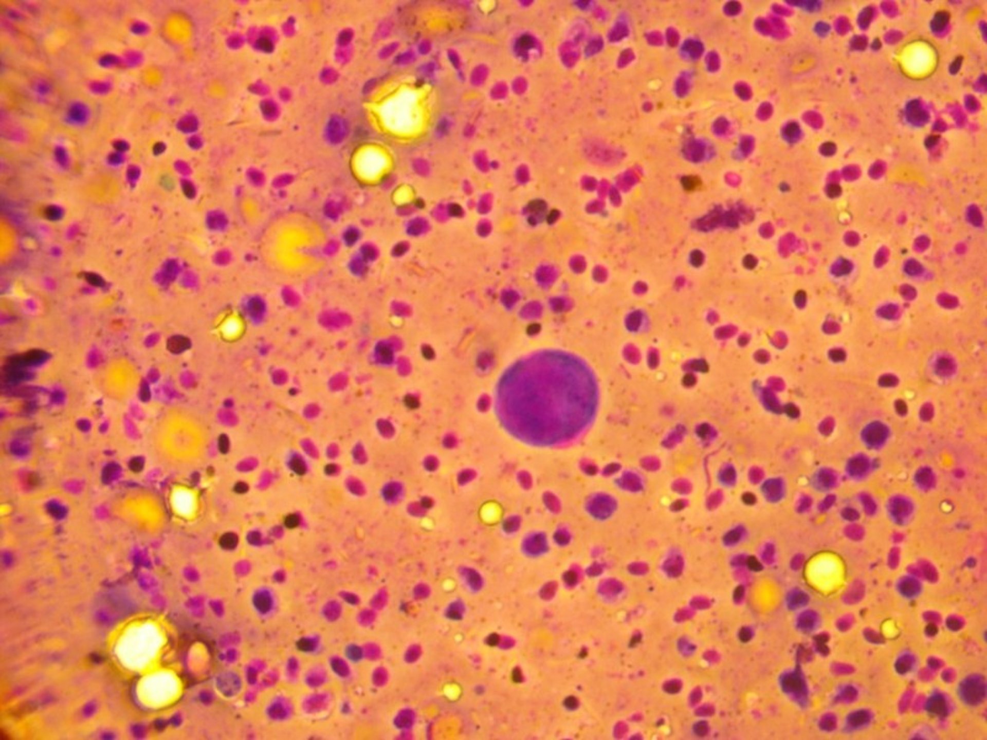
Courtesy of Dr. Denise Petty.
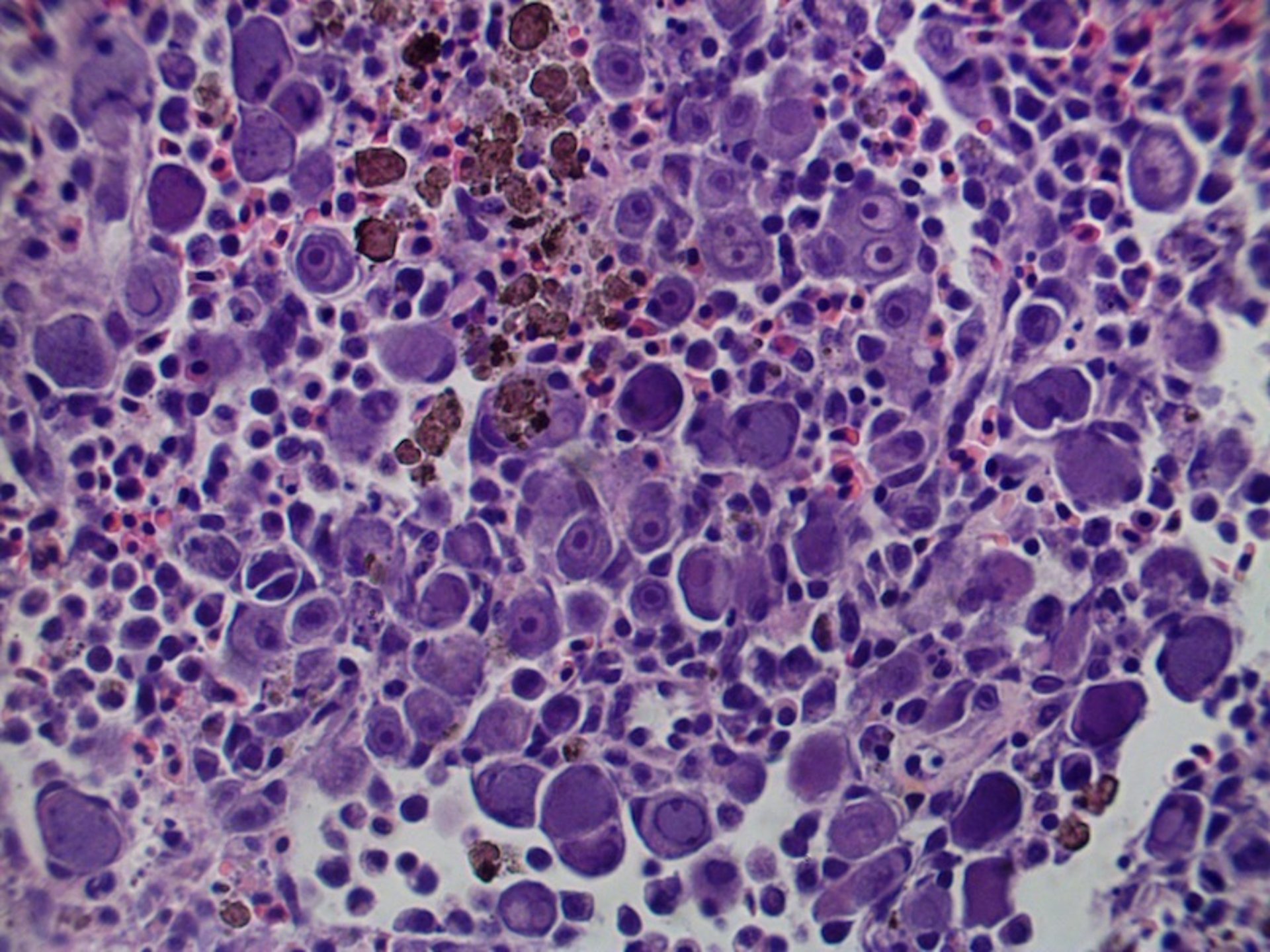
Courtesy of Dr. Denise Petty.
Several other iridoviruses have been described in ornamental fish. The genus Megalocytivirus consists of three major groupings, red sea bream iridovirus (RSIV), infectious spleen and kidney necrosis virus (ISKNV), and turbot reddish body iridovirus (TRBIV). Red sea bream iridovirus has been found in >30 other marine fish so far; many of the affected species include some tropical fish kept in aquaria. Red sea bream iridovirus is a reportable disease. Infectious spleen and kidney necrosis virus includes megalocytiviruses that primarily affect ornamental freshwater fish and some tropical marine ornamental and food fish. Turbot reddish body iridovirus has been reported in freshwater angelfish (Pterophyllum scalare), oscars (Astronotus occellatus) and other cichlids, dwarf gourami (Trichogaster lalius) and is the virus that caused high mortality in farmed gourami T trichopterus and T leeri in Florida in the 1990s.
Both ISKNV and TRBIV are not currently reportable in the US.
Most reports of Megalocytivirus infection are in higher teleosts, but lower teleosts are also susceptible to infection. High mortality is usually seen, and infected fish exhibit non-specific clinical signs. If necropsy is performed, a touch prep of spleen can be prepared and stained with Giemsa. Enlarged basophilic cells are easily seen, but should be confirmed histologically. If hypertrophied basophilic cells are seen on histopathology of a marine fish, fresh or frozen tissues should be submitted for PCR assay to determine if the disease is caused by RSIV, ISKNV, or TRBIV.
Other Viruses
Koi sleepy disease is also known as carp edema virus. Caused by a poxvirus, it was first seen in Japan in 1974 and has since occurred in other countries, including the US. Affected fish appear lethargic, lie on their sides, and have thin body condition. Mortality is usually low but can be variable. Gill hyperplasia is a common histopathologic finding. Infected koi can recover, but it is unknown whether they retain a carrier state.
For More Information
Also see pet health content on routine health care for fish and emergencies of fish.


