The most common pet and research hamster is the golden, or Syrian, hamster (Mesocricetus auratus).
All Syrian hamsters in captivity appear to have originated from a litter of eight hamsters collected in 1930 near Aleppo, Syria. Four of the animals escaped, a male killed one female, and only one male and two females remained. From these three animals, litters were raised that were distributed to Europe and the US for research and subsequently as pets.
In 1971, an additional 12 Syrian hamsters were captured in the field by farmers and imported from Syria to the US.
The head-and-body length of Syrian hamsters is 170–180 mm; the tail length is 12 mm. They range in weight from 110 to 140 g, and females are larger than males. Wild Syrian hamsters have a light, reddish-brown dorsal coat, and the underparts are white. The skin of Syrian hamsters is very loose.
Other hamster species now common as pets are the dwarf hamsters—eg, the Djungarian (Phodopus sungorus) and Roborovsky (Phodopus roborovskii) hamsters. Because of their small body size (< 100 g body weight), these hamsters are more difficult to handle and restrain for physical examinations and treatment. This discussion deals primarily with the Syrian hamster.
Biology of Hamsters
At least 20 mutations affecting coat color in Syrian hamsters are known. Most are simple recessive traits, four are dominant, and two are sex-linked. Five mutations affect the fur, giving rise to longhaired (these animals are also known as "teddy bear" hamsters), rex, and satin coats.
The length of hair in the longhaired Syrian hamster is influenced by testosterone. Starting at the age of sexual maturity, longhaired males have substantially longer hair than females or castrated males, which display fluffy, shorter hair.
Syrian hamsters possess paired flank organs in the costovertebral area that are androgen dependent and consist of sebaceous glands, pigmented cells, and terminal hairs. These glands are larger in males than in females and heavily pigmented in males; they are used for territorial marking.
All hamsters possess enormous cheek pouches that open inside the lips, extend well back of the shoulders, and, when filled with food, more than double the width of the animal’s head and shoulders.
Adult male Syrian hamsters develop large adrenal glands because of enlargement of the zona reticularis, which is 3 times the size of the female zona reticularis. Like gerbils, Syrian hamsters have a high proportion of erythrocytes with polychromasia.
Female Syrian hamsters are heavier than males and generally are aggressive toward other hamsters. Nonestrous females can behave especially aggressively toward young males, sometimes killing them.
The 4-day estrous cycle of hamsters is characterized by a copious postovulatory discharge on the last day. The discharge is creamy white and has a distinctive odor; it fills the vagina and usually extrudes through the vaginal orifice. The stringy nature of this discharge is distinctive, and if touched, it can be drawn out as a thread approximately 10–15 cm (4–6 inches) long.
Estrus in hamsters lasts approximately 1 day, and the gestation period is 16–19 days. Litter sizes range from 2 to 16, with an average of 9.
Cannibalism of young hamsters accounts for nearly all preweaning deaths. Cold ambient temperatures (< 10°C [50°F]), lean diets, and low body weight during pregnancy increase cannibalism.Disturbing the mother by handling the young or nest, as well as not providing adequate nesting material, warmth, food, or water, often results in cannibalism.
Syrian hamsters are prolific breeders, producing as many as 3–5 litters per year. The young are weaned at the age of approximately 20 days and are capable of reproducing at 7–8 weeks old. Syrian hamsters have a lifespan of 2–3 years.
Husbandry of Hamsters
In the wild, Syrian hamsters live on dry rocky steppes or brushy slopes, and they construct shallow burrows. In captivity, deep bedding that is appropriate for burrowing is recommended. Cages with at least 40–80 cm (approximately 15–30 inches) of bedding enhance the welfare of Syrian hamsters.
Wild Syrian hamsters are omnivorous, eating many kinds of green vegetation, seeds, fruit, and meat. Hamsters kept in captivity should be fed a complete, balanced, pelleted diet specifically made for hamsters (certain manufacturers offer a hamster/gerbil diet). Generic seed mixes should be avoided because hamsters selectively pick out seeds and ignore pellets.
Exposure to cold stimulates hamsters to gather food, and they often hibernate at temperatures < 5°C (41°F). Syrian hamsters do not fatten before hibernation and will starve if they do not awaken periodically to eat. Hibernating animals remain sensitive to external stimuli and are usually aroused if handled.
Syrian hamsters have prominent depositions of brown fat beneath and between the shoulder blades, in the axillae, and in the neck and perirenal areas.
Syrian hamsters are active chewers and skillful at escaping from their cages. Glass water tubes are contraindicated for Syrian hamsters because they readily bite through glass. Stainless steel sipper tubes close to the floor are recommended. Because Syrian hamsters have broad muzzles that often preclude eating from feed hoppers, pelleted food should be placed on the floor of their cage. Hamsters are naturally coprophagic.
Both male and female hamsters can be aggressive toward conspecifics. If group housing is desired, individuals should be housed together from an early age to decrease aggression. When housing animals together for breeding, it is recommended that older males be used, and that the animals be observed carefully immediately after introduction to ensure that the female does not injure the male.
Physical Examination of Hamsters
In a physical examination, a hamster's overall appearance and behavior, particularly in relation to its cagemates, should be noted. Sick animals often isolate themselves from others and can show weight loss, hunched posture, lethargy, rough fur, labored breathing, and a loss of exploratory behavior.
Early signs of disease in hamsters include changes in the color, consistency, odor, and volume of urine and feces. Fecal samples can be taken for parasite detection and bacterial culture.
In addition, attention should be directed to the following specific body areas:
The perineal area should be checked for fecal or urine stains or, in females, discharges from the vulva.
The fur and skin should be examined for alopecia, fight wounds, or other trauma, as well as for ectoparasites.
The oral cavity should be checked for overgrown teeth or impacted cheek pouches.
The ears should be examined for discharges or inflammation, and the eyes, for discharges or conjunctivitis.
The feet should be examined for sores and overgrown or broken nails.
The abdomen should be palpated for masses.
Individual Syrian hamsters, separated from conspecifics, are not normally aggressive, but they can be provoked if suddenly startled, awakened, or roughly handled. It might be easier to scoop Syrian hamsters up in a small container than to pick them up directly. Their highly elastic skin should be grasped sufficiently to prevent the animal from biting.
Infectious Diseases of Hamsters
Bacterial Infections in Hamsters
Diarrhea can occur in Syrian hamsters of any age and is known as “wet tail”; this euphemism is frequently used to describe the disease in young hamsters. Proliferative ileitis (see ) is the most consequential intestinal disease of 3- to 10-week-old Syrian hamsters and results in high mortality rates. It is caused by the intracellular bacterium Lawsonia intracellularis.


Gross lesions of proliferative ileitis in a hamster. This stress-related disease resulted in distention of the small intestinal tract and an intestinal intussusception in this hamster.
Courtesy of Dr. Louise Bauck.
Treatment of proliferative ileitis involves correcting life-threatening electrolyte imbalance and dehydration, administering antimicrobials, and syringe-feeding.
Antimicrobials that may be used include the following (1):
doxycycline (5–10 mg/kg, PO, every 12 hours for 5–7 days)
enrofloxacin (10 mg/kg, PO or IM, every 12 hours for 5–7 days)
trimethoprim-sulfamethoxazole (30 mg/kg, PO, every 12 hours for 5–7 days)
Supportive care for persistent diarrhea may include the administration of bismuth subsalicylate. Replacement electrolyte and glucose solutions should be given orally, and electrolyte fluid replacement, such as saline solution or lactated Ringer's solution, should be given at 10 mL/100 g body weight, SC, every 24 hours (2).
Sequelae of proliferative ileitis in surviving Syrian hamsters can include eventual obstruction, intussusception, or rectal prolapse.
Diarrhea in adult Syrian hamsters is associated with Clostridioides difficile enterotoxemia (see ). As in guinea pigs, it might occur 3–5 days after administration of antimicrobials such as penicillin, lincomycin, or bacitracin.


Necropsy photo of gross lesions due to Clostridioides difficile in a hamster. Note the markedly hemorrhagic distal small intestine and the distended cecum.
Courtesy of Dr. J. Glenn Songer.
Tyzzer disease due to Clostridium piliforme occurs in Syrian hamsters and is usually precipitated by stress such as overcrowding, high environmental temperature and humidity, heavy internal and external parasite load, and nutritionally inadequate diet. C piliforme is opportunistic in immunosuppressed hamsters and not found in immunocompetent hamsters.
Bacterial pseudomycetoma producing firm, pus-filled nodules in the skin, has been described in several dwarf hamsters (3, 4). The treatment is excision.
Viral Infections in Hamsters
Hamster polyomavirus (HaPV) is the cause of epizootic lymphoma in young Syrian hamsters and epitheliomas in older, enzootically infected hamsters. When first introduced into a naive population of breeding Syrian hamsters, HaPV results in an epizootic of lymphoma, with an incidence of up to 80% (2).
Lymphomas caused by HaPV in hamsters often arise in the mesentery; they can also arise in the axillary and cervical lymph nodes.
Once lymphoma is enzootic in a hamster population, its occurrence declines to a much lower level. Enzootically infected Syrian hamsters develop HaPV skin tumors rather than lymphoma. HaPV lymphoma–affected Syrian hamsters appear thin, often with palpable masses in the abdomen. Often, they have demodectic mange caused by either Demodex criceti or Demodex aurati (see ).

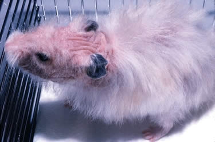
Gross lesions of Demodex infection in a hamster. Note the characteristic hair loss on the head and neck.
Courtesy of Dr. Louise Bauck.
Parasitic Infestations in Hamsters
Fecal smears of Syrian hamsters show an abundance of protozoal organisms. However, the role of these protozoa in enteric disease is speculative, because similar protozoa are found in comparable numbers in both healthy and diseased animals.
D criceti and D aurati are occasionally found on hamsters. Clinical signs of Demodex infestation consist of mild to moderate alopecia, pruritus, and erythema generally on the dorsal region of the body, the hindlimbs, and the face. In addition, crusts and scaling can be found on physical examination. Skin scrapings confirm the presence of a large number of Demodex spp in various stages of development.
Treatment options for Demodex infestation include bathing with 1% selenium sulfide shampoo (once weekly until clinical signs resolve [5]), dipping in lime sulfur dip (diluted 1:40 in water, every 7 days for 6 weeks [1]), applying topical selamectin (15 mg/kg, once [6]), and subcutaneous ivermectin injection (0.2–0.4 mg/kg, SC, every 5–7 days [1]). Hamsters that do not respond to treatment or that relapse often have serious underlying disease and typically die within 3 months.
Historically, there have been reports of pet hamsters infected with the tropical rat mite Ornithonyssus bacoti (5).
Fungal Infections in Hamsters
Spontaneously occurring dermatophytosis (ringworm) is extremely rare in Syrian hamsters.
Neoplasia of Hamsters
In older Syrian hamsters, lymphoma is the most frequently observed neoplasm of the hematopoietic system. It is multicentric and commonly affects lymphatic organs.
Cutaneous lymphoma, resembling mycosis fungoides (an epidermotropic T-cell lymphoma in humans), occurs occasionally in adult Syrian hamsters. Affected animals show anorexia, weight loss, and patchy alopecia. Cutaneous lymphoma can be misdiagnosed as pituitary-dependent hyperadrenocorticism (Cushing disease), because affected hamsters initially show patchy alopecia and dermal hyperpigmentation. However, cutaneous lymphoma progresses rapidly, with a mean time of approximately 10 weeks from presentation to euthanasia.
Adenomas of the adrenal gland commonly occur in Syrian hamsters; however, there are few reports of confirmed Cushing disease causing clinical signs in hamsters.
Melanomas, not only of the flank organ but also of the skin, are common in Syrian hamsters (see ). There is a striking sexual predisposition to melanoma in hamsters, with males 10 times more likely than females to be affected.


Malignant melanoma in a hamster. The flank scent glands on the lateral trunk are pigmented glands that are occasional sites of melanoma formation.
Courtesy of Dr. Louise Bauck.
Djungarian hamsters show a high prevalence of neoplasia (5 times greater than Syrian hamsters). Most tumors are integumentary (eg, mammary tumors, atypical fibromas, and papillomas).
Miscellaneous Disorders of Hamsters
Age-Related Disorders of Hamsters
Atrial thrombosis occurs in aging Syrian hamsters with an incidence of up to 70% (7). Most thromboses develop in the left atrium secondary to heart failure and lead to consumptive coagulopathy. Although the incidence does not differ between sexes near the end of their respective lifespans, atrial thrombosis occurs on average at a younger age in females (13.5 months) than in males (21.5 months).
Older Syrian hamsters with atrial thrombosis show signs of cardiomyopathy, such as hyperpnea, tachycardia, and cyanosis. In untreated Syrian hamsters, death usually follows within a week after these signs are evident.
The incidence of atrial thrombosis in hamsters is influenced by the endocrine status of the animal, especially by the amount of circulating androgens. Thus, the castration of male Syrian hamsters is linked to an increase in the prevalence of atrial thrombosis.
Weight loss occurs in older Syrian hamsters and is often associated with hepatic and renal amyloidosis. Amyloidosis is the principal cause of death in long-term research studies of hamsters. Compared to males, females have a higher incidence (80% among hamsters > 18 months old), increased severity, and earlier age of onset of amyloidosis. There is a correlation between social stress induced by crowding and amyloidosis in laboratory Syrian hamsters. In pet Syrian hamsters, for which overcrowding is not a problem, amyloidosis is infrequently reported.
Degenerative kidney disease also occurs more frequently in older Syrian hamsters, particularly females. Affected kidneys are pale and granular. Microscopically, glomerular changes vary from thickening of the basement membrane to glomerular obliteration. Amyloid deposition occurs frequently as a concurrent event.
Polycystic liver disease occurs in Syrian hamsters > 1 year old (see ). The lesions are due to developmental defects of the bile duct and are not associated with clinical signs. At necropsy, numerous thin-walled cysts might be evident.


Necropsy photograph of an aged, male Syrian hamster with benign polycystic liver disease. Note the light-colored, thin-walled cysts (arrow) arising between the liver parenchyma.
Courtesy of Dr. Jennifer Frohlich.
Zoonotic Risk of Hamsters
Syrian hamsters have a reputation as carriers of lymphocytic choriomeningitis virus (LCMV), a rodent-borne virus that can cause substantial neurological disease in humans, particularly prenatal and immunocompromised people.
The common house mouse, Mus musculus, is the natural host and principal reservoir of LCMV. When apparent, LCMV infection in hamsters is characterized by wasting. Early clinical signs are decreased activity and appetite, and unkempt coat. Later signs include weight loss, hunched posture, blepharitis, seizures, and eventually death.
Humans are typically infected with LCMV via close proximity to wild mice and their droppings. Historically, several large outbreaks of LCMV infection in the US were attributable to hamsters obtained from a single supplier in the late 1970s (8, 9, 10). More recently, individual cases of LCMV infection in organ transplant recipients have been linked to hamsters 11.
Although the risk of hamsters transmitting LCMV to the general population might be overstated, a German survey of people in contact with pet hamsters confirmed an increased risk of LCMV infection (12). Transplacental infection occurs in the fetuses of women who develop viremia during the first and second trimesters.
LCMV acts as a neuroteratogen, causing chorioretinopathy, hydrocephalus, microcephalus, lissencephaly, and potentially fetal death. The relative risk posed by hamsters should be considered among transplant recipients and pregnant women, because infected animals can remain clinically unaffected by LCMV and can transmit the virus for at least 8 months.
Key Points
Diarrhea ("wet tail") in hamsters is commonly caused by bacteria; the top two pathogens are Lawsonia intracellularis and Clostridioides difficile.
Neoplasia, most commonly multicentric lymphoma and malignant melanoma, is frequently reported in hamsters.
Lymphocytic choriomeningitis virus is a zoonotic risk for humans with depressed immune systems, and standard precautions such as handwashing should be discussed with anyone who comes in contact with hamsters.
For More Information
Miwa Y, Mayer J. Hamsters and gerbils. In: Quesenberry KE, Orcutt CJ, Mans C, Carpenter JW, eds. Ferrets, Rabbits, and Rodents: Clinical Medicine and Surgery. 4th ed. Elsevier; 2021:368-384.
Turner PV, Wheler CL, Beaufrère H, Fabian N, Harkness JE, eds. Harkness and Wagner's Biology and Medicine of Rabbits and Rodents. 6th ed. John Wiley & Sons; 2025.
Heatley JJ, Harris CM. Hamsters and gerbils. In: M Mitchell, Tully TN Jr. Manual of Exotic Pet Practice. Saunders Elsevier; 2009:406-432.
Also see pet owner content regarding hamsters.
References
Frohlich J, Mayer J. Rodents. In: Carpenter JW, Harms CA, eds. Carpenter's Exotic Animal Formulary. 6th ed. Saunders; 2023:530-573. doi:10.1016/B978-0-323-83392-9.00009-5
Miwa Y, Mayer J. Hamsters and gerbils. In: Quesenberry KE, Orcutt CJ, Mans C, Carpenter JW, eds. Ferrets, Rabbits, and Rodents: Clinical Medicine and Surgery. 4th ed. Elsevier; 2021:368-384.
Martorell J, Gallifa N, Fondevila D, Rabanal RM. Bacterial pseudomycetoma in dwarf hamster, Phodopus sungorus. Vet Dermatol. 2006;17(6):449-452. doi:10.1111/j.1365-3164.2006.00554.x
Eshar D, Mayer J, Keating JH. Dermatitis in a Siberian hamster (Phodopus sungorus). Bacterial pseudomycetoma. Lab Anim (NY). 2010;39(3):71-73. doi:10.1038/laban0310-71
Miedel EL, Hankenson FC. Biology and diseases of hamsters. In: Fox JG, Anderson LC, Otto G, Pritchett-Corning KR, Whary MT, eds. Laboratory Animal Medicine. 3rd ed. Academic Press; 2015:209-245.
Morrisey JK, Carpenter JW. Formulary. In: Quesenberry KE, Orcutt CJ, Mans C, Carpenter JW, eds. Ferrets, Rabbits, and Rodents: Clinical Medicine and Surgery. 4th ed. Elsevier; 2021:620-630.
McMartin DN. Spontaneous atrial thrombosis in aged Syrian hamsters. I. Incidence and pathology. Thromb Haemost. 1977;38(2):447-456. doi:10.1055/S-0038-1651480
Bowen GS, Calisher CH, Winkler WG, et al. Laboratory studies of a lymphocytic choriomeningitis virus outbreak in man and laboratory animals. Am J Epidemiol. 1975;102:233-240. doi:10.1093/oxfordjournals.aje.a112152
Skinner HH, Knight EH. The potential role of Syrian hamsters and other small animals as reservoirs of lymphocytic choriomeningitis virus. J Small Anim Pract. 1979;20(3):145-161. doi:10.1111/j.1748-5827.1979.tb07023.x
Jay MT, Glaser C, Fulhorst CF. The arenaviruses. J Am Vet Med Assoc. 2005;227:904–915. doi:10.2460/javma.2005.227.904
Amman BR, Pavlin BI, Albariño CG, et al. Pet rodents and fatal lymphocytic choriomeningitis in transplant patients. Emerg Infect Dis. 2007;13:719. doi:10.3201/eid1305.061269
Ackermann R, Stille W, Blumenthal W, Helm EB, Keller K, Baldus O. Syrische Goldhamster als Uberträger von Lymphozytärer Choriomeningitis [Syrian hamster as a vector of lymphocytic choriomeningitis]. Dtsch Med Wochenschr. 1972;97(45):1725-1731. doi:10.1055/s-0028-1107638