Rats are more commonly kept as pets than mice are. Both species are used extensively in research. Although a great deal of information about wild and laboratory rats and mice has been accumulated, very little of this information pertains to these rodents as pets.
The prevalence and types of diseases in mice and rats kept as pets are quite different from those that occur in a research setting. The diagnosis and treatment of pet mice and rats involves evaluation and care of an individual animal from a household, not the health management of rodents from a research colony.
The most common problems in mice and rats are dermatopathies, respiratory infections, and neoplasia.
Biology of Mice and Rats
Male rats are sexually mature by 6–10 weeks old; female rats are sexually mature by 8–12 weeks old. The breeding life of both male and female rats is 9–12 months. Estrous cycle length in female rats is 4–5 days, and estrus lasts 10–20 hours. Female rats ovulate approximately 10–20 eggs. Gestation in rats lasts 21–23 days; pseudopregnancy from sterile matings lasts approximately 12 days. Rats have an average litter size of 8–18 pups. Weaning takes place at approximately 21 days.
Both male and female mice are sexually mature by 6–8 weeks old and have a breeding life of 9 months. Estrous cycle length in female mice is 4–5 days, and estrus lasts 10–20 hours. Female mice ovulate approximately 6–10 eggs. Gestation in mice lasts 19–21 days; pseudopregnancy from sterile matings lasts approximately 12 days. Mice have an average litter size of 5–12 pups. Weaning takes place at approximately 21 days.
Male mice and rats produce a small amount of sperm daily at puberty (eg, 40–50 days old in a rats). Not until rats are 75–100 days (10–14 weeks) old do optimal sperm production and reserve occur. Male rodents show a constant libido after sexual maturity; in contrast, females are receptive to copulation only during estrus. Males cannot fertilize females until 6–8 weeks after reaching puberty.
The average lifespan of mice is 18–24 months; of rats, 18–36 months. Restricting dietary calories without compromising overall nutrition increases the lifespan. Obesity in pet rats and mice is common, and calorie-restricted pets live markedly longer lives.
Husbandry of Mice and Rats
The best cages for pet mice and rats are made of a material that is easy to clean and deodorize and that is indestructible to rodent chewing or digging in the corners. The cage floor can be solid but should be waterproof and easy to clean. Wire-mesh floors should be avoided because rats and mice can trap their feet, and especially hindlimbs, in the openings, resulting in fractures and injuries.
Animal cage bedding is categorized by use (eg, contact, noncontact, enrichment bedding) or by the material from which it is made (eg, wood-based [chips, shavings, peelings, wood-wool, and sawdust], paper-based [cotton and pulp fiber, recycled paper], corn [husks and cobs], cellulose, vermiculite). The purpose of bedding is to keep animals dry and clean.
Pet owners generally choose bedding on the basis of cost and availability; laboratory veterinarians choose bedding on the basis of cost and water-holding capacity. Some pet bedding contains lemon and chlorophyll to give it a pleasant scent. This type of bedding irritates pet rodents, and the coloring agents can stain the coat of white rats or mice.
Historically, owners of pet rats or mice preferred paper and softwood chips, such as kiln-dried pine or aspen, over straw because it requires fewer bedding changes. Wood chip bedding (such as pine and aspen) that has not been heat treated should not be used, because it contains volatile chemicals that can irritate the respiratory tract and activate liver enzymes in rodents (1, 2). Paper is also nontoxic and digestible if ingested, in contrast to wood, which can lead to intestinal tract obstruction if eaten.
Owners must combine frequent bedding changes with good husbandry practices, such as regular cage cleaning, low animal density, and low environmental temperature and humidity. These practices decrease the buildup of toxic or odor-causing gases (such as ammonia) that is due to the breakdown of urea in urine by urease-positive bacteria in the feces. Aquariums are not suitable cages for rats and mice, because air circulation is inadequate and therefore ammonia builds up.
Pearls & Pitfalls
|
Environmental temperature and relative humidity can depend on husbandry and housing design and can differ considerably between the cage and the room the cage is in. Factors that contribute to variation in temperature and humidity include cage material and construction, number of animals in the cage, frequency of bedding changes, and bedding type.
The main factors that determine the type of bedding material for pet mice and rats are size and manipulability. Mice and rats avoid bedding consisting of small particles; they prefer bedding consisting of large, fibrous particles.
Mice and rats are optimally maintained at temperatures of 18–26°C (64–79°F). Relative humidity should also be controlled; however, the range is much wider than for temperature (3). An acceptable range of relative humidity is 30–70%.
Both temperature and humidity regulation are important to prevent ringtail (avascular necrosis of the tail) and exacerbation of respiratory disease. Ringtail occurs primarily in young rats in low-humidity environments. Excess heat and humidity cause heatstroke and indirectly cause decompensation due to chronic respiratory disease, resulting in death.
Owners of pet mice and rats should check the animals' water bottles daily. If mice are deprived of water for only a short time (1–2 hours) and experience dramatic fluctuations in the surrounding temperature, especially > 37°C (99°F), they die. In contrast, healthy rats tolerate water deprivation and temperature fluctuations better. They can survive under conditions of heat exposure of up to 4 hours per day at 37.5°C (99.5°F) for 7 consecutive days without access to water during the heat exposure period (4).
Minimal space allocations for pet mice and rats are as follows:
For rats ≤ 200 g: 58 cm2 (23 square inches) per animal
For rats ≥ 500 g: 152 cm2 (60 square inches) per animal
For mice > 25 g: 38 cm2 (15 square inches) per animal
These are small areas. Veterinarians should advocate that owners provide more space for their pets.
Most rodent enrichment studies focus on modifying the area inside an enclosure rather than the enclosure's size. However, rats prefer larger cages, and experimental studies suggest that rats should have a space large enough to be shared with up to four other rats (5).
The minimum recommended cage height for rats and mice should take into account their typical postures, including standing fully erect on hindlegs and moving vertically, such as stretching upward and possibly climbing. Minimal cage heights of 30–38 cm (12–15 inches) for rats and 18–20 cm (7–8 inches) for mice are recommended.
Environmental enrichment is important for both mice and rats. For example, suspended cloth hammocks are popular with rats, and suspended (plastic or stainless steel) shower hooks fitted into one another can make a swinging chain. Rats use enrichment devices more than mice do; however, they usually stop using the devices after 3–4 days. Rotation of enrichment toys and introduction of novel devices excite their curiosity.
Food treats are also valuable enrichment items. They can range from simple, inexpensive treats (such as a daily piece of breakfast cereal) to formulated nutritious or calorie-free treats. Rats also love chocolate, which can be fed in small amounts (1 chocolate chip occasionally as a treat) without causing toxicosis.
Pet mice and rats accustomed to being handled will eat food treats out of their owner’s hand. This daily routine enables owners to detect subtle changes in their pet’s behavior. Sick rodents effectively hide signs of disease; however, sick rats show less interest in daily treats, and this shift can alert the owner early to disease, when it is still treatable and/or reversible.
Housing male and female rodents together will result in breeding and subsequent litters. Mice and rats experience postparturient estrus, and fertilization can occur. However, implantation of the resulting blastocysts is delayed during lactation and occurs at weaning, ensuring that the next litter is not born until the earlier one has been weaned. Unless opposite-sex rodents housed together are separated or neutered, production of a new litter every 3–5 weeks is possible.
Pearls & Pitfalls
|
Rats and mice are omnivores, eating foods of both plant and animal origin. In the wild, rats and mice eat a wide variety of seeds, grains, and other plant material, as well as invertebrates, small vertebrates, and carrion. Their ability to scavenge partly accounts for their successful colonization of diverse geographical regions.
Formulated pelleted diets for laboratory rodents are convenient and nutritionally balanced diets for early life and reproduction. However, laboratory rodent diets are relatively high in fat and low in fiber, and when provided ad libitum, they cause obesity. Consequently, the daily amount of pelleted diet that owners provide pet mice and rats should be limited.
Diets formulated specifically for pet rodents are now commercially available. Owners should supplement their pet’s diet with feeds high in fiber such as vegetables, limited amounts of fruit, and occasional treats.
Coprophagy is a normal behavior in rats and mice. It is an innate behavior, as indicated by its occurrence in germ-free rodents purposely bred for research. Unlike rabbits, which eat cecal feces from their anus, mice and rats eat fecal pellets on the floor of their caging.
The amount of feces eaten by rodents depends on the species, their age and physiological status (eg, coprophagy increases in pregnancy), and the diet they are fed. On a nutritionally complete diet, rats eat approximately 10% of their feces. When housed together, rats ingest each other’s feces.
The unique odor of a colony is due in part to the ingestion of feces, and the group scent enables the animals to distinguish between members and nonmembers of their colony.
Mice engage in coprophagy approximately 6 times/day. Growing mice show vigorous coprophagous activity, eating 13 pellets/day. However, consumption of feces gradually decreases to 1½ pellets/day by the age of 2 years.
Owners should house different species of rodents separately to prevent interspecies disease transmission. For example, rats carry Streptobacillus moniliformis, a cause of fatal septicemia in mice, as part of the normal nasopharyngeal microbiota.
Rodents of the same species should be housed in such a way that vulnerable animals are protected from more aggressive members of their group. Such protection includes separating young animals from older ones. Female rodents are generally compatible when housed in the same cage, unless one female has lived much of its life alone.
Male rats are generally compatible, especially if raised together. Strange male rats will fight if they are housed in the same cage. Male mice generally fight if housed together, unless they are littermates raised together without females present. Male mice are best housed singly or with female mice.
Rats are social animals, and grooming is a socially affiliative behavior. If left alone without human contact and environmental enrichment, singly housed rats might develop isolated-rat stress syndrome, which includes signs such as increased startle reflex and elevated basal levels of stress hormones (6). Rats reared in isolation typically experience fighting, physical injuries, and weight loss when placed in a colony of socially experienced rats.
Young rats should be housed together to develop social affiliation. Singly housed rats should not be put together. Owners should be advised to encourage enrichment and human contact with pet rats.
Physical Examination of Mice and Rats
Observing the condition of the living quarters of mice and rats provides useful information about the animals. Information obtained from a physical examination is often limited because of the mouse or rat’s size. Activity level, coat condition, general posture at rest, and any blood or bodily discharges in the cage should be noted.
If a mouse or rat exhibits dyspnea or depression, extreme care should be taken when handling the animal, because the stress of a physical examination could result in death of a severely debilitated animal.
Pearls & Pitfalls
|
Pet rats and mice that have been frequently and gently handled usually require only minimal restraint. Less cooperative animals need to be more firmly restrained. A common method for restraint of mice is "scruffing," in which the restrainer grasps the scruff of the mouse between the thumb and forefinger while maintaining a grip on the base of the tail to secure the animal.
A useful restraint method for handling rats for physical examination is a technique sometimes known as the "baseball hold" or "claw hold" (see ).


Commonly used method, often called the "baseball hold," to restrain rats for physical examination and other procedures. The restrainer places the index finger and third finger of one hand on either side of the head, with the palm against the animal's back. The thumb and fourth finger of the restraining hand then go underneath the forelimbs. Care must be taken not to squeeze the thorax too tightly and thus impede respiration. The hindlegs and rump should be supported with the other hand.
Dr. Jennifer Frohlich
Weight measurement is essential to calculate appropriate dosages of medications and provides an opportunity to gauge the rodent’s temperament before beginning the actual physical examination.
Key components of the physical examination of mice and rats include the following:
The head, ears, eyes, and nose should be examined for discharges, and the oral cavity for dentition. Lymph nodes and glands of the head can be observed for size and palpated for consistency. Assessment of the head is probably the most time-consuming part of the examination.
The abdomen should be palpated for consistency and the presence of unusual masses. Animals should not be squeezed too vigorously, because overzealous palpation can result in visceral rupture.
The mammary glands and adjacent areas should be carefully palpated to check for tumors, which are common in rats. Keep in mind that mammary tissues extend the full length of the body in these rodents, so it is important to include axillary regions as well as inguinal and abdominal regions.
The anogenital region should be examined for discharges and staining of the fur or skin.
The limbs should be palpated for tenderness or fractures, with special attention given to the paws, noting the length of the nails and condition of the footpads.
When a rodent is picked up, it generally urinates and defecates. The examiner should have a dipstick ready to perform immediate urinalysis; feces can be caught in a small tube and examined later.
Respiration rate and heart rate in mice and rats are rapid and, therefore, difficult to measure. Instead, signs of dyspnea should be noted before handling. Some respiratory infections, such as mycoplasmosis, are subclinical. Signs of these diseases are more easily heard than seen; abnormal sounds called “snuffling” in rats and “chattering” in mice are noticeable without a stethoscope.
Venipuncture in Mice and Rats
Blood collection can be difficult in small rodents, especially in mice, because of their small body and blood vessel size. In both rats and mice, the saphenous vein or lateral tail vein can be used to collect small amounts of blood for blood smears, blood glucose measurement, etc. See , , and images.


This mouse has been anesthetized for blood collection from the saphenous vein. The fur on the hindleg has been removed to increase visibility of the vessel. Lubricating eye ointment is applied to the skin before blood collection to prevent blood from wicking into the fur. A lancet is being used here to nick the vessel; a small-gauge needle (25 g) can also be used. Once the vessel is nicked, the blood can be collected using a microcapillary tube.
Courtesy of Dr. Jennifer Frohlich.


Specially made device for restraining mice to collect blood from the tail vein. Note the prominent lateral tail vein.
Courtesy of Dr. Jennifer Frohlich.

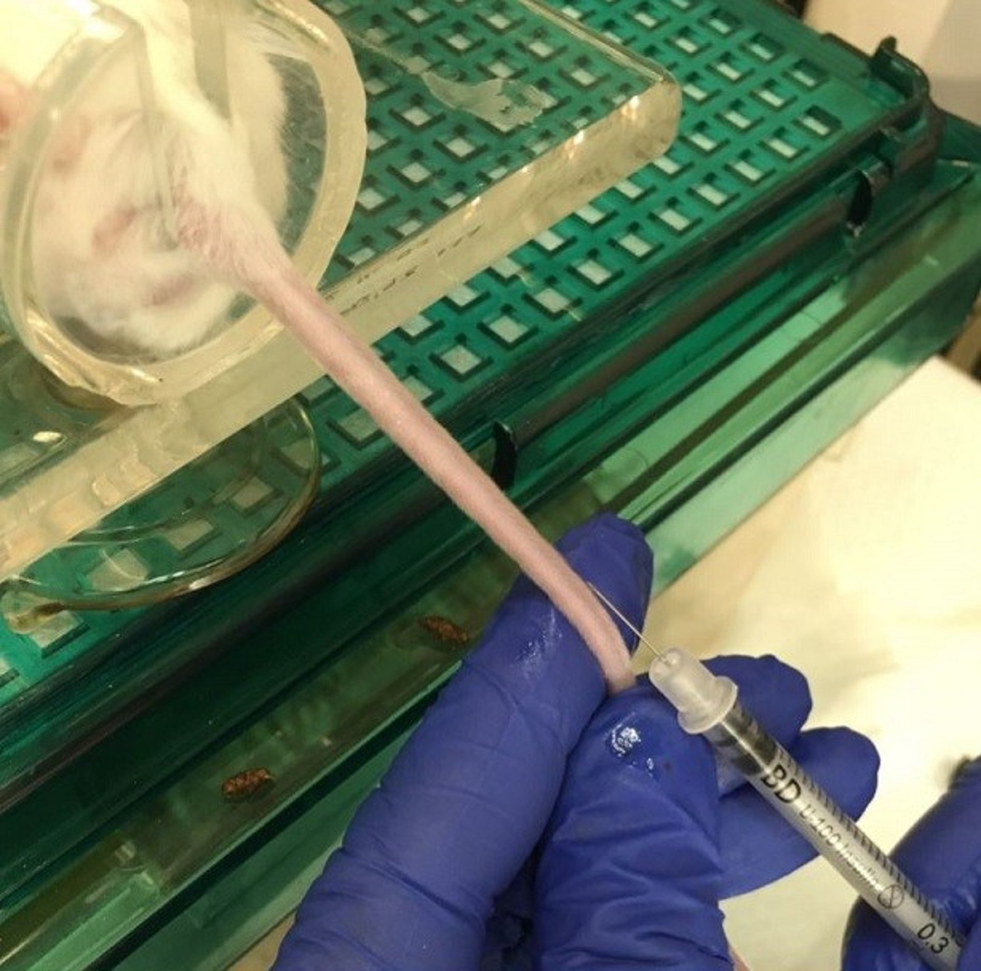
Blood collection from the tail vein of a restrained mouse. The phlebotomist gently holds the tail straight with the nondominant hand and positions an insulin syringe and needle at the distal end of the tail vein for blood collection.
Courtesy of Dr. Jennifer Frohlich.
In gingival vein puncture, blood is drawn from the labialis mandibularis vein located in the gingival papillae region just below the mandibular incisors (see ).


Gingival vein puncture in mice and rats is performed under anesthesia, with the animal in dorsal recumbency. The lower lip is pulled back, and a 28- to 30-g needle is inserted along the midline between the mandibular incisors at approximately 20–25°. The needle should be inserted approximately 2 mm into the gingiva.
In rats, this method can yield approximately 800 mcL of blood; in mice, approximately 100 mcL. The method can also be used to inject boluses (up to 1 mL in rats, up to 150 mcL in mice) intravenously (7).
To collect large amounts of blood from a mouse or a rat, the cranial vena cava can be used; however, the animal must be sedated or anesthetized to accomplish this safely. Other methods to collect larger samples of blood in mice, such as facial vein puncture or retrobulbar sampling, are useful but carry great risk of pain and trauma, making them difficult for use in pet mice.
Infectious Diseases of Mice and Rats
Bacterial Infections in Mice and Rats
Rats are the natural host of the bacterium Streptobacillus moniliformis, a commensal organism of the nasopharyngeal microbiota that does not cause disease in rats. However, humans bitten by rats can become infected with S moniliformis and develop rat-bite fever, a potentially fatal disease.
Mice housed with rats might become infected with S moniliformis and develop fatal septicemia characterized by polyarthritis, cervical lymphadenitis, pneumonia, and splenomegaly.
Respiratory disease caused by infectious agents is the most common health problem in rats. Three major respiratory pathogens cause overt disease: Mycoplasma pulmonis, Streptococcus pneumoniae, and Corynebacterium kutscheri.
Other organisms—such as Sendai virus (a paramyxovirus), pneumonia virus of mice (a paramyxovirus), rat respiratory virus (a hantavirus), Filobacterium rodentium (formerly cilia-associated respiratory bacillus [CARB]), and Haemophilus spp—are minor respiratory pathogens in mice and rats that rarely cause overt disease by themselves. However, the minor respiratory pathogens interact synergistically as copathogens with the major respiratory pathogens to produce two major clinical syndromes: chronic respiratory disease (CRD) and bacterial pneumonia.
CRD, also known as murine respiratory mycoplasmosis, is the best-understood multifactorial respiratory infection in rats. M pulmonis is the major component of CRD. In a retrospective study, rats with dyspnea of bacterial etiology lived 6–44 months (median 24 months) (8).
Clinical signs of CRD in rats are highly variable. Initial infection commonly occurs without any clinical signs; early signs involve both the upper and lower respiratory tracts and can include snuffling, nasal discharge, polypnea, weight loss, hunched posture, ruffled coat, head tilt, and red tears. Dyspnea, the primary sign, is caused by ciliostasis, subsequent buildup of lysozyme-rich inflammatory exudate in airways, bronchiectasis, and bronchiolectasis resulting from inflammatory damage to bronchiolar membranes. Chronic disease often includes middle ear infection (via the eustachian tube).
Treatment with antimicrobials can alleviate clinical signsof CRD in rats; however, it does not eliminate the infection. Severe signs can often be alleviated by either enrofloxacin (10 mg/kg) or doxycycline (5–10 mg /kg), both administered PO every 12 hours for 14 days (9).
Bronchodilators are the primary treatment for the inflammatory aspect of CRD in rats and mice; they can be provided either orally (theophylline at 10 mg/kg, every 12 hours as needed [10]) or as an aerosol (albuterol, also known as salbutamol, at 100 mcg/rat in a small chamber, every 4–6 hours [10]). Additional treatments, such as daily nebulization therapy with 7% hypertonic saline to break down the mucus biofilm in respiratory passages, and daily removal of soiled bedding to decrease ammonia levels in cages, ameliorate the disease.
For chronic cases of CRD in rats and mice, additional supportive care with analgesics or supplemental nutrition might also be required. Although it is not ideal to treat animals with prolonged courses of antimicrobials, chronic low-dose doxycycline (5–10 mg/kg, PO, every 24 hours) helps to prevent acute relapses in rats that relapse with CRD recurrently when taken off antimicrobials (9). Doxycycline is bactericidal for mycoplasma organisms and also has a marked immunomodulatory effect on chronic airway inflammation.
For clinicians, the most important aspect of CRD in rats and mice is that respiratory mycoplasmosis varies greatly in disease expression because of environmental, host, and organism factors that influence the host-pathogen relationship. Examples of such factors include intracage ammonia levels; concurrent infection with Sendai virus, coronavirus (sialodacryoadenitis virus), pneumonia virus of mice, rat respiratory virus, and/or F rodentium; genetic susceptibility of the host; virulence of the Mycoplasma strain; and vitamin A or E deficiency.
Although CRD rarely occurs in laboratory rats, most pet rats have CRD to some degree. One survey of pet ratteries in the northwestern US showed virtually all (95%) were positive for F rodentium and M pulmonis, and approximately half were positive for other viral respiratory agents (11).
Bacterial pneumonia in mice and rats is nearly always caused by S pneumoniae; usually, however, S pneumoniae occurs together with M pulmonis, Sendai virus, or F rodentium. Infection with C kutscheri also results in pneumonia, but only in conjunction with debilitation or immunosuppression. In pet rats, immunosuppression can result from diabetes, neoplasia, or dietary deficiencies. Pneumonia due to C kutscheri is rare in pet rats.
Pneumonia caused by S pneumoniae can have a sudden onset in rats. Young rats are more severely affected than older ones, and often the only sign they exhibit is sudden death. Mature rats might demonstrate dyspnea, snuffling, and abdominal breathing. A purulent exudate might be evident around the nares and on the front paws from wiping of the nostrils.
A tentative diagnosis of bacterial pneumonia in mice and rats is based on the identification of numerous gram-positive diplococci on a Gram stain of the exudate or in a sample submitted for cytological examination. Severe bacteremia is an important consequence of advanced disease and results in multiorgan abscesses and infarction.
Bacterial pneumonia in rats and mice is treated with amoxicillin/clavulanic acid (20 mg/kg, PO, every 12 hours until clinical and radiographic resolution [9]). Beta-lactamase–resistant penicillins such as cloxacillin, oxacillin, and dicloxacillin, administered orally or parenterally, also may be used at empirical dosages to treat bacterial pneumonia in rats and mice; for this indication, no dosages for these drugs have been published (12).
Ulcerative dermatitis caused by Staphylococcus aureus infection in rats and mice results from self-trauma associated with fur mite infestation or, more commonly, from scratching of the skin over an inflamed salivary gland. Rats have a remarkable ability to resist infection with S aureus. Treatment consists of clipping the toenails of the hindfeet, cleaning the ulcerated skin, and applying a topical antimicrobial. Systemic treatment is rarely necessary.
The two most common causes of clinical respiratory disease in mice are Sendai virus and M pulmonis infection. Sendai virus is associated with an acute respiratory infection in which mice display chattering and mild respiratory distress. Neonates and weanlings might die. Adults generally recover within 2 months.
When the expression of respiratory disease does not follow this pattern in mice, the cause is most likely concurrent mycoplasmal infection. M pulmonis is the cause of chronic pneumonia, suppurative rhinitis, and, occasionally, otitis media.Chattering and dyspnea are caused by accumulations of purulent exudate in inflamed and thickened nasal passages. Survivors often develop chronic bronchopneumonia, bronchiectasis, and pulmonary abscesses.
Antimicrobials can alleviate clinical signs of respiratory disease in mice; however, they do not eliminate the infection.
Viral Infections in Mice and Rats
Viral diseases of mice and rats are common. However, most diseases are subclinical and important only in laboratory animals in which they have the potential to have a notable effect on research.
Sialodacryoadenitis virus (SDAV), a coronavirus, causes inflammation and edema of the cervical salivary glands in rats. Owners of infected rats often describe their pets as having mumps.
SDAV infection is highly contagious. It initially causes rhinitis, which is followed by epithelial necrosis (see ) and inflammatory swelling of the salivary and lacrimal glands (see ). Cervical lymph nodes also become enlarged.


Photomicrographs showing (A) marked destruction of the ductal epithelial cells of the parotid salivary gland of a rat 4 days after exposure to rat coronavirus (sialodacryoadenitis virus) and (B) the same area of the parotid salivary gland stained to demonstrate viral antigen confined to the ductal epithelial cells. A, H&E stain; B, fluorescent antibody stain.
Courtesy of Dr. Dean H. Percy.

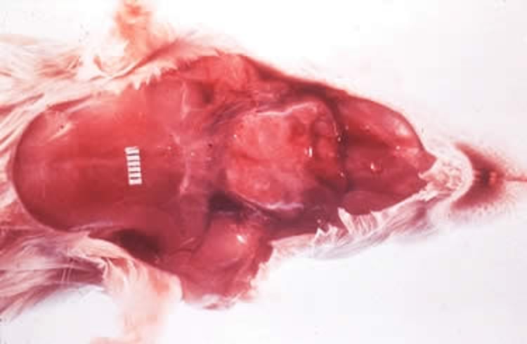
Necropsy photograph showing marked enlargement of the submandibular salivary glands in a rat with acute sialodacryoadenitis.
Courtesy of Dr. Dean H. Percy.
There is no treatment for sialodacryoadenitis. Glandular healing generally occurs within 7–10 days, and clinical signs subside within 30 days, with minimal residual lesions remaining.
During acute inflammation, affected rats are at high risk of anesthesia-related death because of the decreased diameter of the upper respiratory tract lumen.
Ocular lesions such as conjunctivitis, keratitis, corneal ulcers, synechia, and hyphema can develop secondary to lacrimal dysfunction. The eye lesions usually resolve; occasionally, though, they progress to chronic keratitis and megaglobus (buphthalmos or hydrophthalmos).
Parasitic Infestations in Mice and Rats
Protozoa in Mice and Rats
Endoparasites are relatively common in mice. However, only two parasites regularly encountered in the digestive tract—the protozoa Spironucleus muris and Giardia muris—are considered pathogenic, even though they are not associated with clinical signs in immunocompetent hosts.
Diagnosis of parasitic infestation in mice is based on demonstration of characteristic trophozoites in wet mounts of fresh intestinal contents or feces. Treatment is metronidazole (10–40 mg/kg, PO, every 24 hours or 2.5 mg/mL in drinking water for 14 days) (9). However, it does not completely eliminate the infestation.
Nematodes in Mice and Rats
Pinworms are ubiquitous and considered nonpathogenic in mice. Two are commonly encountered in mice: Syphacia obvelata and Aspiculuris tetraptera. Rats can become infested with these two species, as well as with the rat pinworm Syphacia muris (see ).


Syphacia muris egg on a tape test. Note the elongated banana shape of the egg, which is different from the rounder football shape of Aspiculuris tetraptera ova.
Courtesy of RADIL, University of Missouri.
Often, the only indication of pinworm infestation in mice is rectal prolapse due to straining. To establish a diagnosis of S obvelata or S muris infestation, a clear cellophane tape impression of the perianal skin can be made. Adult Syphacia females deposit ova around the anus. A tetraptera does not deposit its ova in this area, and fecal smear or flotation is required to confirm a diagnosis.
Ivermectin (2 mg/kg, PO, administered twice, with a 10-day interval [13]) or fenbendazole (20 mg/kg, PO, every 24 hours for 5 days [9]) eliminates pinworms from mice.
Mites in Mice and Rats
Most infectious causes of alopecia and dermatitis in mice are associated with fur mites. Generalized thinning of the hair occurs, especially on areas that are difficult to groom, such as the head and trunk. The coat often has a greasy appearance, and in cases of heavy infestation, noticeable pruritus and self-inflicted dermal ulceration can occur.
Three mites are common in mice: Myobia musculi, Myocoptes musculinus, and Radfordia affinis. Myobia musculi is the most clinically important mouse mite (see ).

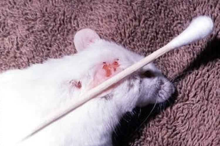
Myobia musculi lesions in a pet white mouse, characteristic of hypersensitivity to the mites. Intense pruritus, often directed at the neck and ears, leads to self-trauma. Early lesions consist of subtle hair thinning on the dorsal neck and shoulders.
Courtesy of Dr. Louise Bauck.
Mite infestations are usually caused by more than one species. Mites are spread by direct contact with infected mice or infested bedding.
Diagnosis of mite infestation in mice is based on identification of adult mites, nymphs, or eggs on hair shafts with the use of a hand lens or stereoscopic microscope. Adults and nymphs appear pearly white and elongate; eggs are oval and are found attached to the base of hairs or inside mature females.
Mite infestations in mice are treated with ivermectin (0.2 mg/kg, SC or PO, twice, with a 10-day interval [14]).Alternatively, a few drops of ivermectin solution (diluted to 1:100 in equal parts water and propylene glycol for three treatments at 10-day intervals) can be placed on the mouse’s head to enable spread by grooming and ingestion (15).
Ectoparasitic infestation is less common in rats than in mice. Occasionally, the fur mite Radfordia ensifera can be found. Although R ensifera infestation produces few ill effects, heavy infestation can lead to self-trauma and ulcerative dermatitis.
Ornithonyssus bacoti (see ), the tropical rat mite or red mite, is a bloodsucking parasitic mite found primarily in wild rats such as the brown rat (Rattus norvegicus) or the black roof rat (Rattus rattus).

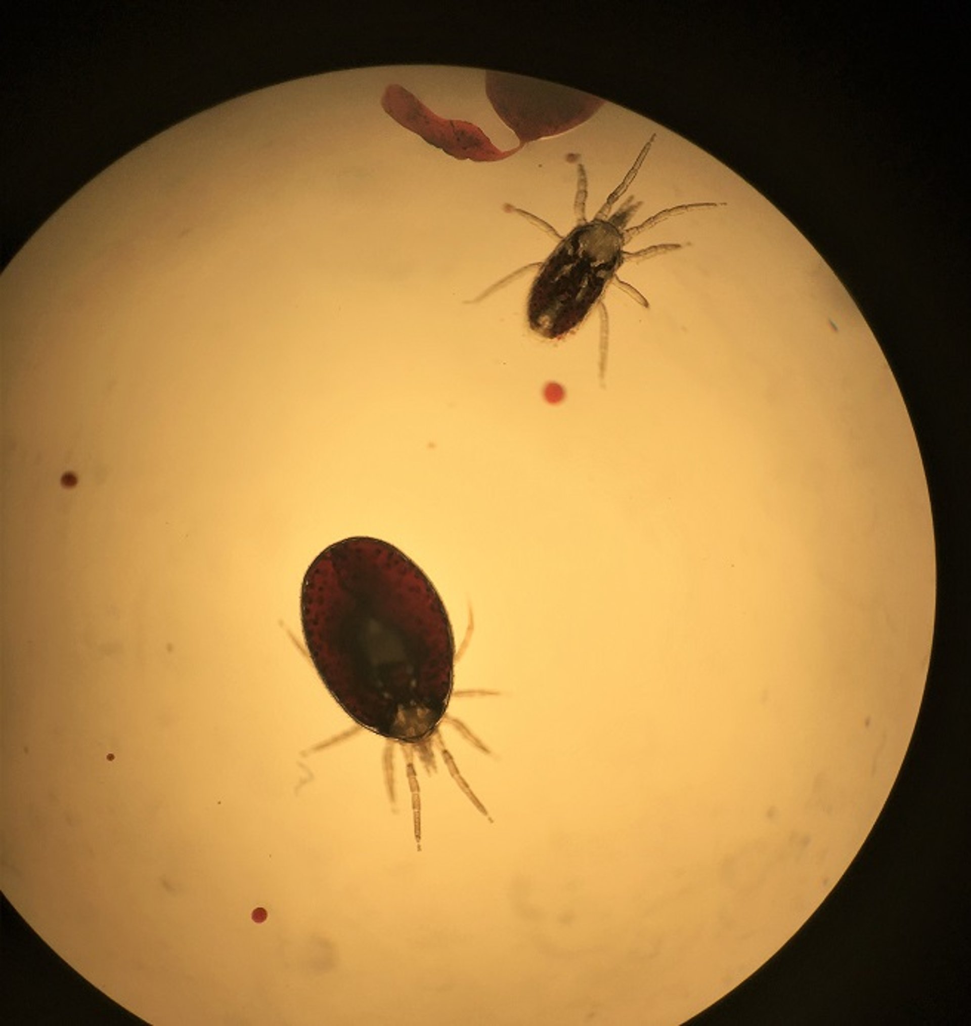
The tropical rat mite (Ornithonyssus bacoti) as viewed under a microscope (4X). Note the difference in appearance and size between the engorged mite (lower left) and the one that has not fully fed (upper right).
Courtesy of Dr. Jennifer Frohlich.
O bacoti has a wide host range and occasionally infects pet hamsters, gerbils, rats, and mice that live in very old buildings or that are exposed to it when building construction or renovations disturb colonies of wild rodents that were on the premises and acting as hosts.
O bacoti can survive for long periods in the environment and travel considerable distances in search of new hosts. If it does not find a suitable rodent host, it will feed on humans. Its common name is a misnomer, because it is found worldwide, in both tropical and temperate climates.
Mites appear white before feeding and become red-brown after engorgement. Heavy infestations on pet rodents resemble fine sawdust within the fur. O bacoti typically does not cause clinical signs in pet rodents; however, heavy infestations cause anemia, debility, weakness, pruritus, and death in small rodents.
As well as causing discomfort, O bacoti is a vector for the rodent filarial nematode Litomosoides carinii and a potential vector of several human pathogens. Most infestations occur in late spring or early summer, when pups are leaving the nest and the mites are searching for another food source.
Treatment of the environment as well as the animal is essential because O bacoti lives primarily in bedding and attaches to the host only for feeding. To combat infestations, host animals should be bathed and/or have topical selamectin applied (15 mg/kg, once, repeated after 21 days if necessary [9]). The pet's cage can be treated with insecticides that are safe for rodents, such as permethrin-impregnated cotton balls (7.4% placed inside the cage weekly for 6 weeks), fipronil spray, or synthetic pyrethroids.
The final step in eradicating an O bacoti infestation is to eliminate the source of the mite by practicing appropriate pest management in the home.
Fungal Infections in Mice and Rats
Dermatophytosis (ringworm) is uncommon in pet mice and rats. It is caused by the fungus Trichophyton mentagrophytes. Lesions, when present, are most common on the face, head, neck, and tail. The lesions have a scurfy appearance, with patchy areas of alopecia and variable degrees of erythema and crusting. Pruritus is usually minimal to absent, and the lesions do not fluoresce under a Wood's lamp.
T mentagrophytes can be isolated from the fur of clinically normal mice but is rare in rats.
Neoplasia of Mice and Rats
The most common subcutaneous tumor in rats is mammary fibroadenoma. The distribution of the mammary tissue is extensive, and the tumors can develop anywhere from the neck to the inguinal region. Tumors can reach 8–10 cm in diameter and occur in both males and females.
The surgical technique for tumor removal in rats is straightforward, and the survival rate after mastectomy has been reported to be good if the tumor is benign. The prevalence of both mammary and pituitary tumors is substantially lower in ovariectomized rats than in sexually intact rats. However, the recurrence of fibroadenomas is common in uninvolved mammary tissue, and often several surgeries are needed.
Spaying female rats before the age of 7 months, before mammary tumor formation, decreases the chance of mammary tumor formation later in life (16). Spaying at the time of mammary tumor excision has not been shown to prevent mass recurrence (17).
Pearls & Pitfalls
|
Deslorelin implant after surgical excision was successful in preventing mass recurrence in one reported case. In addition, in 75% of cases of rats with mammary fibroadenomas, a concomitant pituitary gland tumor was found (17).
Pituitary gland tumors are common in older rats without concurrent mammary gland tumors; clinical signs include hindlimb paresis, hindlimb paralysis, vision changes, and behavioral changes. Some of these tumors can secrete prolactin, resulting in spontaneous lactation. Treatment with cabergoline (0.6 mg/kg, PO, every 72 hours for life) can decrease the mass size and help eliminate clinical signs; however, regrowth is still possible (18). The location of this tumor type precludes surgical intervention.
In contrast to rats, mammary tumors in mice are nearly always malignant and often are not amenable to surgical removal. The most common spontaneous tumors associated with the skin are mammary adenocarcinomas, followed by fibrosarcomas.
The incidence of mammary tumors in mice varies according to the mouse strain and the presence or absence of mouse mammary tumor viruses. The incidence is as high as 70% in some strains (19). In wild and outbred mice, the incidence of fibrosarcomas is 1–6% (20).
Subcutaneous tumors in mice are nearly always malignant and often have ulcerated by the time a diagnosis is made. Tumors can be treated by surgical excision; however, the chance of recurrence is high, and the prognosis is poor.
Neoplasia of lymphoid and hematopoietic tissues is estimated to have a prevalence of 1–2% in mice (21). These tumors can present as lymphoma or leukemia; the most common presentation is lymphocytic leukemia originating in the thymus. Mice are also unusual in that pulmonary tumors occur with relatively high frequency and are usually primary pulmonary tumors, not metastatic from another location.
Miscellaneous and Iatrogenic Disorders of Mice and Rats
Dental Overgrowth in Mice and Rats
Dental problems are common in pet mice and rats because of their continually erupting incisors.Overgrown incisors are the most common problem in rats and mice (in contrast with cheek teeth malocclusion, which is the most common problem in guinea pigs and chinchillas).
Incisor overgrowth is easily treated with a high-speed drill that cuts through the incisors without splitting or splintering them, leaving a clean, smooth surface.
Cutting the teeth with rongeurs does not produce good long-term results, and problems can arise. The incisor might fracture longitudinally; the fracture might reach the apex and cause the animal discomfort. Bacteria can enter the fracture site, migrate down the pulp chamber to the apex, and cause an apical abscess.
Extraction of the incisors is an alternative to trimming; however, this procedure is difficult because of the incisors' long roots.
Age-Related Disorders of Mice and Rats
Chronic progressive nephrosis is a common disease in older rats. The kidneys are enlarged, pale, and have a pitted, mottled surface that often contains pinpoint cysts. Lesions consist of a progressive glomerulosclerosis and widespread tubulointerstitial disease, primarily involving the proximal convoluted tubule.
Rats with nephrosis often have proteinuria with daily urine losses of protein > 10 mg. The disease occurs earlier and is more severe in males than in females. Dietary factors appear to play an important role in the progression of kidney disease. Restricting calories, feeding diets low in protein (4–7%), and limiting the source of dietary protein decrease the incidence and severity of the disease. Treatment is supportive.
Avascular necrosis of the tail, or ringtail (see ), occurs primarily in young rats, and occasionally in young mice, kept in low-humidity environments. If ringtail is diagnosed, treatment involves amputation of the tail below the necrotic annular constriction.


This adult rat lost a portion of its tail as a young animal. Raising young rats under conditions of low humidity can cause annular constrictions that can result in loss of the distal tail.
Courtesy of Dr. Louise Bauck.
Skin Diseases of Mice
Most diseases that occur in pet mice are associated with the skin, accounting for > 25% of all cases (22). Behavioral disorders, husbandry-related problems, and microbiological or parasitic infections are relatively straightforward to diagnose and treat. However, many skin diseases characterized by chronic or ulcerated skin (often secondarily colonized by bacteria) are diagnosed as idiopathic. Mice with diseases in this group usually do not respond to treatment, topical or systemic, and affected mice are often euthanized.
In mice, idiopathic skin disease is characterized by ulcerative dermatitis with pruritus that is negative for primary ectoparasitic, bacterial, or mycotic infections. Histological examination and immunofluorescent microscopy of selected inbred strains of mice have revealed an underlying vasculitis attributed to immune complex deposition on dermal vessels (23).
Dietary factors and dysregulated fatty acid metabolism have been implicated in the development of ulcerative dermatitis in these mice. A recent systematic literature review revealed that dietary caloric restriction was associated with decreased risk of developing ulcerative dermatitis, but more research is needed to elucidate the epidemiological aspects of this disease (24).
Various topical treatment regimens have been tried for skin disease in mice: 0.2% cyclosporine in 2% lidocaine gel, neutral buffered 0.015% hypochlorous acid, triple antibiotic ointment, combination antimicrobial and steroid ointment, and dilute 0.005% sodium hypochlorite solution, to list a few. Additional systemic therapies include dietary vitamin E supplementation, ibuprofen, and maropitant citrate.
None of these treatments have a 100% success rate. Topical treatment should be combined with regular trimming of the toenails, especially those of the hindfeet, to prevent further damage to ulcerated skin.
Barbering and fighting are manifestations of social dominance in mice, a form of behavior relating to the social rank and dominance status of an individual mouse in a group. Barbering is a unique behavior of group-housed mice in which the dominant mouse nibbles off the whiskers and hair around the muzzle, eyes, or other body parts of its cagemates. There are no other lesions, and only one mouse, the dominant individual, retains all of its fur.
Removal of the dominant mouse stops barbering; however, another mouse might assume the dominant role. Barbering is common in female mice caged together. Male mice, except littermates raised together from birth, are more likely to fight, often very savagely, and inflict severe bite wounds on one another, especially over the rump, tail, and shoulders.
Mechanical abrasion resulting from self-trauma on cage equipment is a form of husbandry-related alopecia in mice. Small patches of alopecia appear on the lateral surfaces of the muzzle. They result from chafing on metal feeders, poorly constructed watering device openings, and metal cage tops. Unlike hair loss due to barbering, alopecia can be accompanied by dermatitis. Treatment consists of replacing the poorly constructed equipment.
Mice housed alone can display aberrant stereotypical behavior, such as polydipsia and bar chewing, that results in mechanical abrasion and alopecia. In such cases, replacing the cage equipment does not help. Instead, environmental enrichment such as running wheels or hollow tubes should be provided.
Nursing mice often have ventral abdominal and thoracic alopecia; this condition is normal and is nearly always associated with extensive distribution of mammary glands.
Sometimes a pet mouse shows clinical signs of mite infestation but no evidence of mites or known history of recent exposure to other animals. Biopsy samples can be useful in these cases to distinguish active acariasis from dermal hypersensitivity to mites or other allergens, such as wood chip bedding.
Dermal hypersensitivity is well described in certain inbred strains of mice and is characterized by severe pruritus, the presence of fine dandruff all over the body, and, occasionally, ulcerative dermatitis.
Skin swellings in mice are usually tumors or abscesses. Needle biopsy often reveals the nature of the contents and enables diagnosis.
Three opportunistic pathogens—Staphylococcus aureus, Pasteurella pneumotropica, and Streptococcus pyogenes—are isolated frequently in the skin of mice and can cause abscesses in other organs. Antimicrobial treatment with penicillins or cephalosporins, concurrent with drainage and debridement of the abscess, is effective.
Chromodacryorrhea in Rats
The Harderian glands of rats are located behind the eyes and secrete porphyrins that increase in response to stress and disease, coloring the tears red (see ).
Pearls & Pitfalls
|

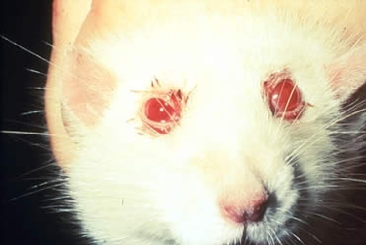
Chromodacryorrhea ("red tears") in a rat. Note the typical encrustations around the eyes due to the release of porphyrins from the Harderian lacrimal glands—a response that frequently indicates the animal has been subjected to some form of stress (eg, chronic respiratory disease, changes in environment). These porphyrins fluoresce pink under UV light.
Courtesy of Dr. Dean H. Percy.
When porphyrin-enriched tear fluid dries around the eyes and external nares, it resembles crusts of blood. Owners often describe their pet rats as hemorrhaging from the eyes and nose. Porphyrins can be readily differentiated from blood with a Wood’s lamp, because they fluoresce under UV light.
Although chromodacryorrhea is not pathological, it is a consequence of acute-onset stress such as that caused by pain, disease, or restraint. It usually indicates a chronic underlying disease.
Zoonotic Risk of Mice and Rats
A 2020 survey of rat-bite cases revealed that children and young adults (ages 0–19) accounted for > 50% of the cases of rat-bite fever in the US (25).
Rat-bite fever, caused by infection with Streptobacillus moniliformis, manifests in humans as bacteremia and septicemia characterized by fever, chills, myalgia, arthralgia, headache, and vomiting. A petechial rash develops over the extremities, especially the palms and soles, but sometimes is present all over the body. The incubation period is 3–10 days (average 5 days).
Infants and children with rat-bite fever experience severe diarrhea resulting in weight loss. Death occurs in 7–13% of untreated patients. After infection, polyarthritis develops in 50–75% of patients (26). The prognosis is good when the disease is treated early with parenteral penicillin. Fatalities are associated with late reporting or late recognition of the disease.
The nonspecific initial presentation of rat-bite fever, combined with difficulties in culturing Streptococcus moniliformis, can result in a substantial risk of delay or failure in diagnosis. Rats are not recommended as pets for children. Adults with pet rats should practice regular handwashing and avoid hand-to-mouth contact when handling rats or cleaning rat cages.
Pearls & Pitfalls
|
There are sporadic but persistent reports of dermatitis from the tropical rat mite, Ornithonyssus bacoti, in humans, acquired from pet rodents, that manifests as an erythematous papular rash. Affected patients often excoriate because of intense pruritus. Occasionally, vesicles, urticarial plaques, diffuse erythema, or hemorrhage occurs. Affected areas of the skin are those usually covered by clothing (such as arms and torso); the face and webs of fingers are usually spared.
Considerable evidence suggests that O bacoti carries and has the potential to transmit several human pathogens (27, 28). Experiments have shown that O bacoti transmits the following pathogens:
coxsackievirus (hand-foot-and-mouth disease)
Francisella tularensis (tularemia)
Rickettsia akari (which causes rickettsialpox)
Trypanosoma cruzi (Chagas disease)
Yersinia pestis (plague)
Researchers have also documented O bacoti specimens containing the following pathogens (29, 30):
Bartonella spp
Borrelia spp (Lyme disease)
Coxiella burnetii (Q fever)
Rickettsia spp
As inapparent carriers of dermatophytes, pet mice are a zoonotic risk for dermatophytoses in their owners, especially children.
Key Points
Appropriate husbandry is key to maintaining the health of pet mice and rats. Use of appropriate bedding and frequent cleaning of cages help prevent respiratory diseases that can be exacerbated in an environment with increased ammonia levels.
Because of the small size of mice and rats, handling methods must be adapted to minimize stress while achieving appropriate restraint for clinical procedures. Care must be taken, especially with mice, not to withdraw too much blood during blood collection.
Older rats commonly have tumors, usually mammary fibroadenomas. If a rat has a mammary tumor, a concomitant pituitary tumor should always be considered. Treatment recommendations include surgical excision followed by deslorelin implant to prevent recurrence. Spaying females before the age of 7 months decreases the chance of mammary tumor formation.
For More Information
Frohlich J. Rats and mice. In: Quesenberry KE, Orcutt CJ, Mans C, Carpenter JW, eds. Ferrets, Rabbits, and Rodents: Clinical Medicine and Surgery. 4th ed. Elsevier; 2021:345-367.
Turner PV, Wheler CL, Beaufrère H, Fabian N, Harkness JE, eds. Harkness and Wagner's Biology and Medicine of Rabbits and Rodents. 6th ed. John Wiley & Sons; 2025.
References
Vesell ES. Induction of drug-metabolizing enzymes in liver microsomes of mice and rats by softwood bedding. Science 1967:157(3792):1057-1058. doi:10.1126/science.157.3792.1057
Nevalainen T, Vartiainen T. Volatile organic compounds in commonly used beddings before and after autoclaving. Scand J Lab Anim Sci. 1996;23(2). doi:10.23675/sjlas.v23i2.793
National Research Council, Institute for Laboratory Animal Research, Division on Earth and Life Studies, Committee for the Update of the Guide for the Care and Use of Laboratory Animals. Guide for the Care and Use of Laboratory Animals. 8th ed. National Academies Press; 2011. https://grants.nih.gov/grants/olaw/guide-for-the-care-and-use-of-laboratory-animals.pdf
Barney CC, Kuhrt DM. Intermittent heat exposure and thirst in rats. Physiol Rep. 2016;4(8):e12767. doi:10.14814/phy2.12767
Patterson-Kane EG. Cage size preference in rats in the laboratory. J Appl Anim Welf Sci. 2002;5(1):63-72. doi:10.1207/S15327604JAWS0501_5
Weiss IC, Pryce CR, Jongen-Rêlo AL, Nanz-Bahr NI, Feldon J. Effect of social isolation on stress-related behavioural and neuroendocrine state in the rat. Behav Brain Res. 2004;152(2):279-295. doi:10.1016/j.bbr.2003.10.015
de Oliveira DT, Souza-Silva E, Tonussi CR. Technical report: gingival vein punction: a new simple technique for drug administration or blood sampling in rats and mice. Scand J Lab Anim Sci. 2009;36(2):109–113. doi:10.23675/sjlas.v36i2.174
Fouriez-Lablée V, Vergneau-Grosset C, Kass PH, Zwingenberger AL. Comparison between thoracic radiographic findings and postmortem diagnosis of thoracic diseases in dyspneic companion rats (Rattus norvegicus). Vet Radiol Ultrasound. 2017;58(2):133-143. doi:10.1111/vru.12459
Morrisey JK, Carpenter JW. Formulary. In: Quesenberry KE, Orcutt CJ, Mans C, Carpenter JW, eds. Ferrets, Rabbits, and Rodents: Clinical Medicine and Surgery. 4th ed. Elsevier; 2021:620-630.
Meredith A. BSAVA Small Animal Formulary: Part B: Exotic Pets. 9th ed. British Small Animal Veterinary Association; 2015.
Graham JE, Schoeb TR. Mycoplasma pulmonis in rats. J Exot Pet Med. 2011:20(4);270-276. doi:10.1053/j.jepm.2011.07.004
Donnelly TM. Disease problems of small rodents. In: Quesenberry KE, Carpenter JW. Ferrets, Rabbits, and Rodents: Clinical Medicine and Surgery. 2nd ed. Saunders; 2004:299-315. doi:10.1016/B0-72-169377-6/50031-9
Flynn BM, Brown PA, Eckstein JM, Strong D. Treatment of Syphacia obvelata in mice using ivermectin. Lab Anim Sci. 1989(5);39:461-463.
Frohlich J, Mayer J. Rodents. In: Carpenter JW, Harms CA, eds. Carpenter's Exotic Animal Formulary. 6th ed. Saunders; 2023:530-573. doi:10.1016/B978-0-323-83392-9.00009-5
Baumans V, Havenaar R, van Herck H, Rooymans TP. The effectiveness of Ivomec and Neguvon in the control of murine mites. Lab Anim. 1988;22:243-245. doi:10.1258/002367788780746368
Planas-Silva MD, Rutherford TM, Stone MC. Prevention of age-related spontaneous mammary tumors in outbred rats by late ovariectomy. Cancer Detect Prev. 2008;32(1):65-71. doi:10.1016/j.cdp.2008.01.004
Vergneau-Grosset C, Keel MK, Goldsmith D, Kass PH, Paul-Murphy J, Hawkins MG. Description of the prevalence, histologic characteristics, concomitant abnormalities, and outcomes of mammary gland tumors in companion rats (Rattus norvegicus): 100 cases (1990–2015). J Am Vet Med Assoc. 2016;249(10):1170-1179. doi:10.2460/javma.249.10.1170
Mayer J, Sato A, Kiupel M, DeCubellis J, Donnelly T. Extralabel use of cabergoline in the treatment of a pituitary adenoma in a rat. J Am Vet Med Assoc. 2011;239(5):656-660. Erratum in: J Am Vet Med Assoc. 2011;239(8):1089. doi:10.2460/javma.239.5.656
Squartini F, Pingitore R. Tumours of the mammary gland. In: Turusov VS, Mohr U, eds. Tumours of the Mouse. 2nd ed. International Agency for Research on Cancer; 1994:47-100.
Heider K, Eustis SL. Tumors of the soft tissues. In: Turusov VS, Mohr U, eds. Tumours of the Mouse. 2nd ed. International Agency for Research on Cancer; 1994:611-631.
Whary MT, Baumgarth N, Fox JG, Barthold SW. Biology and diseases of mice. In Fox JG, Anderson LC, Otto G, Pritchett-Corning KR, Whary MT, eds. Laboratory Animal Medicine. 3rd ed. Academic Press; 2015:43-149. doi:10.1016/B978-0-12-409527-4.00003-1
Krogstad AP, Franklin CL, Besch-Williford CL. An epidemiological and diagnostic approach to murine skin lesions [abstract]. In: Proceedings of the 52nd American Association for Laboratory Animal Science National Meeting, Baltimore; 2001:94.
Andrews AG, Dysko RC, Spilman SC, Kunkel RG, Brammer DW, Johnson KJ. Immune complex vasculitis with secondary ulcerative dermatitis in aged C57BL/6NNia mice. Vet Pathol. 1994;31(3):293-300. doi:10.1177/030098589403100301
Sargent JL, Koewler NJ, Diggs HE. Systematic literature review of risk factors and treatments for ulcerative dermatitis in C57BL/6 mice. Comp Med. 2015;65(6):465-472. https://pmc.ncbi.nlm.nih.gov/articles/PMC4681240
Kache PA, Person MK, Seeman SM, McQuiston JR, McCollum J, Traxler RM. Rat-bite fever in the United States: an analysis using multiple national data sources, 2001-2015. Open Forum Infect Dis. 2020;7(6):ofaa197. doi:10.1093/ofid/ofaa197
Center for Food Security and Public Health. Rat bite fever [factsheet]. Updated May 2021. Accessed April 27, 2026. https://www.cfsph.iastate.edu/Factsheets/pdfs/rat_bite_fever.pdf
d'Ovidio D, Noviello E, Santoro D. Prevalence and zoonotic risk of tropical rat mite (Ornithonyssus bacoti) in exotic companion mammals in southern Italy. Vet Dermatol. 2018;29(6):522-e174. doi:10.1111/vde.12684
Schwab M, Allen B, Sulkin SE. The tropical rat mite (Liponyssus bacoti) as an experimental vector of Coxsackie virus. Am J Trop Med Hyg. 1952;1(6):982-986. doi:10.4269/ajtmh.1952.1.982
Reeves WK, Loftis AD, Szumlas DE, et al. Rickettsial pathogens in the tropical rat mite Ornithonyssus bacoti (Acari: Macronyssidae) from Egyptian rats (Rattus spp.). Exp Appl Acarol. 2007;41(1-2):101-107. doi:10.1007/s10493-006-9040-3
Lopatina IuV, Vasil'eva IS, Gutova VP, et al. [An experimental study of the capacity of the rat mite Ornithonyssus bacoti (Hirst, 1913) to ingest, maintain and transmit Borrelia]. Article in Russian. Med Parazitol (Mosk). 1999;2:26-30. https://pubmed.ncbi.nlm.nih.gov/10703202