


The ocular fundus consists of the upper tapetal fundus (if present), ventral and surrounding nontapetal fundus, retinal vasculature, and optic disc. Histologically, the posterior segment, from superficial to deep, consists of the following structures: 1) posterior sclera; 2) choroid, which contains pigmented cells, blood vessels to support the high metabolic needs of the outer retina, and the tapetum lucidum to enhance vision in dim light (tapetum cellulosum in carnivores and tapetum fibrosum in herbivores); 3) retina, which consists of the nine layers of neurosensory retina and the outer retinal pigment epithelium; and 4) the optic disc, where the retinal ganglion axons leave the eye through a weak and fenestrated scleral lamina cribrosa to synapse in the lateral geniculate body (vision) or middle brain (pupillary light reflex [Edinger-Westphal nucleus] or midbrain and rostral colliculi [dazzle reflex]). Whereas dogs, cats, horses, cattle, sheep, goats, and many other species have an upper tapetal fundus, pigs and rabbits typically lack a tapetal fundus.
Disorders of the ocular fundus may be primary or may be manifestations of systemic diseases. Inherited abnormalities may be congenital or appear later, and they are important in the pathogenesis of retinopathies in dogs and cats. Trauma, metabolic disturbances, systemic infections, neoplasms, blood dyscrasias, hypertension, and nutritional deficiencies are possible underlying causes of retinopathies in all species.
Inherited Retinopathies
Collie eye anomaly is a congenital, recessively inherited (CEA-CH mutation) ocular defect with variable expression in rough- and smooth-coated Collies. It also occurs in Shetland Sheepdogs, Border Collies, Australian Shepherds, Lancashire Heelers, longhaired Whippets, Boykin Spaniels, and Nova Scotia Duck Tolling Retrievers. The basic lesion is an area of choroidal or chorioretinal hypoplasia that appears as a focal, variable-sized, pale, pink area lateral to the optic disc. More severely affected dogs (10%–20%) can have additional colobomatous lesions of the optic papilla or peripapillary region and occasional retinal detachments (2%–5%). Intraocular hemorrhage may occur. Vision is not appreciably affected unless retinal detachment develops. With recent availability of the CEA-CH DNA test, ophthalmoscopic diagnosis of Collie eye anomaly can be confirmed in additional dog breeds that have the same mutation, as well as in potential carrier dogs within affected breeds before breeding.

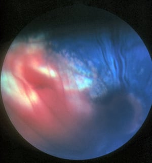
Early retinal detachment and hemorrhage developing lateral to the optic disc in the eye of a Collie puppy.
Early retinal detachment and hemorrhage developing lateral to the optic disc in the eye of a Collie puppy.
Courtesy of K. Gelatt.
Early retinal detachment and hemorrhage developing lateral to the optic disc in the eye of a Collie puppy.
Early retinal detachment and hemorrhage developing lateral to the optic disc in the eye of a Collie puppy.
Courtesy of K. Gelatt.

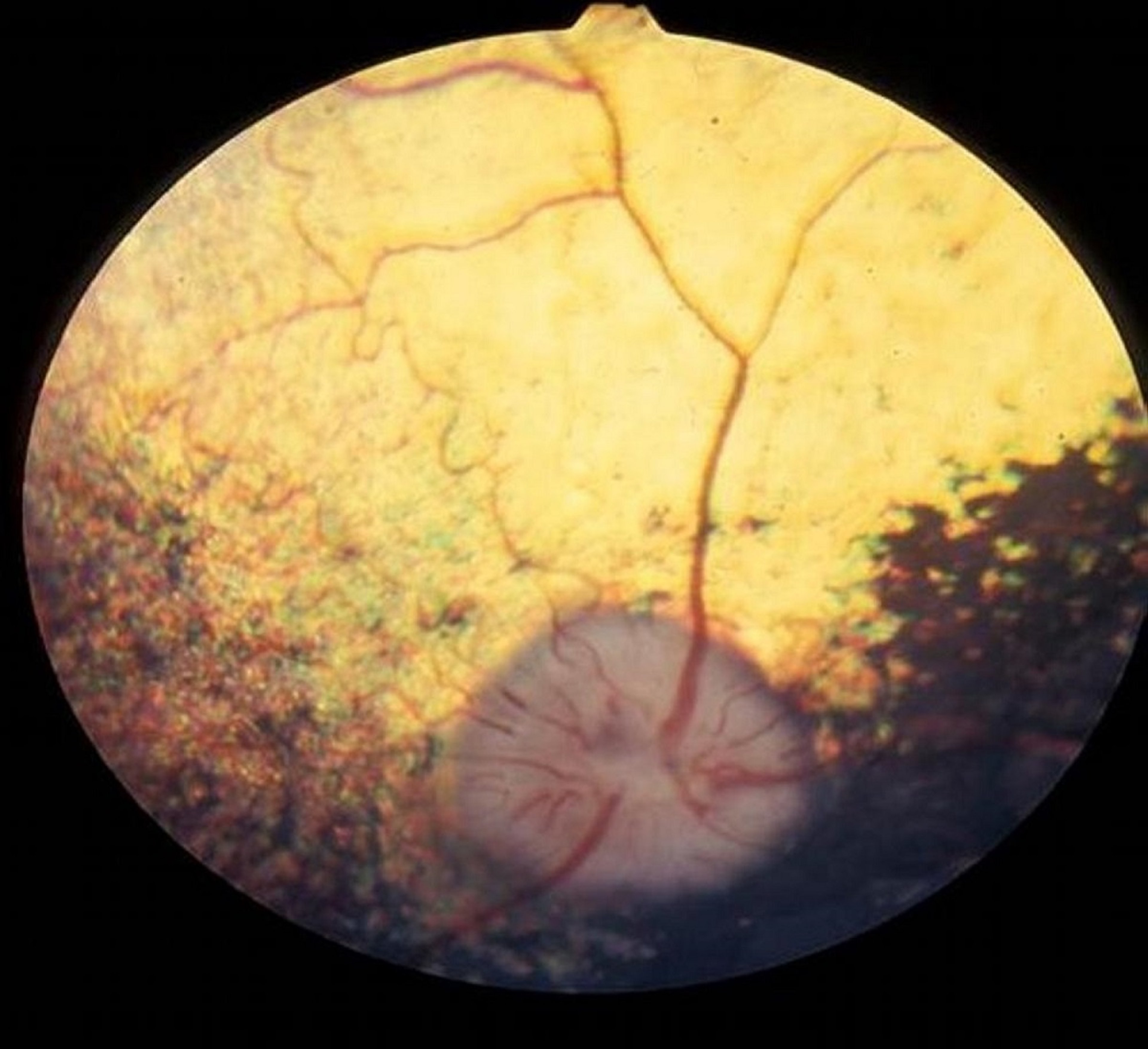
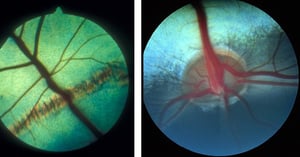
Both choroidal hypoplasia and optic nerve coloboma are evident in this photograph of a sable Rough Collie's eye. The choroidal hypoplasia is lateral to the optic nerve. The optic nerve coloboma is in the central half of the optic disc, where the blood vessels on the optic disc are out of focus.
Both choroidal hypoplasia and optic nerve coloboma are evident in this photograph of a sable Rough Collie's eye. The ch
Courtesy of K. Gelatt.


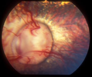
Choroidal hypoplasia (pink to white area lateral to the optic disc) and a peripapillary coloboma (arrow)—typical characteristics of Collie eye anomaly.
Choroidal hypoplasia (pink to white area lateral to the optic disc) and a peripapillary coloboma (arrow)—typical charac
Courtesy of K. Gelatt.
Early retinal detachment and hemorrhage developing lateral to the optic disc in the eye of a Collie puppy.
Early retinal detachment and hemorrhage developing lateral to the optic disc in the eye of a Collie puppy.
Courtesy of K. Gelatt.
Early retinal detachment and hemorrhage developing lateral to the optic disc in the eye of a Collie puppy.
Early retinal detachment and hemorrhage developing lateral to the optic disc in the eye of a Collie puppy.
Courtesy of K. Gelatt.
Both choroidal hypoplasia and optic nerve coloboma are evident in this photograph of a sable Rough Collie's eye. The choroidal hypoplasia is lateral to the optic nerve. The optic nerve coloboma is in the central half of the optic disc, where the blood vessels on the optic disc are out of focus.
Both choroidal hypoplasia and optic nerve coloboma are evident in this photograph of a sable Rough Collie's eye. The ch
Courtesy of K. Gelatt.
Choroidal hypoplasia (pink to white area lateral to the optic disc) and a peripapillary coloboma (arrow)—typical characteristics of Collie eye anomaly.
Choroidal hypoplasia (pink to white area lateral to the optic disc) and a peripapillary coloboma (arrow)—typical charac
Courtesy of K. Gelatt.

English Springer Spaniel's eye with areas of retinal dysplasia (white to gray and dark patches) affecting the central retina immediately dorsal to the optic disc.
Courtesy of K. Gelatt.

Multiple linear, hyporeflective retinal folds in the tapetal fundus are evident.
Courtesy of Dr. Ralph Hamor.
Retinal dysplasia is a congenital, focal, or generalized maldevelopment of the retina that may arise from trauma, genetic defect, or prenatal viral infection. It occurs as circular to multifocal areas of focal retinal detachment dorsal to the optic nerve. Most forms of retinal dysplasia in dogs are inherited, and many DNA mutations have been reported. Maternal viral infections, especially during early fetal development, can result in multiple ocular anomalies with retinal dysplasia in kittens (feline panleukopenia), lambs (bluetongue), puppies (canine herpesviral infection), and calves (bovine viral diarrhea). Breeds of dogs with focal, geographic, and generalized retinal dysplasia thought to be inherited as an autosomal recessive trait include Beagles, Cocker Spaniels, Labrador Retrievers, Rottweilers, and Yorkshire Terriers. Focal areas of retinal maldevelopment may be asymptomatic, but if they are large, hyperreflective, and pigmented, they can interfere with central vision. Generalized retinal dysplasia with retinal detachment, visual impairment, or blindness is inherited in English Springer Spaniels, Bedlington Terriers, Sealyham Terriers, Labrador Retrievers, Doberman Pinschers, and Australian Shepherds. Other ocular anomalies, including microphthalmia and congenital cataracts, often accompany the generalized forms. In Labrador Retrievers and Samoyeds, retinal dysplasia may be associated with skeletal dysplasia (shortening) of the forelegs.

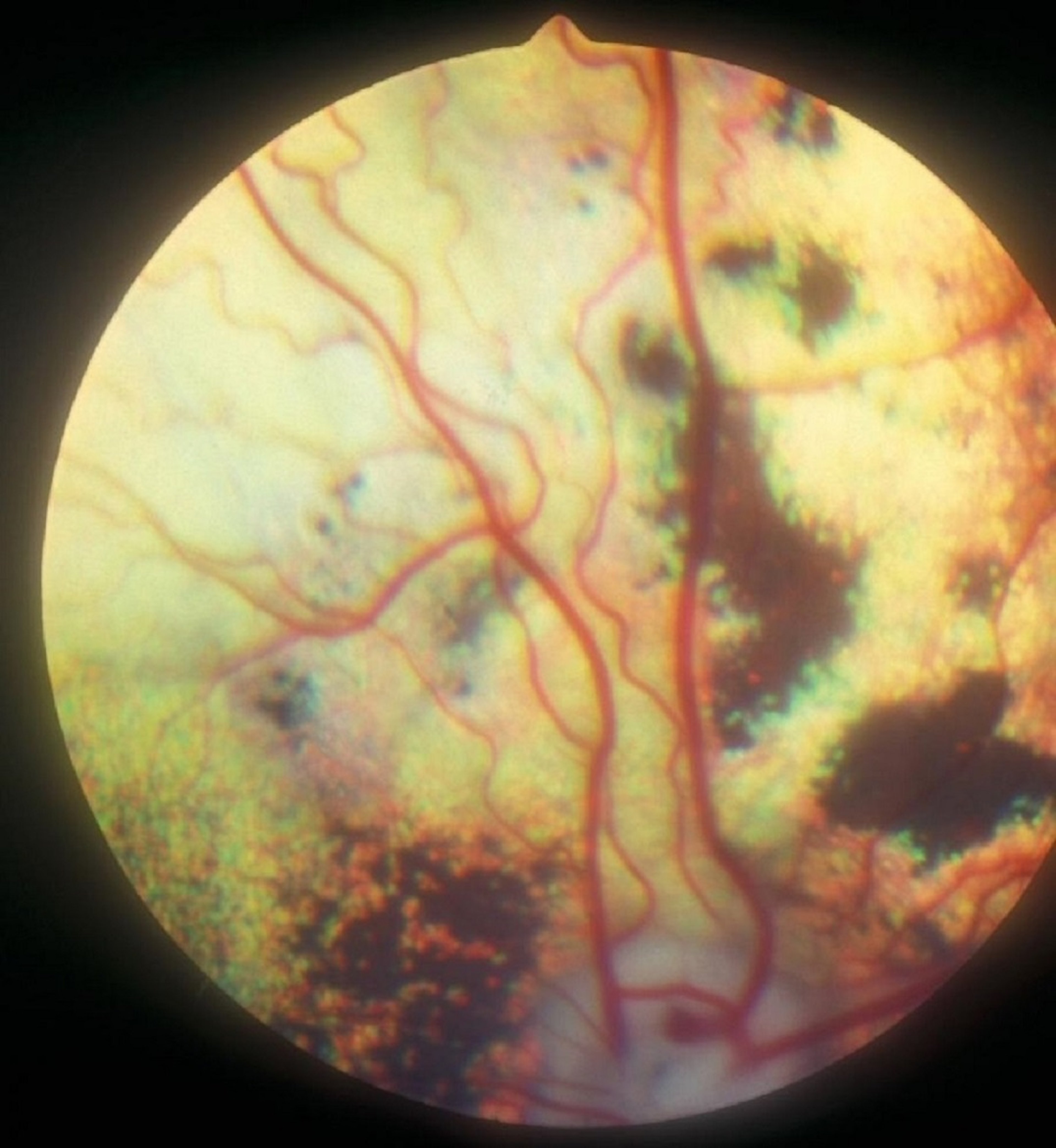
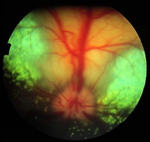
Early progressive retinal atrophy in a Collie's eye: increased tapetal reflectivity and a decreased number of retinal vessels.
Courtesy of K. Gelatt.

The increased tapetal reflectivity, decreased retinal vascularity, and early optic nerve atrophy evident in this photograph of a Miniature Poodle's eye are consistent with a diagnosis of progressive retinal atrophy.
Courtesy of K. Gelatt.
Progressive retinal atrophy (PRA) is a group of degenerative retinopathies consisting of inherited photoreceptor dysplasia and other degenerations that have a similar clinical appearance. A large number of mutations and DNA tests have been reported for these diseases that vary by dog breed. The photoreceptor dysplasias inherited as autosomal recessive traits in which clinical signs develop in the first year occur in Irish Setters, Collies, Norwegian Elkhounds, Miniature Schnauzers, and Belgian Sheepdogs. The photoreceptor degenerations inherited as autosomal recessive traits in which clinical signs develop at 3–5 years occur in Miniature and Toy Poodles, Cocker Spaniels and English Cocker Spaniels, Labrador Retrievers, Tibetan Terriers, Tibetan Spaniels, Papillons, English Springer Spaniels, miniature longhaired Dachshunds, Akitas, and Samoyeds. In Siberian Huskies, PRA is inherited as an X-linked trait, whereas in Mastiffs and Bullmastiffs, PRA is inherited as an autosomal dominant trait. Many other breeds of dogs are also suspected of having inherited PRA. In Abyssinian cats, PRA occurs as both photoreceptor dysplasia and degeneration. Typically, night blindness (nyctalopia) is noted early and progresses to total blindness over months to years, because most inherited retinopathies affect rod photoreceptors first. Ophthalmoscopic lesions are a bilateral symmetrical increase in reflectivity of the tapetal fundus, decreased pigmentation of the nontapetal fundus, attenuation and a decrease in the number of retinal vessels, and eventual atrophy of the optic disc. Electroretinography is often used to investigate and diagnose the condition. Cortical cataracts are common late in the course of PRA in many breeds and may mask the underlying retinopathy. No effective treatment is currently available. For many dog breeds, blood and buccal-mucosa-based DNA marker and specific gene tests have been developed to detect carrier and affected animals before clinical signs develop. The list of breeds affected with inherited retinal degenerations and causative genes continues to increase; for current information consult the recent literature.

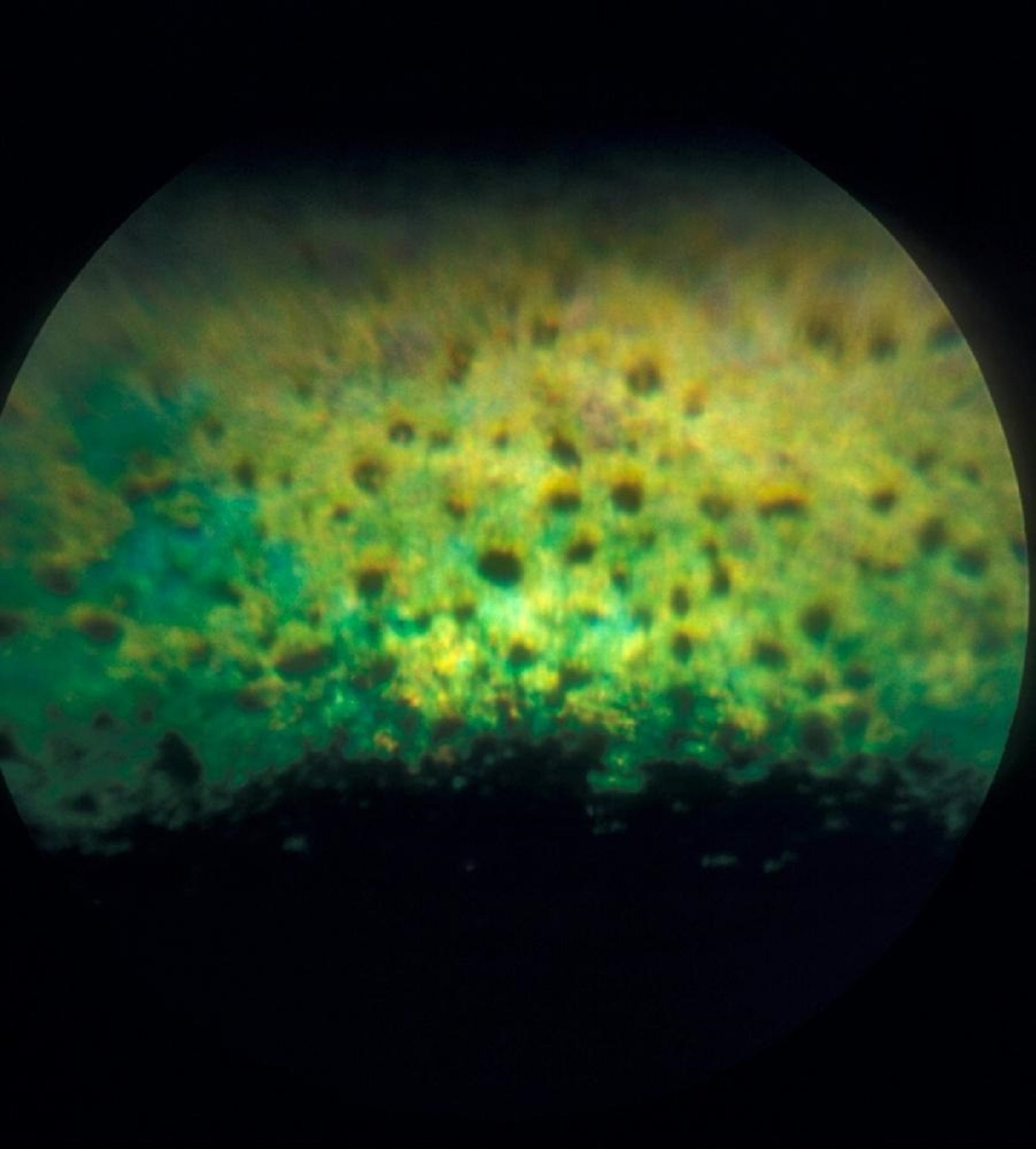
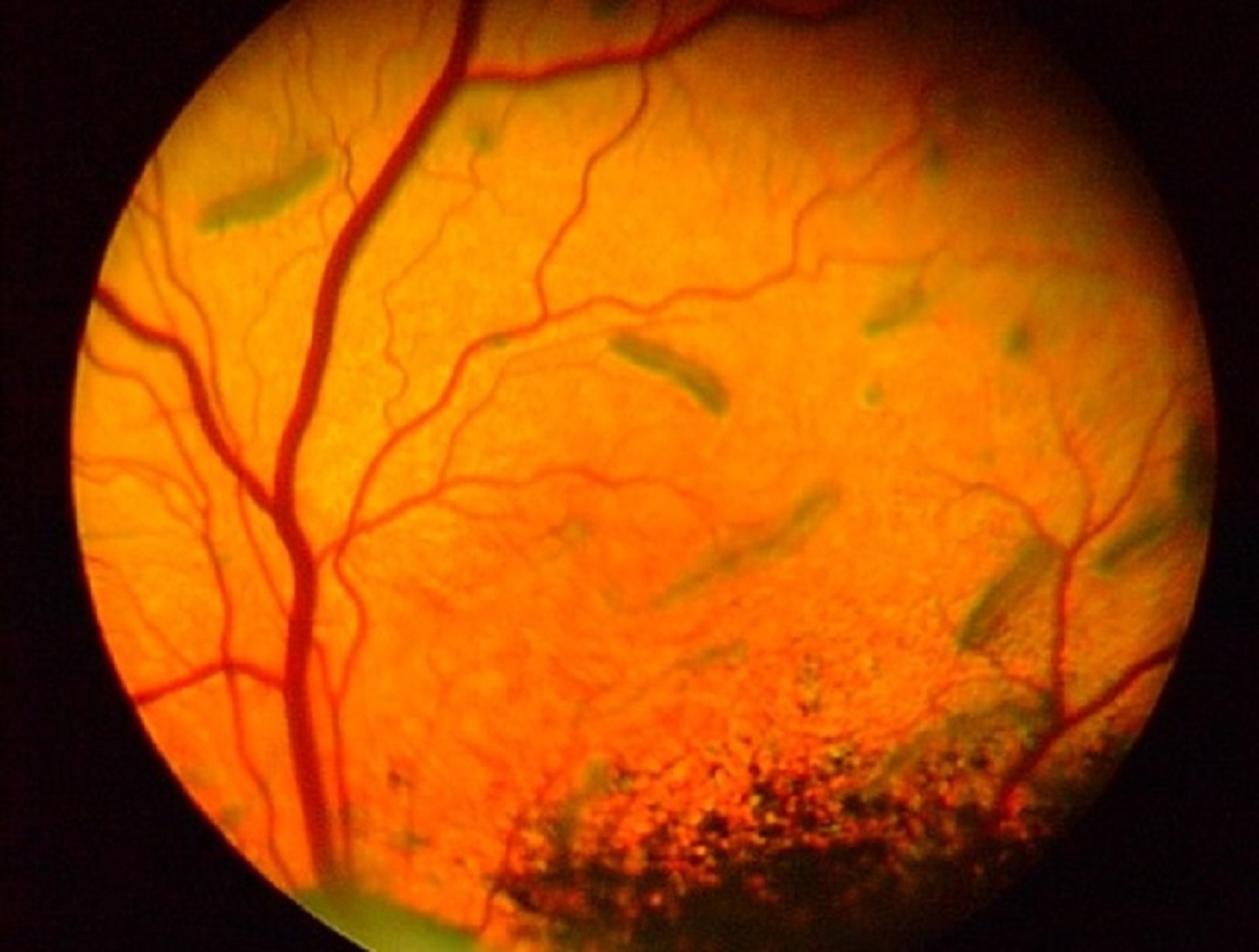
Spots caused by retinal pigment epithelium dystrophy in the eye of a Labrador Retriever that has chronic disease. Note the large, pigmented foci and diffuse retinal thinning (tapetal hyperreflectivity).
Courtesy of K. Gelatt.
Retinal pigment epithelial dystrophy (central progressive retinal atrophy) occurs in Labrador Retrievers, smooth and rough Collies, Border Collies, Shetland Sheepdogs, and Briards. The condition may be inherited in Labrador Retrievers as a dominant trait with variable penetrance. Early ophthalmoscopic findings (often before clinical signs are apparent) are small foci of irregular pigmentation (consisting of lipofuscin) in the tapetal fundus, which eventually coalesce and fade as reflectivity of the tapetal fundus increases. The pigmented nontapetal fundus becomes mottled, the retinal vasculature gradually decreases, and the optic disc atrophies. Progressive visual impairment occurs gradually over several years. Cataracts form late in the disease. There is no treatment. Studies in English Cocker Spaniels suggest that vitamin E disorders may also be important in the pathogenesis of this disease complex. A similar condition in horses, equine motor neuron disease, has similar focal, yellow-brown areas scattered throughout the tapetal fundus and has also been associated with vitamin E deficiency.
Chorioretinitis
Chorioretinitis is frequently a manifestation of systemic infectious disease; it is important as both a convenient diagnostic clue and a prognosticator of visual function. Unless the lesions are generalized or involve the optic nerve, they may not cause a notable visual deficit. Scars may be differentiated from active lesions by the haze and ill-defined borders of the latter. Retinal scars are hyperreflective and may have increased pigmentation, whereas active lesions are hyporeflective. Routine ophthalmoscopic examinations of all animals with systemic diseases often permit rapid diagnosis of many specific diseases. Chorioretinitis may be present with canine distemper, systemic mycoses in dogs and cats, algaemia, feline toxoplasmosis, tuberculosis, bacterial septicemias in young animals, feline infectious peritonitis (FIP), thromboembolic meningoencephalitis in cattle, bovine malignant catarrhal fever, classical swine fever, and leptospirosis and onchocerciasis in horses. Treatment is directed at the systemic disease.

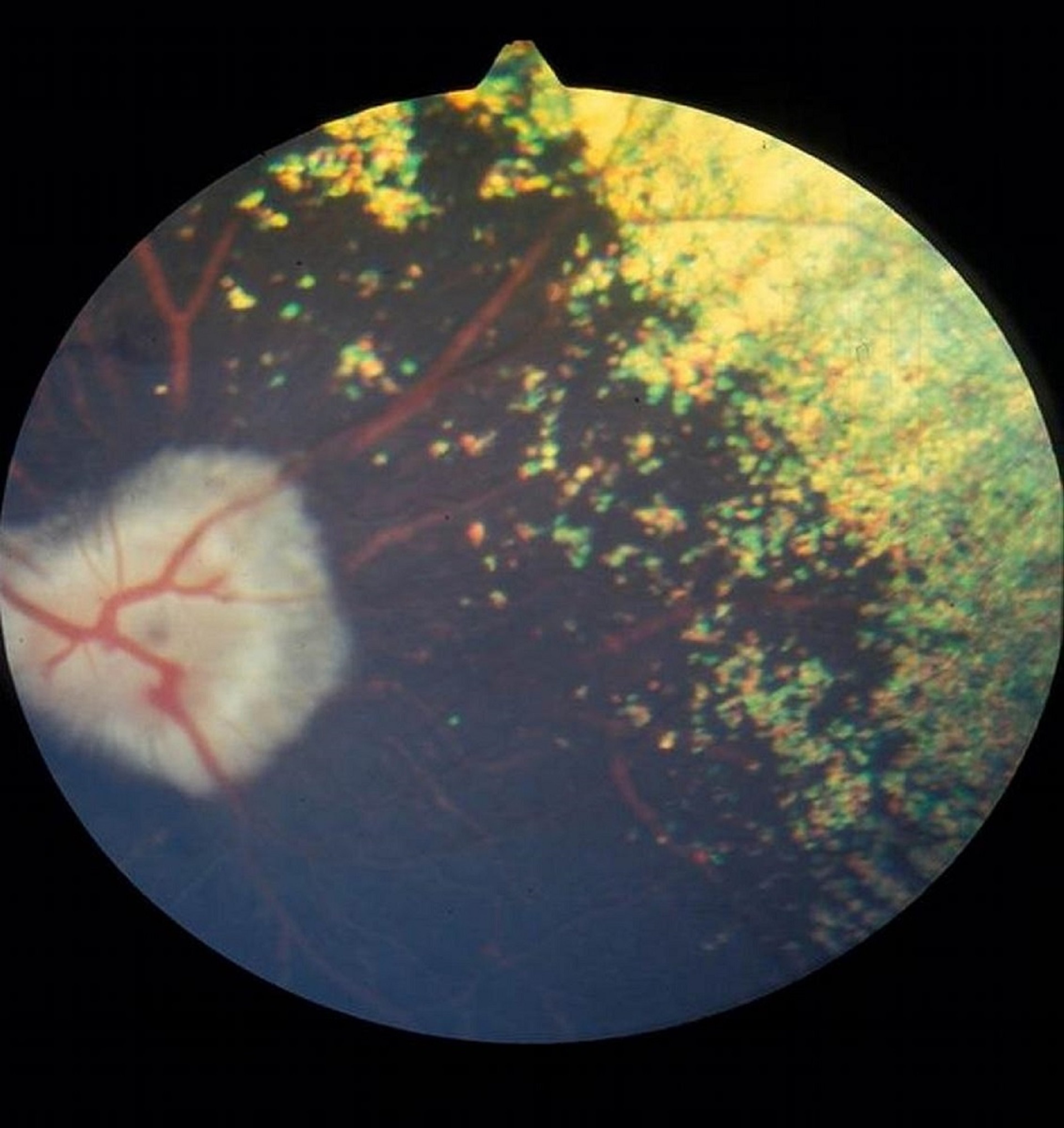
Left: Active chorioretinitis in the eye of a cat. The normally transparent retina is infiltrated with edema, inflammatory exudates, and cells appearing as raised, translucent, or cloudy areas (hyporeflective lesions). This lesion is consistent with a fungal infection such as histoplasmosis. Right: Normal cat fundus.
Left: Active chorioretinitis in the eye of a cat. The normally transparent retina is infiltrated with edema, inflammato
Left image courtesy of K. Gelatt. Right image courtesy of Dr. Ralph Hamor.

Left: Lesion (tan, linear, hyper-reflective lesion with cross-hatches) associated with previous bacterial septicemia in the eye of an adult Guernsey cow. Pigmentation of the inflamed tissues and changes in reflectivity of the tapetal fundus are evident. Right: Normal cow fundus.
Left: Lesion (tan, linear, hyper-reflective lesion with cross-hatches) associated with previous bacterial septicemia in
Left image courtesy of K. Gelatt. Right image courtesy of Dr. Ralph Hamor.

Large, yellow, hyporeflective lesion in most of the tapetum, secondary to systemic blastomycosis.
Large, yellow, hyporeflective lesion in most of the tapetum, secondary to systemic blastomycosis.
Courtesy of Dr. Ralph Hamor.

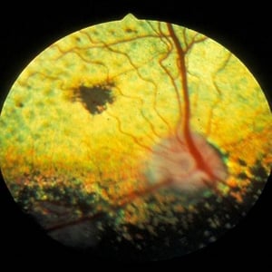
An inactive lesion (black area in the central tapetal fundus) of the choroid and retina is evident in this photograph showing the eye of a hunting dog affected with testicular blastomycosis.
An inactive lesion (black area in the central tapetal fundus) of the choroid and retina is evident in this photograph s
Courtesy of K. Gelatt.
Left: Active chorioretinitis in the eye of a cat. The normally transparent retina is infiltrated with edema, inflammatory exudates, and cells appearing as raised, translucent, or cloudy areas (hyporeflective lesions). This lesion is consistent with a fungal infection such as histoplasmosis. Right: Normal cat fundus.
Left: Active chorioretinitis in the eye of a cat. The normally transparent retina is infiltrated with edema, inflammato
Left image courtesy of K. Gelatt. Right image courtesy of Dr. Ralph Hamor.
Left: Lesion (tan, linear, hyper-reflective lesion with cross-hatches) associated with previous bacterial septicemia in the eye of an adult Guernsey cow. Pigmentation of the inflamed tissues and changes in reflectivity of the tapetal fundus are evident. Right: Normal cow fundus.
Left: Lesion (tan, linear, hyper-reflective lesion with cross-hatches) associated with previous bacterial septicemia in
Left image courtesy of K. Gelatt. Right image courtesy of Dr. Ralph Hamor.
Large, yellow, hyporeflective lesion in most of the tapetum, secondary to systemic blastomycosis.
Large, yellow, hyporeflective lesion in most of the tapetum, secondary to systemic blastomycosis.
Courtesy of Dr. Ralph Hamor.
An inactive lesion (black area in the central tapetal fundus) of the choroid and retina is evident in this photograph showing the eye of a hunting dog affected with testicular blastomycosis.
An inactive lesion (black area in the central tapetal fundus) of the choroid and retina is evident in this photograph s
Courtesy of K. Gelatt.
Retinal Detachments
Retinal detachment, or separation of the neurosensory retina from the retinal pigment epithelium, occurs in most species. In dogs, retinal detachment is associated with congenital retinal disorders (retinal dysplasia and Collie eye anomaly), chorioretinitis, systemic hypertension, penetrating trauma, intraocular surgery, and posterior segment neoplasia. In cats, retinal detachments occur most commonly with chorioretinitis associated with infectious disease and systemic hypertension. In horses, the most frequent causes are trauma, intraocular surgery, and recurrent uveitis.
Retinal detachments are divided clinically into nonrhegmatogenous (serous, exudative, hemorrhagic, secondary to vitreal syneresis) and rhegmatogenous (with retinal breaks [hole or tear]). Clinical signs include mydriasis, anisocoria, vision impairment, and intraocular hemorrhage. Methods used for diagnosis are ophthalmoscopy and, in eyes with an opaque cornea or lens, ocular ultrasonography.
Nonrhegmatogenous serous retinal detachments are usually treated medically, with treatment directed at the primary disease. Retinal reattachment occurs with resolution of the subretinal exudates and hemorrhage. Variable retinal degeneration may follow in the detached areas. Rhegmatogenous retinal detachments with retinal breaks generally require surgical correction.


