

Cryptococcosis is a systemic fungal disease that often affects the respiratory tract (especially the nasal cavity), CNS, eyes, and skin. The causal fungi exist in tissues in an encapsulated yeast form and likely in the environment in a filamentous form or an unencapsulated yeast form.
Infection occurs worldwide from Cryptococcus neoformans and C gattii, among others. The fungi are found in soil and bird (especially pigeon) feces. Transmission is by inhalation of spores or contamination of wounds. In avian feces, the fungus may occur in a nonencapsulated form as small as 1 mcm, which can be inhaled deep into the respiratory tract.
Cryptococcosis is most common in cats but also occurs in dogs, cattle, horses, sheep, goats, birds, and wildlife. In humans, many cases are associated with a defective cell-mediated immune response. Multiple serotypes of Cryptococcus exist with each serotype being assigned to one or more species; insufficient information is available to separate the pathogenicity of each serotype or species.
Clinical Findings and Lesions in Cryptococcosis in Animals

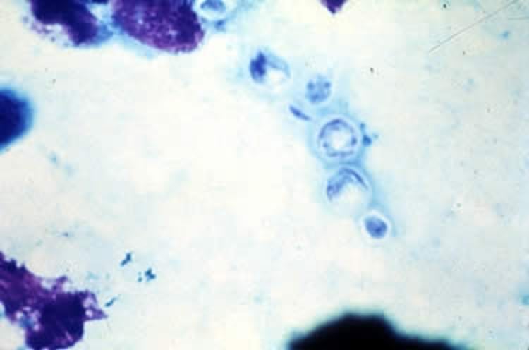
Cryptococcus neoformans, tissue smear, Wright stain, high power. Note the daughter cell budding from the large yeast. The typically large capsule is not visible with this stain.
Courtesy of Dr. John Prescott.
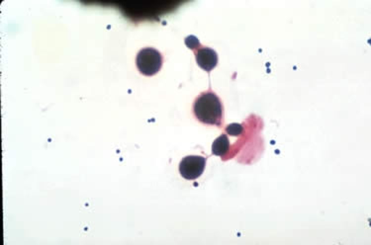
Cryptococcus neoformans, Gram stain, high power. Note the large size of these budding yeasts compared with the small gram-positive cocci bacteria present. The large capsule of the yeasts is barely visible.
Courtesy of Dr. John Prescott.

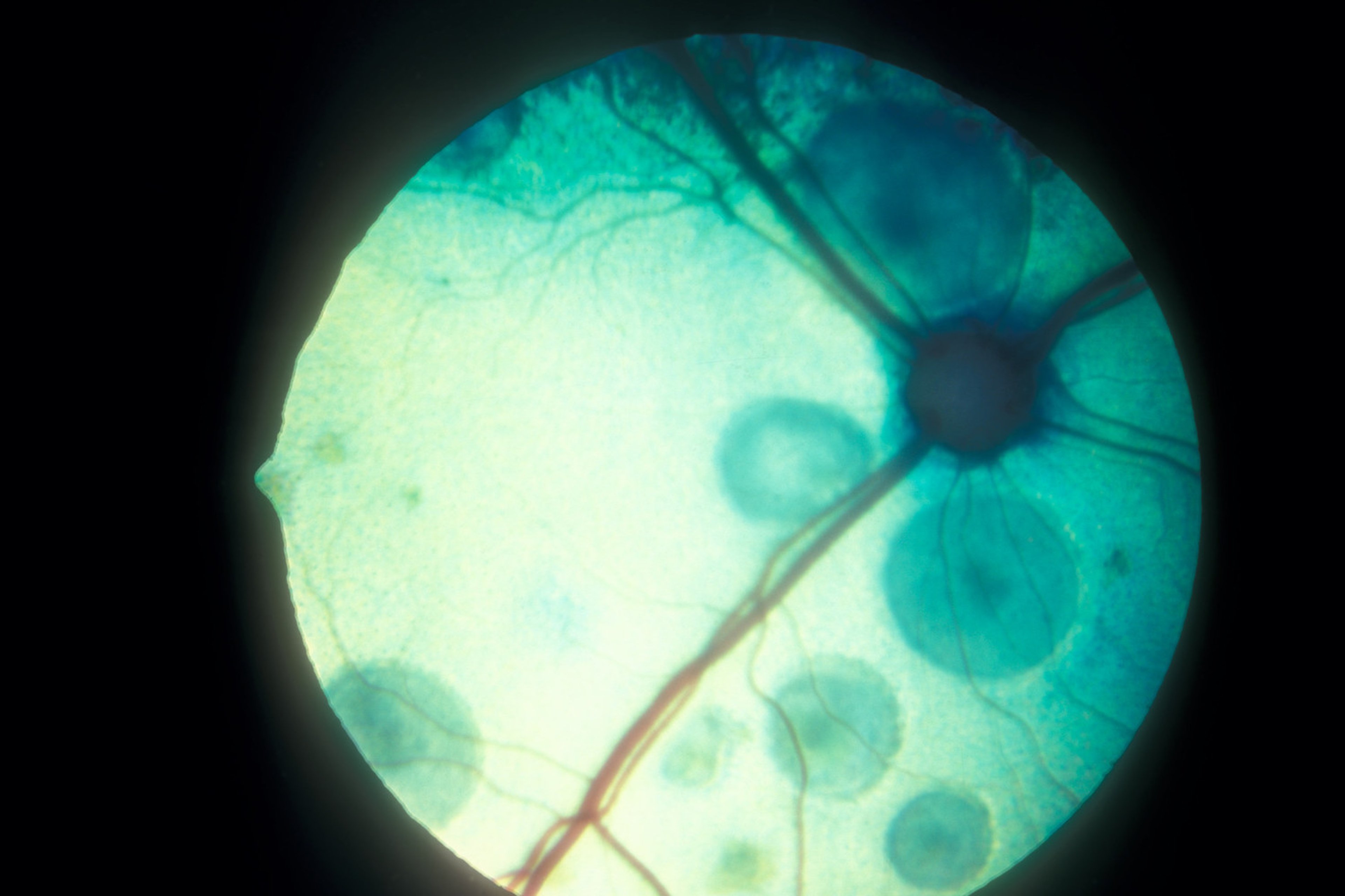
Cryptococcosis, characterized by neuroretinitis and focal retinal detachment, in a cat.
Courtesy of Dr. Kirk N. Gelatt.
In cats, chronic cryptococcosis is common, usually limited to the upper respiratory tract. Clinical signs secondary to nasal cavity infection are most common and include sneezing; mucopurulent, serous, or hemorrhagic unilateral or bilateral chronic nasal discharge; polyplike mass(es) in the nostril; and/or a firm, subcutaneous swelling over the bridge of the nose. Cutaneous lesions are also common and are characterized by papules and nodules that are fluctuant to firm.
Larger lesions tend to ulcerate, leaving a raw surface with a serous exudate. Neurologic signs associated with cryptococcosis of the CNS may include depression, changes in temperament, seizures, circling, paresis, and blindness. Ocular abnormalities may also develop, including dilated unresponsive pupils and blindness due to exudative retinal detachment, granulomatous chorioretinitis, panophthalmitis, and optic neuritis.
If disseminated disease occurs, lesions may also be found in bone and/or lymph nodes.
In contrast to cats, more than 50% of dogs may have disseminated disease with CNS or ocular involvement. Clinical signs are often related to meningoencephalitis, optic neuritis, and granulomatous chorioretinitis.
Lesions in the nasal cavity of many dogs have been reported; however, they are usually not the primary finding or reason for presentation. Approximately 50% of dogs have lesions in the respiratory tract, usually the lungs, and most have granulomas present in multiple systems. Structures often involved in order of decreasing frequency are CNS, respiratory tract, skin, kidneys, lymph nodes, spleen, liver, thyroid, adrenals, pancreas, bone, GI tract, muscle, myocardium, prostate, heart valves, and tonsils.
Bovine cryptococcosis has been associated only with cases of mastitis, and many cows in a herd may be infected. Affected cows have anorexia, decreased milk production, swelling and firmness of affected quarters, and enlarged supramammary lymph nodes. The milk becomes viscid, mucoid, and gray-white, or it may be watery with flakes. The disease in horses almost invariably manifests as obstructive masses within the nasal cavity.
The incubation period may range from 2–13 months. Lesions associated with cryptococcosis vary from a gelatinous mass consisting of numerous organisms with minimal inflammation to granuloma formation. The lesion is usually composed of aggregates of encapsulated organisms within a connective tissue framework.
The cellular response is primarily macrophages and giant cells with a few plasma cells and lymphocytes. Epithelioid giant cells and areas of caseous necrosis are less common than with the other systemic mycoses.
Diagnosis of Cryptococcosis in Animals
Cytology with Gram stain
Yeasts are 5–10 mcm ovoid structures with narrow-based budding
The most rapid method of diagnosis of cryptococcosis is cytologic evaluation of nasal exudate, skin exudate, CSF, or samples obtained by paracentesis of the aqueous or vitreous chambers of the eye or by impression smears of nasal or cutaneous masses. Gram stain is most useful; the organism retains the crystal violet, whereas the capsule stains lightly red with safranin.
India ink may also be used to visualize the organism, which appears unstained and silhouetted against a black background. It is not as definitive as Gram stain unless budding is present, because lymphocytes, fat droplets, and aggregated India ink particles may be confused with the organism. Wright stain has been used most often in diagnosing canine and feline cases; however, this stain can cause the organism to shrink and the capsule to become distorted. New methylene blue and periodic acid-Schiff (PAS) stains are better than Wright stain for this reason.
Because cytologic evaluation is rapid, impression smears or potassium hydroxide preparations should always be made of suspected cryptococcal lesions. Histopathologic examination with fungal stains or immunohistochemistry may be useful if no organisms are evident on impressions.
Detection of cryptococcal capsular antigen in serum, urine, or CSF is a useful, rapid method of diagnosis in those suspected cases in which the organism is not identified. A latex agglutination test is commercially available in kit form. The antigen titer can also be used to help determine response to treatment. A point-of-care immunochromatographic assay for serum also has high sensitivity and specificity.
The organism can be cultured from exudate, CSF, urine, joint fluid, and tissue samples if a large enough sample volume is available. Sabouraud agar with antibiotics is used if bacterial contamination is likely.
Treatment of Cryptococcosis in Animals
Fluconazole for cats; dogs with disseminated disease should add amphotericin B
Treat until antigen titer is repeatedly negative
Fluconazole (10 mg/kg, PO, every 12 hours) and itraconazole (5–10 mg/kg every 24 hours) are considered the treatments of choice. Amphotericin B may be added for patients, particularly dogs, with disseminated disease. Consult a formulary for the various formulations and dosing protocols for amphotericin B for a given species.
Flucytosine can be used alone; however, drug resistance may develop, so combination treatment with amphotericin is recommended. Hepatic enzyme activity should be monitored frequently during treatment. Flucytosine should not be used in dogs because of the development of toxic epidermal necrolysis. Voriconazole is usually not tolerated in cats or dogs.
Short-acting glucocorticoid treatment may be necessary for the first few days to weeks of treatment in patients with CNS cryptococcosis; neurologic deterioration during the first week of treatment is common and is not a negative prognostic indicator.
Treatment should continue until at least two cryptococcal antigen tests are negative, which may be months to years. Horses may be treated with debulking of the nasal mass and topical azole treatment followed postsurgically by systemic azole treatment. There is no labeled treatment for cryptococcal mastitis in cattle.
Key Points
Cryptococcosis is a worldwide disease most commonly affecting cats and humans.
Disease is readily diagnosed using cytology.
Treatment is usually fluconazole for an extended period.


