


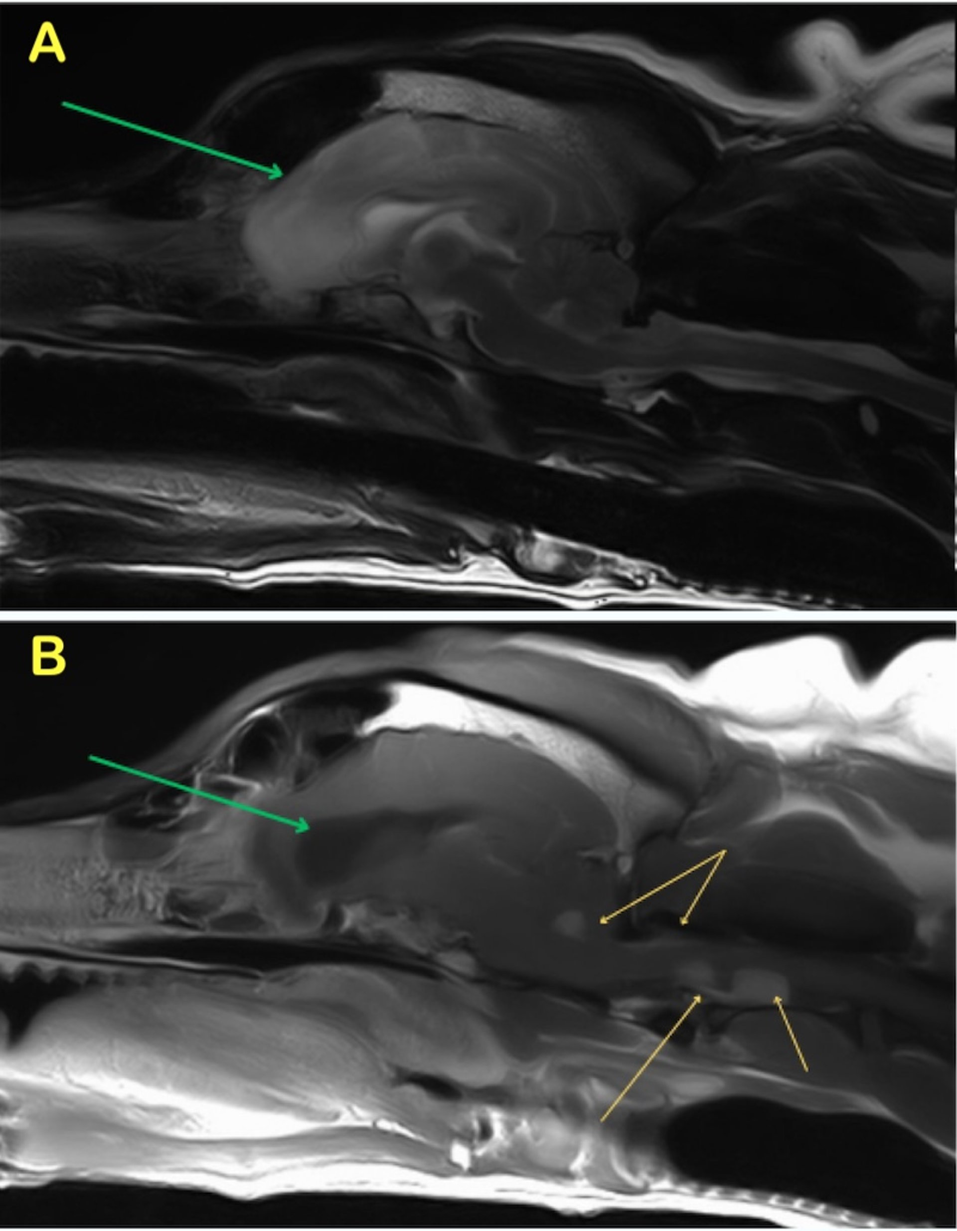
Choroid plexus tumor, MRI, dog
Presumptive choroid plexus tumor with drop metastases and obstructive hydrocephalus. T2W sagittal (A) and T1W postcontrast sagittal (B) images of the brain and cranial cervical spine of an 8-year-old pit bull–type dog that developed seizures. Green arrows denote the enlarged lateral ventricles and intracranial edema indicating obstructive hydrocephalus. Yellow arrows on the postcontrast sagittal image indicate the strongly contrast-enhancing tumor within the fourth ventricle as well as multiple metastases and meningeal involvement within the cranial cervical spine.
Courtesy of Dr. Baye Williamson.
In these topics