

After reproductive failure, lameness is typically the most important reason for removing breeding stock from the swine herd. Lameness has the following negative impacts on breeding-herd efficiencies:
higher rate of breeding stock replacement, and thus increased risk of disease introductions
inability to maintain a breeding schedule because of an unreliable pool of breeding pigs, which affects pig numbers and flow in the growing-finishing area
increased cost of maintaining additional breeding stock
poorer reproductive performance due to regular replacement of lame sows with gilts
poorer growth and feed efficiency in progeny pigs due to a higher proportion of pigs produced by first-parity sows
lower number of pigs born alive, because of higher stillborn rates in lame sows and decreased subsequent litter size due to poor lactation feed intake
increased preweaning mortality due to clumsy, lame sows that tread or lie on baby pigs
decreased weaning weights of pigs due to decreased feed intake of sows in lactation
decreased salvage value of culled sows due to increased lactation weight loss and sow mortality
decreased fertility in sound boars that are overworked while others are lame or being replaced
In sum, the impact of breeding-herd lameness on the biological and economic performance of a farm can be substantial, in addition to posing a serious risk to pig welfare.
Many diseases that affect growing-finishing pigs (see Lameness in Pigs in Growing-Finishing Areas) can also affect young gilts and boars selected as breeding stock.
Arthritis caused by Mycoplasma hyosynoviae and acute or chronic erysipelas can cause an incapacitating lameness.
Polyarthritis and polyserositis can be caused by many of the same opportunistic organisms and mechanisms that affect younger pigs; most cases are initiated by trauma.
Glaesserella parasuis infection can develop as an acute epidemic disease in gilts sourced from herds free of the bacteria after entering a herd that is endemically infected.
If rickets or skeletal weakness was a problem in the growing phase, pigs that could have been affected should not be retained as breeding stock. Ambulation should be assessed as a component of breeding stock selection. Gilts with conformational abnormalities of their limbs or restricted or abnormal ambulation should be culled.
The feet of pigs should be evaluated for uniformity among and angulation of the digits and for integrity of the wall, sole, and heel. If any problems are identified, including abnormal traits such as overgrowth of the major or secondary digits in a particular line of pigs, these pigs also should be culled.
Osteomalacia and Osteoporosis in Breeding Gilts, Sows, and Boars
The metabolic bone disease syndromes osteomalacia and osteoporosis can affect older pigs, with various clinical outcomes.
Osteomalacia is characterized by unmineralized or poorly mineralized osteoid that forms as bone remodeling occurs (or does not occur). Hence, osteomalacia is the component of rickets that affects the growth plate and is described in younger pigs.
Osteoporosis develops when established bone loses minerals and mass by a process of osteolysis, a pathogenesis different from that of either rickets or osteomalacia.
It is worth noting that most pigs, including breeding stock, are slaughtered before their skeleton has fully matured. Nonetheless, inadequate or inadequately balanced amounts of calcium, phosphorus, or vitamin D may precipitate osteomalacia or osteoporosis.
The amounts of phytases, if included in rations to increase phosphorus availability, should be carefully calculated. Any trend toward relative hypocalcemia is further compounded when the sow farrows, because calcium is secreted in the milk. A first-parity sow may soon draw on her skeletal reserves and become osteoporotic.
Because sows can become pregnant within 7 days of weaning, there is little time for recovery of skeletal mass between one breeding cycle and the next, so the skeleton weakens progressively.
Limited exercise may also exacerbate calcium mobilization and bone loss. Consequently, in sows late in gestation, during lactation, or soon after weaning, bones that have become weak are susceptible to fractures (osteoporosis). Therefore, many first- and second-litter sows are culled because of fractures and lameness.
Factors that may lead to fractures in sows include entrapment of a limb in or under the bars of a farrowing crate, activity as sows are moved from their farrowing crates, and fighting as new groups of weaned sows reestablish a social order in the breeding or gestation area in group housing conditions. Sows mounted by other sows that are in estrus are also prone to injury.
The most frequent sites of fractures are femurs, humeri, lumbar vertebrae, and occasionally ribs. No matter what precipitates a fracture, affected sows are in pain and are either severely lame and unwilling to move or paraplegic.
Diagnosis of fracture due to osteomalacia or osteoporosis is based on a history of acute lameness or paraplegia in pregnant, lactating, or recently weaned gilts or sows. Sometimes crepitus can be detected in affected limbs. A neurological examination can help locate spinal lesions if a sow is paralyzed in the pelvic limbs.
Affected sows should be culled or euthanized after an early diagnosis. Adequate nutrition and exercise for gilts and sows prevents the problem.
Osteomyelitis and Spinal Abscesses in Breeding Gilts, Sows, and Boars
In addition to the causes discussed in the section on growing-finishing pigs, osteomyelitis may develop secondary to a vertebral fracture or an epiphyseal separation. It is reasonable to assume that occasional “showering” with organisms from superficial wounds, abscesses, or the respiratory or GI tracts can be a source of infection.
Trueperella pyogenes is the most frequent cause of the suppuration and abscesses (see ).
Osteomyelitis of the ulnar epiphysis in young boars and sows has been reported. Vertebral osteomyelitis and epidural abscesses can cause a variety of signs, including nonspecific lameness, hypermetria, ataxia, or bilateral flaccid paralysis of the pelvic limbs.
Except for the temporal nature of the infectious process, clinically it is difficult to differentiate a destructive or space-occupying abscess from a fracture.
Regardless of underlying cause, recovery is unlikely, and pigs with osteomyelitis should be culled.

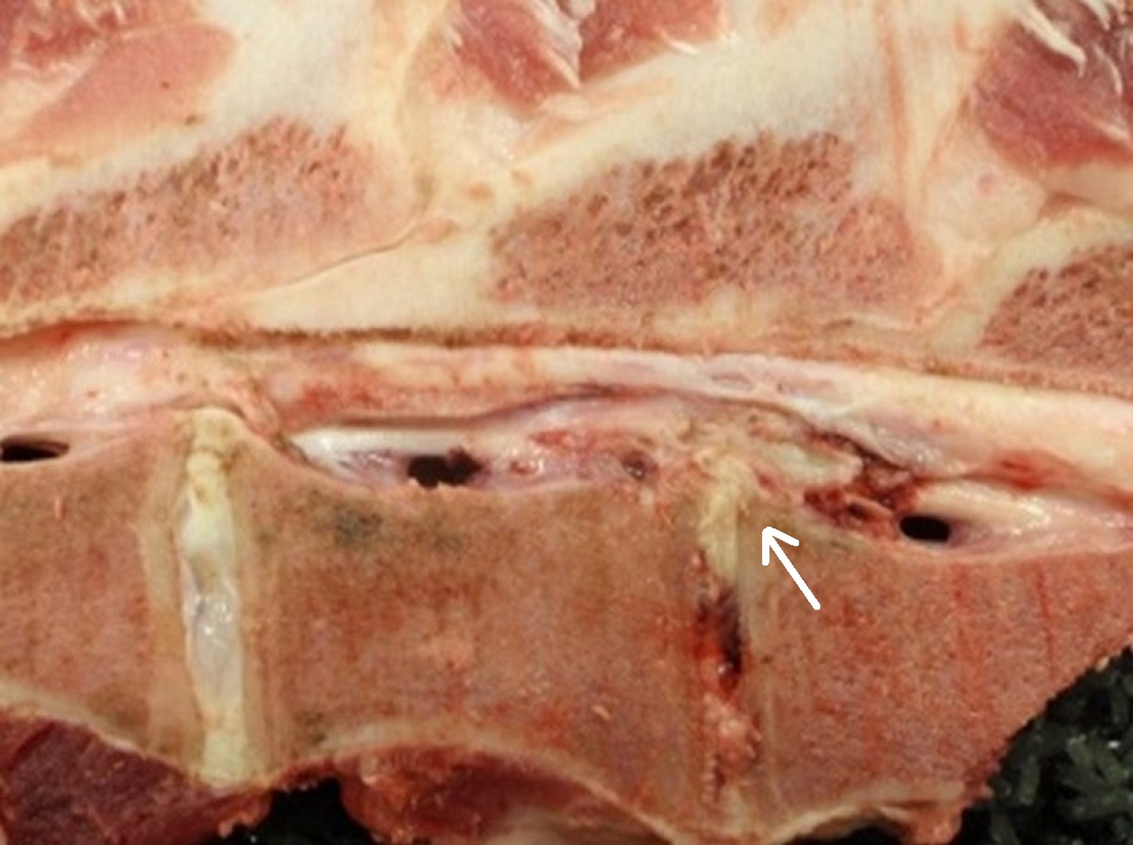
Courtesy of the Iowa State University Veterinary Diagnostic Laboratory photo archive.
Osteochondrosis and Leg Weakness Syndrome in Breeding Gilts, Sows, and Boars
"Degenerative joint disease" (DJD) and "leg weakness syndrome" are generic terms for a syndrome that is a major cause of lameness and culling for lameness in swine breeding stock.
Although the conditions are more often investigated in purebred stock, they can cause major losses in commercial pig herds. Given the increased scale of production in many herds and the shift toward pigs that grow faster, are more muscular, and finish heavier, DJD and leg weakness are critical issues.
Osteochondrosis is a specific developmental condition that is the major cause of DJD. Osteochondrosis is a defect in the development of cartilage of the growth plates or articular cartilage in growing pigs (see ).

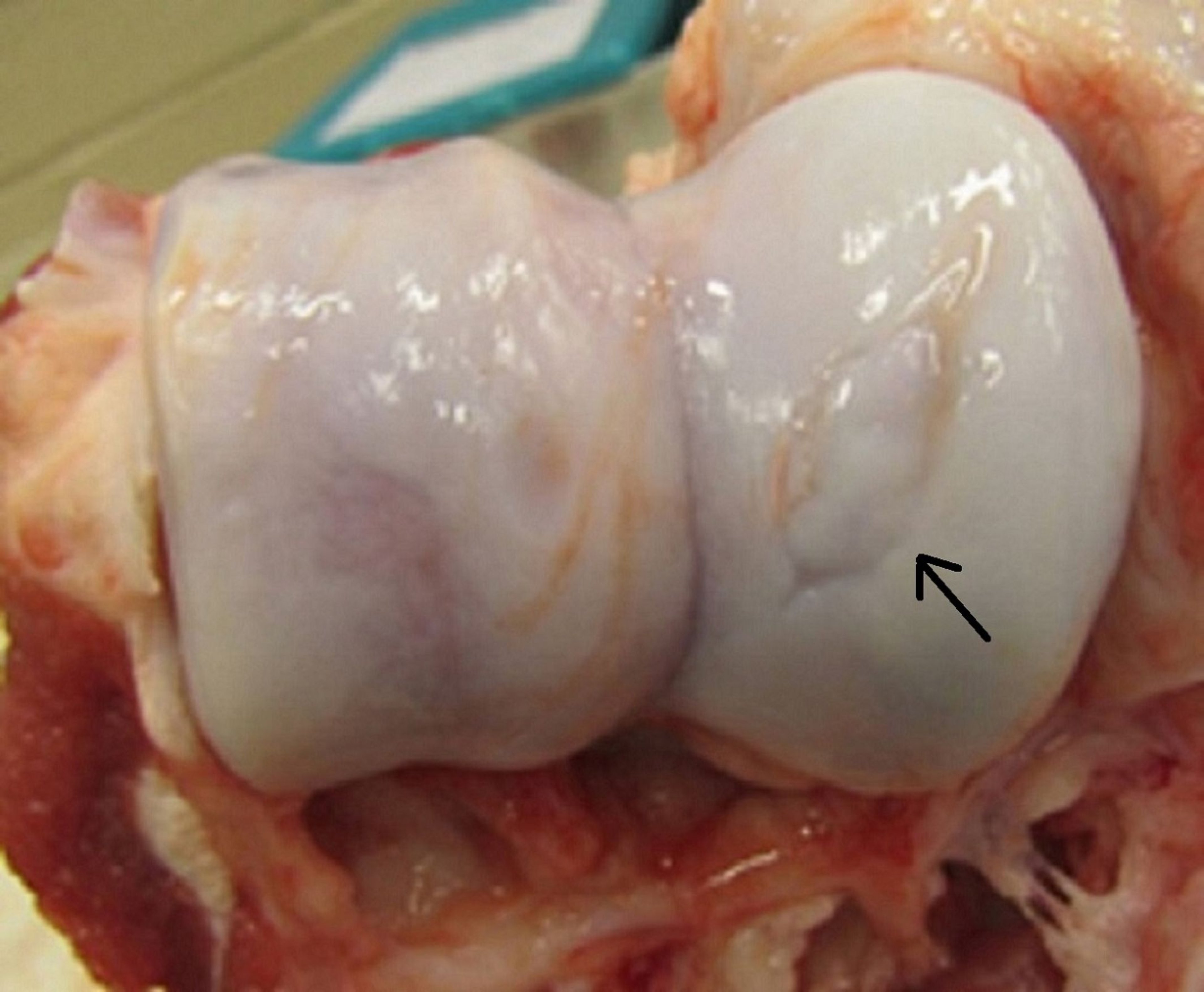
Courtesy of the Iowa State University Veterinary Diagnostic Laboratory photo archive.
In osteochondrosis, growth plates are more prone to fracture because of areas of retained hypertrophic cartilage that focally thicken and weaken the cartilage. Lesions develop when articular cartilage on the interior aspect of the joint surface becomes necrotic, ostensibly because of the loss of vascular supply.
The necrotic cartilage interferes with the advancing ossification front, and the resulting irregular ossification underlying the weight-bearing cartilage surfaces is prone to clefting (creating cartilage flaps—a condition also known as osteochondritis dissecans), exposing the endochondral bone and causing pain and lameness.
Developmental osteochondral lesions are highly prevalent in young pigs; however, they mostly resolve with age and further development.
Osteochondrosis occurs in all the major breeds of purebred and commercial hybrid pigs.
Dyschondroplasia results in deformed long bones, particularly the ulna.
Pigs that have valgus deformity or permanently flexed carpi tend to be unsuitable for sale as breeding stock and may be lame.
In addition, epiphysiolysis and epiphyseal separation may be precipitated by weakening of the underlying growth plates and cause an incapacitating lameness.
Although microscopic lesions that precede or develop into DJD or result in limb deformities are present in pigs < 6 weeks old, clinical signs are often not apparent until pigs are > 4–8 months old.
Frequently, the fastest-growing, most muscular, and heaviest pigs are the ones affected by DJD. Given time, some pigs recover from episodes of lameness; however, deformities remain.
Clinical signs of DJD vary with the site and extent of lesions and can range from stiffness to shortened stride, weight-shifting lameness, stride affected by an angular limb deformity, three-legged lameness, or an inability to stand. Usually, these animals have a weight-bearing, shifting lameness because of bilateral lesions that affect multiple joints in the same pig.
Certain gaits may indicate the source of lameness.
Pigs that ambulate "on their knees" (ie, on flexed carpi) usually have severe DJD in the elbows.
Pigs that tuck their pelvic limbs under their abdomen in a stance that resembles a circus elephant balancing on a ball often have DJD that affects stifles, tarsi, or intervertebral joints.
Pigs in which epiphyseal separation of the femoral head has occurred have difficulty standing and initially will not use the affected limb.
Pigs that have unilateral separation of the ischial tuberosity also have difficulty standing and a tendency to slip. If both ischial tuberosities are affected, the pig has a hopping gait for a few steps after being lifted, and then collapses.
The severity of clinical signs in any of these conditions varies, and joints with less extensive lesions appear to be protected by the pig's gait if they are more painful than other degenerating joints.
Severe joint lesions also have been reported in pigs that did not appear to be lame.
In pigs that have limb deformities (eg, dyschondroplasia affecting the distal ulnar growth plate), thickened, irregular growth plates are evident on radiographic images or at necropsy.
In degenerating joints, there is an excess of yellow synovia, and synovial villi may have proliferated. There are various irregularities of the articular surface, including folds in the cartilage, clefts into the cartilage, flaps of cartilage, and in severe cases, craters and exposed subchondral bone.
In chronic DJD, osteophytes develop, detached fragments of cartilage become embedded in the synovium and start to ossify, and craters fill with fibrocartilage.
If vertebral joints are affected, vertebrae eventually fuse. The growth plates most severely affected by dyschondroplasia are those of the distal part of the ulna and the ribs. Joints most often affected by DJD include the elbow, stifle, and hock, or the intervertebral synovial joints.
Many potential causes of DJD and osteochondrosis have been investigated. A genetic component is suspected because within breeds, specific boars have been identified to have progeny with a higher incidence of osteochondrosis. Breeds and lines of pigs that are heavy and well muscled, particularly in the hams, are commonly affected.
The fastest-growing pigs in a group seem to have a greater propensity for osteochondrosis to develop; however, once slower-growing pigs reach the body weight of their faster-growing peers, lesions are comparable. Growth hormone may affect chondrocyte metabolism and thereby influence the onset of articular lesions.
Research into manipulating the energy, protein, and mineral concentrations of the ration in an attempt to influence the development of osteochondrosis in pigs has been inconclusive thus far. None of the imbalances or deficiencies of nutrients typically associated with lesions of cartilage or bone (calcium, phosphorus, and vitamins A, C, and D) seem to exacerbate osteochondrosis. Deficient or excess zinc and manganese may play a role.
The stress of mixing pigs appears to have little impact on the frequency of osteochondrosis; however, trauma from handling or housing conditions has been found to affect clinical osteochondrosis.
Although pigs with DJD may improve clinically when moved to dirt lots or pasture, they remain potential carriers of heritable joint disease within the herd.
Because osteochondrosis and DJD interfere with production efficiency, the prognosis for affected pigs is poor. At best, the following practices are recommended:
selecting against replacement pigs that are lame or have poor conformation
providing adequately fortified rations in developing gilts for the growth of a strong skeleton
housing gilts in pens with ≥ 1.1 square meter per animal
promoting exercise on nonslip floors
In problem herds, strategic gilt development is encouraged. Measures include the following:
purchasing gilts at < 75 kg live weight
restricting the feed intake of gilts to slow their growth rate
providing ≥ 1.1 square meter per animal in pens with solid or only partially slatted floors
waiting to breed gilts until they are 8–10 months old
housing gilts in pens until they farrow
If replacements are purchased, suitable breeding stock must be found and inferior pigs rejected at the time of arrival at the farm.
Lameness Due to Foot Disorders in Breeding Gilts, Sows, and Boars
Foot lesions can be quite prevalent and severe among sows and boars, and they have been shown to cause lameness and decrease the productivity and longevity of breeding stock. These lesions vary in severity and can include heel erosions, separation along the white line, toe erosions, sole erosions, vertical hoof wall cracks, deep necrotic ulcers, sinuses at the coronary band, and chronic fibrosis.
As with finishing pigs, floor type and condition are important factors in the development of lesions in sows and boars. Housing type and management are also important risk factors for various types of foot lesions. Nutrition, including water quality, can affect the growth rate and quality of the horn wall and heel epithelium.
Bacterial infections of the foot can develop in pigs of any age; in breeding pigs, however, they cause serious losses. These infections are often sequelae of foot lesions that allow bacteria to penetrate into underlying sensitive foot structures. Foot infections occur in both confinement and semiconfinement systems, with a morbidity of 20%–68%.
Foot infections often affect a single limb, and lameness progresses to the point that the pig is three-legged lame. Lesions usually develop gradually, and the foot becomes swollen.
A mix of organisms has been isolated from foot lesions or identified in smears from lesions and tissue sections in breeding pigs. These include Trueperella pyogenes, Fusobacterium necrophorum, Borrelia suilla, and a mixture of gram-negative and gram-positive cocci and rods.
Foot lesion diagnoses in breeding pigs are based on clinical signs and a thorough evaluation of the feet. Ideally, the whole foot should be examined in a recumbent or suitably restrained pig. If there is a herd problem, all sows in crates or pens should be examined. Whenever possible, feet of pigs from affected herds should be evaluated at the slaughterhouse.
Historically, apparent foot infections in breeding pigs have commonly been treated with systemic penicillin. However, the effectiveness of this treatment is unproved, and the rate of success decreases with chronicity of the lesion.
Prevention is a more productive longterm strategy to combat foot infections in pigs. Preventive measures include the following:
improving the nature and cleanliness of the flooring
decreasing moisture
resurfacing rough, abrasive areas
ensuring nutritional adequacy for hoof health
As replacement gilts mature, supplementation with biotin and trace minerals is recommended because it enhances the quality and strength of the hoof.
Lameness Due to Trauma in Breeding Gilts, Sows, and Boars
Trauma associated with overexertion can cause detachment of tendons and a proliferative osteitis on the medial humeral epicondyle and the greater trochanter of the femur in sows.
Mixing gilts or sows before or after breeding or at weaning commonly results in injury as they physically reestablish a social order. Traumatic injuries can also result from moving groups to and from farrowing facilities, especially moving gilts into farrowing areas. Gilts typically are more anxious and flighty than older sows.
For a heavily pregnant sow, walking over potentially slippery flooring already poses a high risk of injury without the addition of other stressors. Movement should be calm and deliberate, with small groups of gilts and sows, to minimize potential injuries.
Sows housed in stalls with concrete slats may tear their dewclaws when they attempt to stand. Treatment with appropriate antimicrobials, protection of the wound with a dressing, and isolation in a hospital pen that has clean, deep bedding should enable a lesion to heal. Prevention by trimming elongated dewclaws is a prudent and simple management practice.
Early and aggressive treatment and nursing care of lame sows and boars is essential for them to remain productive. Prevention by improving the nature and cleanliness of the flooring; decreasing moisture; resurfacing rough, abrasive areas; and ensuring nutritional adequacy for hoof health is the most productive longterm control strategy.
Key Points
Osteochondrosis and degenerative joint disease are major causes of lameness in breeding pigs. Because genetics contributes to these diseases, culling is an important herd management tool to mitigate the problem.
Breeding sows may be at particular risk for metabolic bone disease because they expend calcium during gestation and lactation and have little time to recover between breeding cycles.
Foot disorders and skeletal trauma are common causes of lameness in breeding pigs in particular, because of their greater size and body weight at this stage of production.
For More Information
Karriker L. Identifying, Treating and Preventing Lameness in Sows. National Pork Board; 2013.
Zurbrigg K. 2006. Sow shoulder lesions: Risk factors and treatment effects on an Ontario farm. In: Proceedings of the Annual Meeting of the American Association of Swine Veterinarians. American Association of Swine Veterinarians; 2006:427-431.
Sow lameness. Iowa State University, College of Veterinary Medicine, Research, Disease Topics. Accessed August 4, 2023.
Forseth AK, Karriker LA, Millman ST, et al. Validation of standing and locomotion scoring, behavioral assessments, and mechanical nociceptive threshold testing on naturally occurring sow lameness. Animals. 2023;13(11):1801. doi:10.3390/ani13111801


