

Bumblefoot (Infective Bulbar Necrosis, Heel Abscess) in Sheep
Bumblefoot is a necrotizing or purulent infection involving the distal interphalangeal soft tissues and sometimes the joint. The incidence is usually sporadic, but as much as 25% of the flock may be affected.
The two organisms most consistently recovered from bumblefoot are Fusobacterium necrophorum and Trueperella pyogenes. Foot abscesses may develop as a complication of ovine interdigital dermatitis by extension of the necrotic process into the subcutis and then into the distal interphalangeal joint. They also commonly develop after penetration of the interdigital skin by sharp objects (eg, crusted snow or frozen or stiff stubble of alfalfa and grain), bruising of the foot and injury of the skin when slipping on frozen rocks, or even careless paring of the hoof. This joint is vulnerable to infection on the interdigital aspect where the joint capsule protrudes above the coronary border as the dorsal and volar pouches. At these sites, the joint capsule is protected only by the interdigital skin and a minimal amount of subcutaneous tissue.
Foot abscesses often develop when the soil and pastures are wet or frozen. The disease causes an acute lameness that is usually restricted to one foot, which the sheep will not place on the ground. It may be possible to express necrotic material through an opening in the interdigital skin caused by the bacterial invasion, but more commonly, the swollen sinuses break open and drain at one or more points above the coronet. If this does not occur, the swelling will have to be lanced. In some instances, movement of the affected digit is exaggerated, indicating that the ligaments about the distal interphalangeal joint have ruptured. Displacement of the digit during locomotion and permanent deformity are likely in those cases.
Clinical signs include acute lameness, with the sheep often non–weight-bearing on the affected limb. Swelling of one digit and discharging abscesses distinguish foot abscesses from footrot.
Early treatment with parenteral, long-acting antimicrobials is sometimes effective and may prevent joint infection. The aim of treatment is to maintain the integrity of the joint ligaments by draining the abscess and applying an antimicrobial preparation and a self-adhesive bandage. This reduces stress on the ligaments, keeps the lesion out of the mud, and counters the microbial infection. Although the prognosis for complete recovery is poor, in most cases the foot heals sufficiently to allow adequate locomotion. Once the infection becomes established in the joint, conservative treatment is not effective. However, a toe can be surgically removed (if the other digit is healthy) with relatively good success.
Control depends on early treatment and moving the sheep to avoid conditions that lead to ovine interdigital dermatitis or other causes of infection. Although F necrophorum vaccines are available, they have not proved to be very effective.
Septic Laminitis (Toe Abscess, Lamellar Supparation) in Sheep

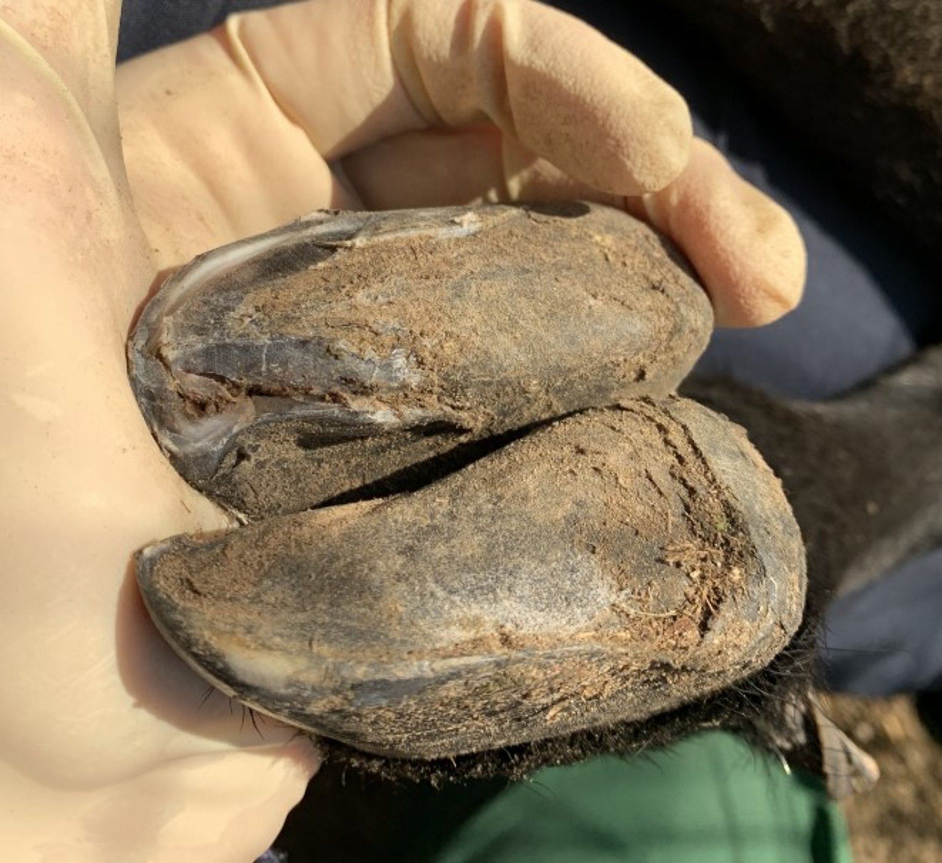
An abscess at the toe and axial wall of the medial claw of a forelimb of a sheep with septic laminitis. Purulent material could be expressed from the fissure, and the sheep had signs of pain on palpation of the claw. The sole was undermined and unattached to surrounding structures.
Courtesy of Dr. Evelyn MacKay.

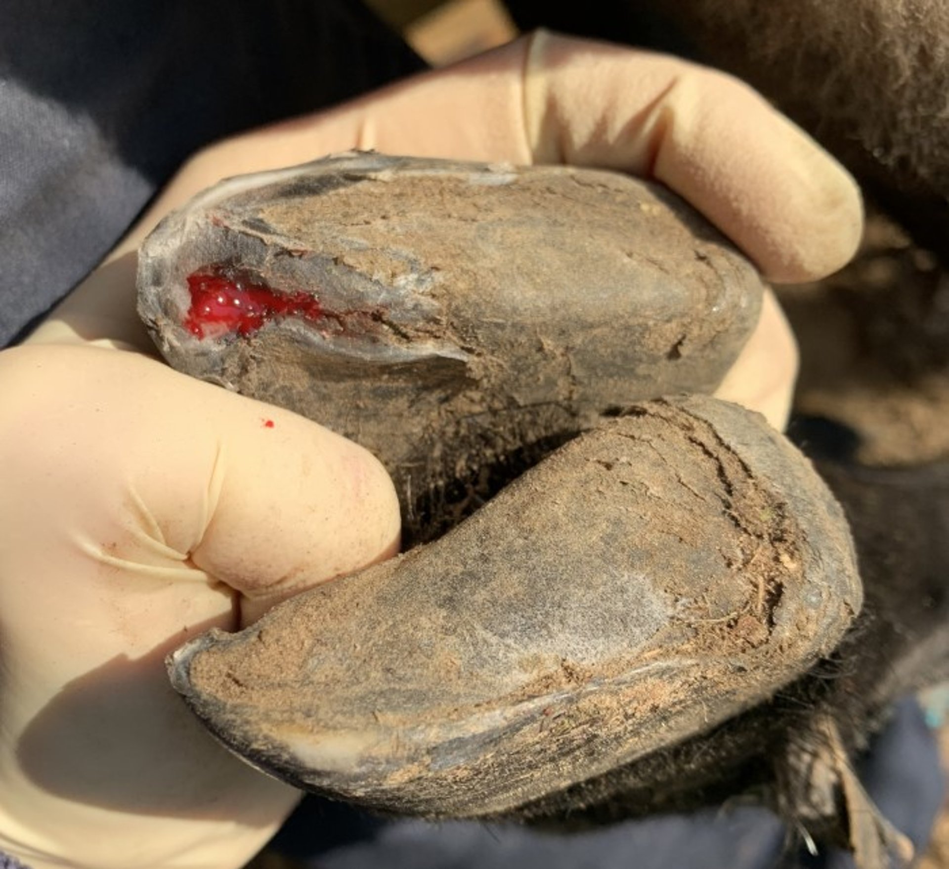
Abscess in the sheep in the above photograph after debridement. The hoof has been trimmed with removal of undermined and unattached sole to allow drainage, with some exposure of sensitive tissues.
Courtesy of Dr. Evelyn MacKay.
Septic laminitis is an acute bacterial infection of the laminar matrix of the hoof that is usually restricted to the toe and abaxial wall. The disease is sporadic and the cause variable, but cases due to Fusobacterium necrophorum and Trueperella pyogenes usually are more severe and extensive than those involving streptococci or other organisms. The organisms probably enter through fissures between the wall and sole and through vertical and horizontal fractures of wall horn. Sometimes, infection is enhanced by impaction with sand, mud, or feces; by overgrowth of the hoof; or by separation of the wall after laminitis.
Clinical signs of septic laminitis include acute, severe lameness. There is typically no swelling of the soft tissue structures; however, the sheep shows signs of pain on palpation of the affected claw. If left untreated, there may be a draining sinus above the lesion at the coronet, or an abscess may be found above the sole on the bottom of the foot. Applying pressure to various sections of the sole with the thumbs will elicit signs of pain and help locate the pocket of infection. Affected sheep usually recover rapidly after paring of the horn to provide dependent drainage. If signs of pain persist after paring of the hoof, a small wooden block can be glued to the healthy claw to reduce weight-bearing on the affected claw.


