


Developmental myelin disorders of the nervous system can be broadly categorized into myelination that is decreased or absent (hypomyelinogenesis) or abnormal (dysmyelination—eg, leukodystrophy, myelinolysis). Myelin disorders can affect the entire nervous system or be localized to the central nervous system or the peripheral nervous system. Developmental myelin disorders should not be confused with demyelination, in which previously normal myelin breaks down and is lost. Signature pathological processes of nerves undergoing demyelination include Wallerian degeneration, axonal degeneration, and segmental and diffuse myelin degeneration. Often there is no treatment for degenerative conditions; however, supportive care may counter underlying causes of acquired disorders of myelin. Genetic testing is available for some degenerative conditions.
Central Myelinopathy in Animals
Hypomyelinating and dysmyelinating diseases are characteristic CNS myelin disorders in young animals.
In pigs, six myelin disorders have been defined. Inherited disorders include the sex-linked type A-III in Landrace, and autosomal recessive type A-IV in Saddlebacks. Clinical signs develop in pigs 2–3 days after birth. Saddlebacks show abnormal myelination; Landrace pigs have decreased numbers of oligodendrocytes.
Hypomelination
CNS hypomyelination primarily involves defects of the oligodendrocytes, which are decreased in number or are unable to produce functional myelin. Whole-body tremors are a common clinical sign.
Hypomyelination has been reported in a number of canine breeds (eg, English and Welsh Springer Spaniels, Weimaraners, Chow Chows, Samoyeds, and Bernese Mountain Dogs as well as lurcher hounds). In Springer Spaniels, a hypomyelination disorder (shaking pup syndrome) is sex-linked and has a variant in the PLP1 gene, which encodes for myelin proteolipid protein, a major protein necessary for myelination. Myelin is abnormally formed; oligodendrocytes are decreased and immature.
Hypomyelination also has been reported in two Siamese cat littermates. Although normal at initial ambulation, affected cats developed action-related myoclonus at 4 weeks of age.(1) Histopathologic findings included diffuse hypomyelination in the spinal cord.
Dysmyelination
The term "dysmyelination" refers to defective myelin synthesis or function that cannot be maintained. Dysmyelination can be subclassified into the categories of leukodystrophy and myelinolytic diseases.
Leukodystrophies are a group of an inherited conditions in which myelin synthesis or function is defective. Leukodystrophies have been described in Dalmatians, Labrador Retrievers, Scottish Terriers, Bullmastiffs, Bernese Mountain Dogs, and Miniature Poodles, as well as in Merino sheep and Charolais cattle. Globoid cell leukodystrophy (named for accumulation of lipid-laden macrophages) lysosomal sphingolipidosis that has been described in West Highland White Terriers, Cairn Terriers, Beagles, Poodles, Basset Hounds, Blue Tick Hounds, Pomeranians, Irish Setters, and Australian Kelpies; domestic shorthair cats and domestic longhair cats; and Dorset sheep.
Clinical signs of leukodystrophies develop at 3–6 months of age. The clinical course of disease is characterized by general proprioceptive ataxia and upper motor neuron paraparesis that progresses to nonambulatory tetraparesis. Cerebellar signs and seizures also may occur. Charolais cattle with leukodystrophy develop progressive paraparesis that leads to tetraparesis and recumbency by 24 months of age. Histopathologic findings include myelin degeneration with replacement by severe astrogliosis or Rosenthal fibers (astrocytic processes) widespread throughout the brain and spinal cord.
Myelinolysis refers to the degeneration of initially normally formed spinal cord myelin. Canine breeds affected include the Leonberger, Afghan Hound, and Miniature Poodle. Clinical signs include acute-onset paraparesis with progressive to the thoracic limbs. Histopathologic findings include bilateral and symmetrical myelin loss in all funiculi; axons are preserved.
The more general term leukoencephalomyelopathy is used for degenerative spinal cord disorders in which the type of myelin degeneration cannot be determined. Leukoencephalomyelopathies have been described in the Rottweiler and Nederlandse Kooikerhondje (Dutch Kooiker dog). The age at the onset of signs varies. The rate of clinical progress vary. Rapid deterioration occurs in the Nederlandse Kooikerhondje, and progression is insidious in the Rottweiler. Histopathologic findings include myelin degeneration in all funiculi; axons are spared.
Spongy Degeneration
Spongy degeneration is a nonspecific term for tissue vacuolation involving myelin or neurons.
The spongy state in white matter can be associated with reactive changes that are symmetrical or asymmetrical(edema, demyelination, and Wallerian degeneration).
Spongiform leukoencephalomyelopathy has been described in the Australian Cattle Dog, Shetland Sheepdog, and Labrador Retriever.
Clinical signs begin between 2 and 9 months of age and progress as cerebellar ataxia, seizures, and opisthotonos.
Histopathologic findings include widespread myelin degeneration in the spinal cord, brainstem, and cerebellum. In the Labrador Retriever, myelin degeneration includes the cerebral white matter.
In Australia, Murray Grey calves with spongiform leukoencephalomyelopathy show ataxia and paresis from birth, and the brainstem and spinal cord exhibit vacuolated lesions.
Etiology of Myelin Disorders in Animals
Canine Myelin Disorders With Identified Gene Mutations
Disease | Breeds | Gene Mutation |
|---|---|---|
Hypomyelination | Weimaraner, Chow Chow | FNIP2 (delay or failure in maturation of oligodendrocytes) |
Springer Spaniel | PLP (altered myelination of early myelinating structures) | |
Leukodystrophy | Standard Schnauzer | TSEN54 (defect in tRNA splicing endonuclease that leads to myelin destruction) |
Spongy degeneration | Labrador Retriever | GFAP (gain of function that leads to cytoskeletal collapse and protein aggregation) |
Leukoencephalomyelopathy | Rottweiler, Great Dane, Leonberger | NAPEPLD (lipid metabolism defect) |
Globoid cell leukodystrophy | Cairn Terrier, West Highland White Terrier, Irish Setter, Australian Kelpie | GALC (loss of function in galactosylceramidase that leads to storage disease) |
Peripheral neuropathy (Schwann cell defect) | Alaskan Malamute, Greyhound | NDRG1 (disruption of myelinating signal in Schwann cells) |
Polyneuropathy | Leonberger (LPN1),a Saint Bernard | ARHGEF10 (regulation of phosphorylation pathways in neural morphogenesis) |
Leonberger (LPN2)a | GJA9 (connexin dysfunction at gap junctions between folds of Schwann cell membranes) | |
Alaskan Malamute | NDRG1 (N-myc downstream-regulated gene) | |
Demyelinating neuropathy | Miniature Schnauzer | MTMR13 (neuropathy with myelin outfoldings) |
aLPN, Leonberger polyneuropathy. | ||
Hypomyelination and dysmyelination can be inherited in some species and breeds. For identified gene mutations that cause hypomyelination and dysmyelination in dogs, see the table Canine Myelin Disorders With Identified Gene Mutations.
In utero infection can result in congenital hypo- and dysmyelination. The viruses of classical swine fever, border disease, and bovine viral diarrhea have been incriminated; however, the mechanisms responsible for the hypomyelination have not been defined. The pestiviruses that cause these three diseases are closely related members of the family Togaviridae and are transmitted both vertically and horizontally.
The inflammatory neuraxial disorders in domestic animals that involve demyelination are canine distemper, maedi-visna, and caprine arthritis encephalitis.
In swine, types A-I and A-II are due to classical swine fever (hog cholera) and an unknown viral agent, respectively.
Lambs with Border disease have a characteristic hairy fleece; thus, they are called "hairy shaker lambs." Lambs are infected in utero by a pestivirus and remain persistently viremic. Lambs with clinical signs of hypomyelination have skeletal deformities.
Calves infected in utero with bovine viral diarrhea virus at 100 days gestation develop cerebellar dysplasia and hypomyelination.
Toxins can affect myelin synthesis, cause demyelination, or lead to edema within myelin, resulting in spongiform changes. Trichlorfon is an organophosphate with a unique toxicity that causes types A–V of porcine congenital tremor syndrome. Pregnant sows treated with trichlorfon during mid and late gestation (days 45–77) produce litters in which up to 90% of the piglets develop a marked tremor syndrome secondary to cerebellar hypoplasia and hypomyelinogenesis.
Toxins associated with intramyelinic edema without damage to the oligodendrocytes include bromethalin, chlorinated hydrocarbons, hexachlorophene, triethyl tin, and isoniazid. Organophosphates, copper chelators, and pyridoxine cause secondary demyelination in the peripheral nervous system (PNS). Lead toxicosis can result in dysmyelination by disturbing oligodendrocyte differentiation. Some plants in Europe associated with spongiform encephalopathy in ruminants include Helichrysum argyrosphaerum and Stypandraspp.
Avian vacuolar myelinopathy has been associated with Aetokthonos hydrillicola, a cyanobacterium growing on aquatic hydrilla plants (Hydrilla verticillata).
Vitamin and mineral deficiencies can alter myelin synthesis or lead to demyelination. A primary myelin degeneration due to an acquired methionine deficiency from feeding a diet composed of rumen has been described in foxes, Beagles, and Harrier hounds. Microscopic findings were most severe the thoracic spinal cord; vacuolated myelin sheaths were prominent in the ventral and lateral funiculi.
Primary demyelination in the brain and spinal cord has been described in cats that were experimentally fed irradiated feed with decreased amounts of vitamin A. Vitamin B12 deficiency has been described in cats infected with feline leukemia virus that have spinal cord myelinopathy (due to alteration of a methylation pathway in myelin synthesis).
Copper deficiency is a cause of hypomyelination in animals.
Clinical Findings of Myelin Disorders in Animals

Pure demyelinating or cerebellar disorders are not characterized predominantly by weakness; however, they may feature tremors of the limbs or whole body. Clinical signs associated with hypomyelination and dysmyelination are usually noticed in the first few weeks of life or when the affected animal begins to walk.
In various canine breeds, shaking pup syndrome involves an apparently diffuse, coarse tremor (having a large amplitude) that makes them bounce up and down rhythmically when they stand or walk. The tremor is not present during sleep. The movement disorder is more severe in the pelvic limbs than the thoracic limbs. Depending on the severity of the tremor, affected animals may be unable to walk.
Clinical signs of dysmyelination are progressive and initially are manifested most severely in the pelvic limbs; however, they reflect the anatomical location that is pathologically involved. The clinical course is often acute and rapidly progressive, resulting in paraplegia and eventually affecting the thoracic limbs. In some myelin disorders, the neuron or myelin must reach a critical threshold of dysfunction to trigger acute onset of the disease.
Because pathways projecting to the cerebellum are heavily myelinated, this anatomical location is particularly sensitive to disorders that affect myelin. Clinical signs of cerebellar dysfunction include cerebellar ataxia, intention tremor, and limb dysmetria. Cerebellar signs are often the first clinical signs because of the complex integration of the fast-conducting sensory and motor pathways.
When the cerebrum is affected, clinical signs manifest with seizures, central blindness, and cognitive dysfunction. Affected animals may have other cranial nerve dysfunction, with signs of abnormal vestibular nystagmus. These neurologic deficits may be so severe in some animals that euthanasia is warranted.
Lesions
In CNS hypomyelination, gross pathological findings include pallor of the white matter of the brain and spinal cord, and possibly a gelatinous appearance. In PNS hypomyelination, the gross changes are minimal and there is no evidence of CNS involvement.
CNS Hypomyelination
The microscopic changes in CNS hypomyelination include absent myelin (the lack is usually severe but not absolute), fewer oligodendrocytes, astrocytes outnumbering oligodendrocytes, oligodendrocytes that differ in appearance from those in healthy animals, and abnormal types of glial cells.
With diffuse myelin disease related to secondary edema, such as metabolic and toxic encephalopathies, primary findings include narrowing of sulci and flattening of gyri. Histopathologic findings include diffuse spongiform degeneration of white matter, as well as myelin vacuolation. Lesions, in addition to cerebral edema, include intramyelinic edema, myelin splitting, and axonal swelling, all of which manifest as white matter spongiosis.
PNS Hypomyelination
The microscopic changes in PNS hypomyelination consist of a paucity of myelinated fibers, fibers with inappropriately thin myelin sheaths relative to the caliber of their enclosed axons, occasional fibers with poorly compacted myelin, Schwann cells with larger-than-normal cytoplasmic volume, and increased numbers of Schwann cell nuclei.
For Schwann cell and myelin diseases, teased nerve fiber studies help to characterize myelin pathology.
Diagnosis of Myelin Disorders in Animals
It is important to distinguish clinical signs of developmental or inherited myelination from signs of congenital cerebellar disease. Although primary myelin disorders are often confused with intention tremor, animals with primary myelin disorders do not display the dysmetria that is observed with cerebellar dysfunction.

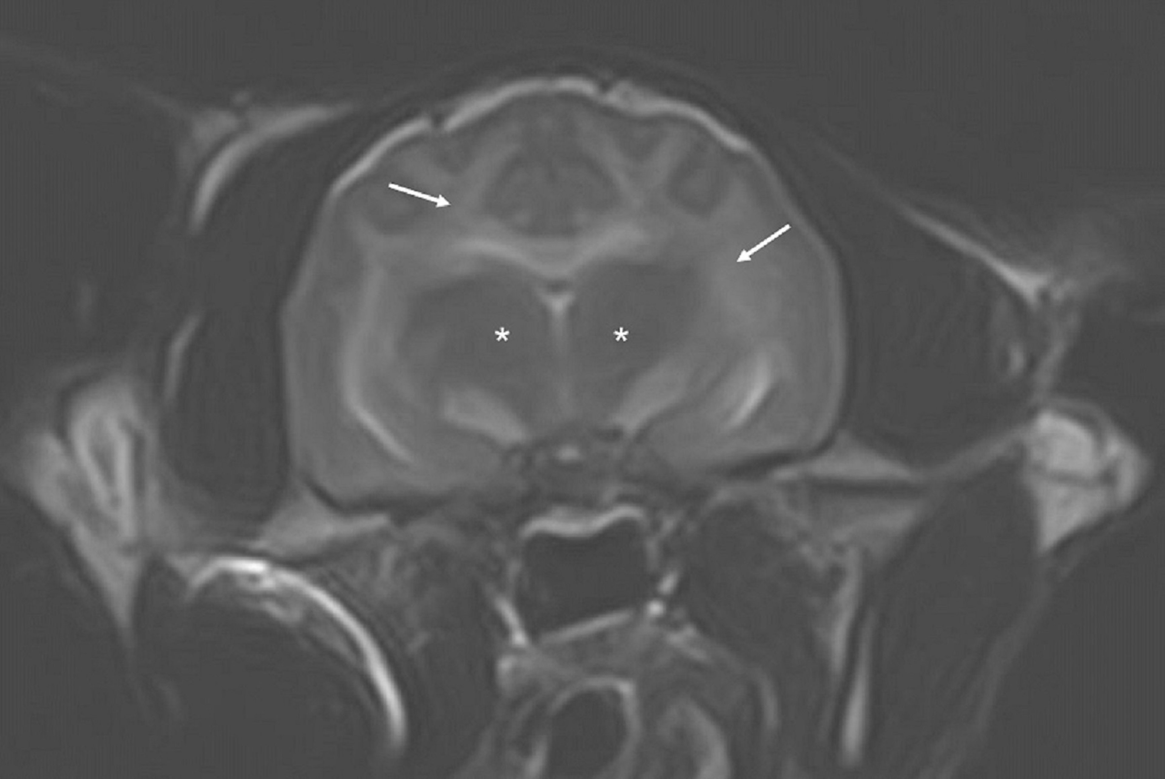
Transverse T2-weighted MRI of the brain of a cat with bromethalin toxicosis. Widespread bilaterally symmetrical hyperintensity of the entire white matter (arrows point to the corona radiata and internal capsule) is evident at the level of the thalamus (asterisks).
Courtesy of Dr. Joan Coates.
Central Nervous System
Clinical evaluation
Histologic examination
Pedigree evaluation and genetic testing
Antibody staining and virus isolation
MRI
The diagnosis of CNS hypomyelination and dysmyelination is based primarily on the spectrum of neurologic deficits and signs, and on the early age of onset. Histopathologic confirmation is the only definitive method of diagnosis.
In cases that have a heritable basis, pedigree evaluation and genetic testing may be helpful. In cases that have a viral cause, confirmation may involve immunofluorescent antibody-staining techniques or virus isolation from nervous tissue (or both).
Brain MRI can show a distinctive widespread T2-weighted hyperintensity in white matter of the brain and spinal cord, indicating differential diagnoses of toxic, metabolic, and degenerative leukoencephalomyelopathies.
Peripheral Nervous System
Electrodiagnostic testing
Histologic examination
A diagnostic assessment of myelinopathy in the PNS requires electrodiagnostic testing. Peripheral nerve conduction studies reflect the integrity of the myelin, and the amplitude and duration of compound action potentials reflect the integrity of the axons. Thus, a diagnosis of demyelination is based on slow nerve conduction velocity, conduction block, and increased temporal dispersion of compound motor action potentials.
Definitive diagnosis of PNS myelinopathy is based on biopsies of proximal and distal portions of the nerves and on detailed histologic examination performed at laboratories with expertise in neuromuscular pathology.
Treatment, Control, and Prevention of Myelin Disorders in Animals
No treatment
To control and prevent heritable syndromes: selective breeding and genetic testing
To control and prevent viral syndromes: immunization
There is no specific treatment for hypomyelination and dysmyelination. The only means of control and prevention is selective breeding and genetic testing (for heritable syndromes) and prevention of infection (for viral syndromes).
In some hypomyelinating myelinopathies, clinical signs improve with age; however, assistance with feeding may be necessary. In Springer Spaniels with sex-linked hypomyelination, males do not show improvement; the less affected carrier females, though, show considerable improvement with age. Chow Chows and Weimaraners improve to near normal with maturity. Sheep, pigs, and cattle with hypomyelination also show recovery.
Key Points
Distinctive whole-body tremor is a clinical sign of pure myelin disease.
Myelin diseases can occur because of direct damage to the myelin sheath or indirectly as a result of genetic disease or damage to the myelin-producing cell (oligodendrocyte or Schwann cell).
References
Stoffregen DA, Huxtable CR, Cummings JF, de Lahunta A. Hypomyelination of the central nervous system of two Siamese kitten littermates. Vet Pathol. 1993;30(4):388-391. doi:10.1177/030098589303000412
For More Information
Vandevelde M, Higgins RJ, Oevermann A. Veterinary Neuropathology Essentials of Theory and Practice. Wiley-Blackwell; 2012.
Granger N. Canine inherited motor and sensory neuropathies: an updated classification in 22 breeds and comparison to Charcot-Marie-Tooth disease. Vet J. 2011;188(3):274-285. doi:10.1016/j.tvjl.2010.06.003
Duncan ID, Radcliff A. Inherited and acquired disorders of myelin: the underlying myelin pathology. Exp Neurol. 2016;283(Pt B):452-475. doi:10.1016/j.expneurol.2016.04.002


