Atrophic rhinitis is an infectious disease of swine characterized by atrophy of the turbinate (conchal) bones. The most severe, progressive form of the disease is caused by infection with toxigenic Pasteurella multocida. Clinical signs include coughing, sneezing, nasal discharge, and, in severe cases, nasal bleeding, deformation of the snout, and poor growth. Diagnosis is based on clinical signs and confirmation of the causative agent. Vaccination with bacterins and improved management have led to a considerable decrease in incidence in most swine herds.
Atrophic rhinitis is characterized by sneezing, followed by atrophy of the turbinate bones, sometimes accompanied by distortion of the nasal septum and shortening or twisting of the upper jaw.
The importance of this disease has declined substantially, and it is no longer considered a major health risk to swine herds.
The term “bullnose” is typically used for Fusobacterium necrophorum infection (necrotic rhinitis) but is also sometimes used to refer to atrophic rhinitis.
Etiology of Atrophic Rhinitis in Pigs
Atrophic rhinitis in pigs has been divided into two forms:
Nonprogressive atrophic rhinitis, caused by Bordetella bronchiseptica, is mild and transient and generally has little effect on growth and performance.
Progressive atrophic rhinitis, caused by toxigenic strains D and A of Pasteurella multocida (with or without concurrent B bronchiseptica infection), is severe, permanent, and usually accompanied by poor growth.
Outbreaks of atrophic rhinitis usually follow either the introduction of infected pigs into a herd or the mixing of pigs from different sources. Piglets can be affected at any age, especially by P multocida, which also can infect mature animals.
Crowding, inadequate ventilation, mixing and moving, and other concurrent diseases are important contributory factors that intensify atrophic rhinitis in pigs.
P multocida is also an important cause of pneumonia in pigs (see Pneumonic Pasteurellosis) and is recognized as part of the porcine respiratory disease complex.
P multocida is zoonotic and is associated with a spectrum of human disease (1, 2).
Clinical Findings of Atrophic Rhinitis in Pigs
Acute signs of atrophic rhinitis, which usually appear in pigs at the age of 3–8 weeks, include sneezing, coughing, and inflammation of the lacrimal duct. In more severe cases, nasal hemorrhage can occur. The lacrimal ducts can become occluded, and tear stains then appear below the medial canthi of the eyes.
Some pigs severely affected with atrophic rhinitis develop lateral deviation or shortening of the upper jaw; others might suffer some degree of turbinate atrophy with no apparent outward distortion. The extent of distortion can be judged from the relationship of the upper and lower incisors, if breed variations are considered.
Outbreaks of atrophic rhinitis in pigs frequently impair their growth rate and feed conversion.
The severity of atrophic rhinitis in a swine herd depends largely on the presence of toxigenic strains of P multocida, management practices, and the immune status of the herd. The latter is related to both vaccination status and the parity distribution of the sow herd, because compared with older, multiparous sows, younger sows tend to shed more organisms and produce less lactogenic immunity for their nursing piglets.
Lesions of Atrophic Rhinitis in Pigs
The extent of atrophy and distortion in cases of atrophic rhinitis in pigs is best assessed by postmortem examination of a transverse section of the nasal cavity (see ) at the level of the second premolar tooth (the first cheek tooth) up to the age of 7–9 months; some clinicians recommend additional parallel sections.

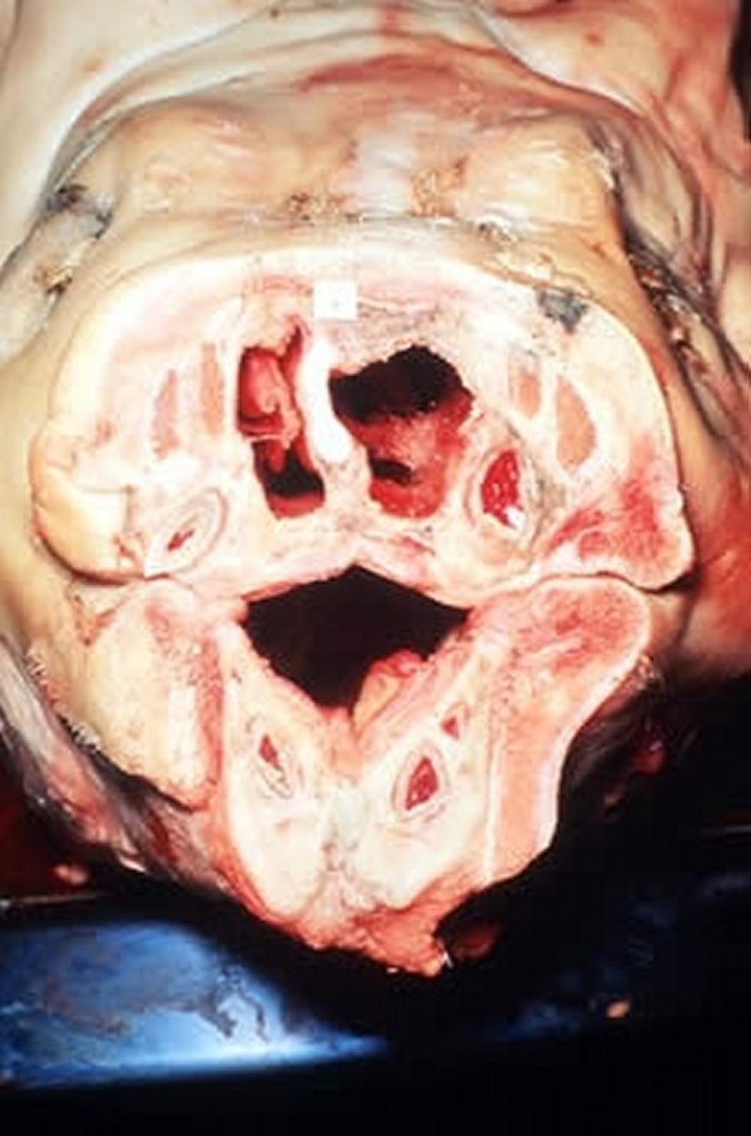
Necropsy photograph showing a transverse section of the snout of a pig with atrophic rhinitis caused by natural infection with type D toxigenic Pasteurella multocida. Note that the nasal turbinate bones have been completely destroyed.
Courtesy of the Department of Pathobiology, University of Guelph.
In active stages of atrophic rhinitis–related inflammation, the mucosa has a blanched appearance, and purulent material might be present on the surface. In later stages, the nasal cavities might be clear; however, there can be variable degrees of softening, atrophy, or grooving of the turbinates; deviation of the nasal septum; and asymmetrical distortion of the surrounding bone structure.
Diagnosis of Atrophic Rhinitis in Pigs
Clinical signs and postmortem lesions
Determination of causative agent
Turbinate atrophy evaluation
Clinical signs (eg, disfigured snout) and characteristic lesions (atrophied turbinates) are commonly the basis for diagnosis of atrophic rhinitis in pigs.
PCR assays can be used to detect B bronchiseptica, as well as both toxigenic and nontoxigenic P multocida isolates. The toxigenicity of P multocida isolates can be also determined by assessing cytotoxic effects in cultured cells or via a commercially available toxin-specific ELISA.
In some breeding swine herds, routine monitoring is done by measuring the extent of turbinate atrophy.
Atrophic rhinitis in pigs must be differentiated from other causes of rhinitis, such as porcine cytomegalovirus infection (inclusion body rhinitis) and F necrophorum infection (necrotic rhinitis), as well as other causes of turbinate atrophy. Necrotic rhinitis is readily differentiated from atrophic rhinitis by the bulging type of facial distortion in the former. Inclusion body rhinitis, caused by porcine cytomegalovirus, does not cause turbinate atrophy.
In cases of nonprogressive atrophic rhinitis caused by B bronchiseptica, the affected pigs' turbinate bones heal and regenerate. Progressive atrophic rhinitis induced by toxigenic P multocida persists until slaughter.
Congenital abnormal shortness of the maxilla (brachygnathia superior) should be distinguished from shortening of the maxillary bone secondary to atrophy and deviation of nasal turbinates.
Control of Atrophic Rhinitis in Pigs
Good management and biosecurity practices
Vaccination
Because of elimination programs of toxigenic P multocida at the seed stock level, progressive atrophic rhinitis is rarely observed in contemporary swine production systems. Good management practices, such as strict all-in all-out procedures, environmental management, and biosecurity measures are important to prevent and control atrophic rhinitis in pigs.
Bacterins against toxigenic P multocida and B bronchiseptica have been developed. Both toxoid vaccines and bacterin-toxoid mixtures against P multocida are available. Although both provide satisfactory results in most herds, infection is best prevented with bacterin-toxoid mixtures.
Typically, sows are vaccinated against both P multocida and B bronchiseptica 4 and 2 weeks before farrowing; young pigs are vaccinated at the ages of 1 week and 4 weeks. However, vaccination schedules recommended by the manufacturer should be followed. A high level of colostral immunity is acquired by piglets that are nursing vaccinated sows.
An intranasal vaccine using modified live strains of B bronchiseptica is also available for young pigs.
Key Points
Toxigenic Pasteurella multocida is the primary pathogen associated with progressive atrophic rhinitis.
The incidence of progressive atrophic rhinitis, once considered an important cause of respiratory disease in swine, has decreased substantially as a result of eradication programs.
References
Henderson SR, Shah A, Banford KB, Howard LS. Pig trotters lung—novel domestic transmission of Pasteurella multocida. Clin Med (Lond). 2010;10(5):517-518. doi:10.7861/clinmedicine.10-5-517
Wilson BA, Ho M. Pasteurella multocida: from zoonosis to cellular microbiology. Clin Microbiol Rev. 2013;26(3):631-655. doi:10.1128/CMR.00024-13