

Oomycosis is due to pathogens in the class Oomycetes. These organisms are not true fungi but are aquatic pathogens, predominantly of plants, that are found in warm, still, fresh water.
Oomycetes are more closely related to algae than fungi, but oomycosis, the disease they cause, closely resembles fungal disease. These organisms, unlike true fungi, produce motile zoospores that are the agents of infection.
Organisms of significance in veterinary medicine include various species of the following:
Saprolegnia spp(eg, S diclina) and Achyla, which are the common agents of cutaneous disease in fish
Pythium insidiosum, the cause of a cutaneous and subcutaneous oomycosis in horses (swamp cancer, leeches); a cutaneous, subcutaneous, and GI disease in dogs; and a cutaneous and paranasal disease of cats and cattle
Lagenidium spp, the cause of cutaneous and systemic lesions and large-vessel aneurysms in dogs
Pythiosis is a common disease of domestic animals in some tropical and subtropical areas of the world. In dogs, pythiosis is most often encountered in southeast Asia, eastern coastal Australia, South America, and the US, especially along the Gulf coast. In the US, the disease most often occurs in fall and winter months.
Clinical Findings and Lesions of Oomycosis in Animals
In horses, lesions are large, roughly circular, granulomatous, ulcerated, fistulated nodules, or subcutaneous swellings with yellow-gray necrotic mineralized masses intralesionally ("kunkers"). The lesions are most common on the legs (especially the lower limbs), abdomen, chest, lips, and genitalia. Distribution of lesions is attributable to the aquatic nature of the organism.

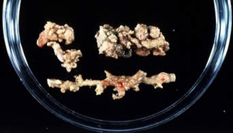
Courtesy of Dr. Corrie Brown.

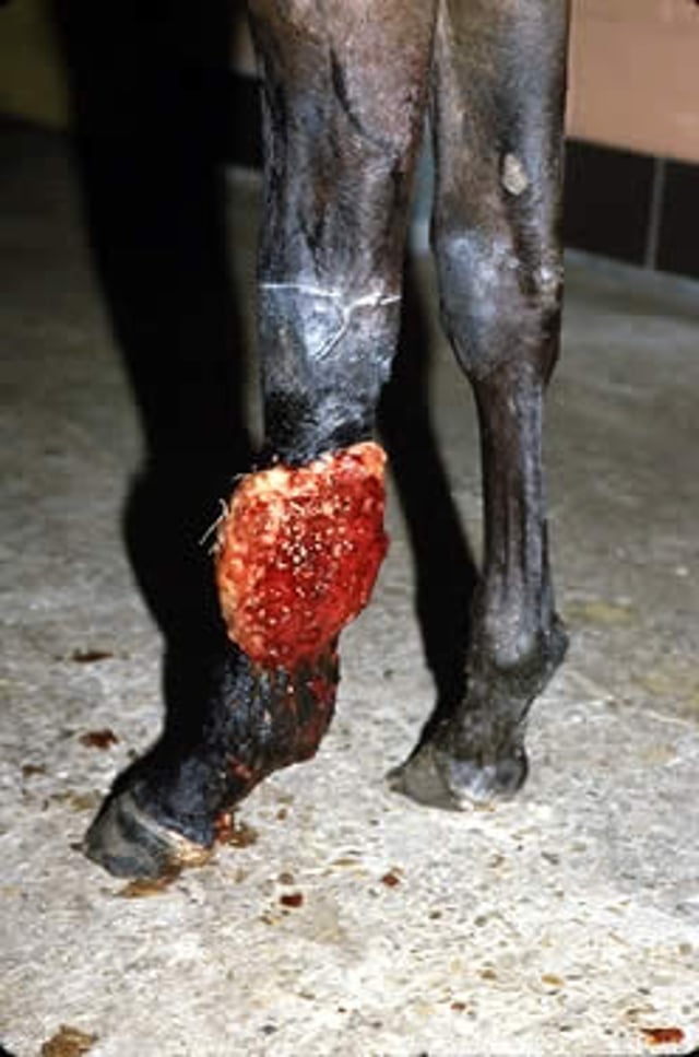
Courtesy of Dr. Corrie Brown.
The lesions are extremely pruritic, discharge a copious mucosanguineous exudate, and often are self-traumatized. The granulomas contain broad, branching aseptate hyphae and are 1–10 mcm in diameter. Bone involvement may be a feature of chronic pythiosis. Enteric pythiosis in horses is characterized by fibrosing and stenotic GI lesions containing intralesional foci of caseous material and fungal hyphae.
Specimens removed at surgery or necropsy consist of fibrous tissue with irregularly spaced, firm, focal areas of necrosis that vary in size and color. Microscopically, alterations vary from foci of acute exudative inflammation with numerous eosinophils to a granulomatous reaction with sequestered areas of necrosis and a framework of hyphae that are thick walled, branching, and irregular in width.
GI and cutaneous forms of pythiosis occur in dogs and cats and are characterized by severe granulomatous and eosinophilic inflammation. P insidiosum infection occurs most often in the GI tract of young adult dogs. The stomach, proximal small intestine, and ileocolic junction are affected most commonly; however, any part of the intestine, esophagus, or colon can be diseased.
Clinical signs include vomiting, weight loss, and anorexia. The weight loss can be severe, but affected dogs usually do not appear systemically ill until late in the disease. The lesions are typically characterized by severe transmural thickening of the gastric or intestinal wall, with mesenteric lymphadenopathy in which the lymph nodes are embedded in a large, firm granulomatous mass involving the surrounding mesentery.
Bowel ischemia, infarction, or acute hemoabdomen may develop because of extension of disease into mesenteric vessels. Enteric pyogranulomas typically consist of necrotic foci infiltrated and surrounded by neutrophils, eosinophils, epithelioid macrophages, plasma cells, and multinucleated giant cells. Etiologic agents may not be apparent on sections stained with H&E. Sections stained with Gomori methenamine silver show branching, rarely septate hyphae.
Cutaneous pythiosis in small animals is typified by nonhealing wounds, invasive masses, and ulcerated nodules with draining tracts. The extremities, tail head, ventral neck, and perineum are affected most commonly. Pythiosis in cats is rare and usually consists of either cutaneous or nasopharyngeal lesions.
Lagenidiosis/paralagenidiosis is an oomycotic infection of dogs characterized by progressive multifocal cutaneous and subcutaneous lesions, most often affecting the extremities, mammary region, perineum, or trunk. Regional lymphadenopathy is common.
Lagenidiumgiganteum var caninum causes more aggressive cutaneous infection with systemic involvement, whereas Paralagenidium spp tend to cause more slowly progressive cutaneous disease. Cutaneous lesions are characterized as firm dermal or subcutaneous nodules or as ulcerated, thickened, edematous areas of deep cellulitis with regions of necrosis and numerous draining tracts.
In contrast to the clinical course of cutaneous pythiosis, in dogs with Lagenidium infection involvement at distant sites often occurs. Thoracic and abdominal lymph nodes, lungs, and especially great vessels may be affected. Patients with great vessel or sublumbar lymph node involvement typically have cutaneous or subcutaneous lesions on the hindlimbs and often develop hindlimb edema. Great vessel aneurysms may acutely rupture, resulting in hemoabdomen and sudden death.
Diagnosis of Oomycosis in Animals
Culture and/or molecular methods necessary to confirm diagnosis
ELISA for Pythium insidiosum antibodies
In horses, lesions of oomycosis, commonly referred to as 'pythiosis', are similar to those of entomophthoromycosis or mucormycosis and may be confused with cutaneous habronemiasis, excessive granulation tissue, and certain equine neoplasms such as squamous cell carcinoma or sarcoids. Oomycosis lesions have necrotic cores (kunkers) that are distinct from the surrounding tissue and may be digitally extracted, and copious mucosanguineous discharge from the lesion is prominent. These two features often permit presumptive diagnosis. Histologically, the lesions contain irregular, branching (at right angles), rarely septate hyphae, 4–8 mcm in diameter.
In dogs, diagnosis can be made by isolation of P insidiosum from infected tissues. Culture identification or PCR assay has been used. An ELISA for detection of anti-P insidiosum antibodies in dogs is available and appears to be both sensitive and specific; this test may also be used in cats but has lower sensitivity and specificity. This ELISA may also be useful in monitoring response to treatment.
Immunoblot serologic testing for detection of anti-Lagenidium antibodies in canine serum can provide a presumptive diagnosis of lagenidiosis but must be interpreted in conjunction with results of serologic testing for P insidiosum infection because of the potential for cross-reactivity in serum from dogs with pythiosis.
The histologic features of lagenidiosis are similar to those of pythiosis, entomophthoromycosis and mucormycosis. However, Lagenidium hyphae are usually much larger and visible on H&E-stained tissues. Definitive diagnosis of lagenidiosis and pythiosis is best made by culture and PCR identification.
Treatment of Oomycosis in Animals
Wide surgical resection, including amputation, for cutaneous or GI pythiosis
In unresectable lesions, itraconazole + terbinafine + tapering prednisone
The prognosis for pythiosis or lagenidiosis is poor if wide surgical excision cannot be done. Complete surgical excision is the treatment of choice (5 cm margins + 2 fascial planes); however, the disease is often too extensive at the time of diagnosis to allow complete resection. In patients with lesions limited to a single distal extremity, amputation is recommended.
Medical treatment for pythiosis should include itraconazole (10 mg/kg every 24 hours) and terbinafine (5–10 mg/kg every 24 hours) plus a tapering dose of prednisone for the first month. Treatment with amphotericin B lipid complex can also be attempted but is rarely successful.
Approximately 20% of dogs with pythiosis respond to longterm antifungal treatment. An experimental immunotherapy product may be available; this product has shown some efficacy in horses but little effect in dogs. Mefenoxam, an agricultural fungicide, has been experimentally used in dogs but has anecdotally been ineffective and is not recommended.
Lagenidiosis appears to be poorly responsive to medical treatment. In horses, the prognosis is guarded, and timely recognition and treatment are essential for successful management. Factors that influence prognosis include size and site of lesion and duration of infection. Small lesions of short duration that have not invaded critical structures usually respond best to treatment. Surgical excision, immunotherapy, or a combination of both may be effective.
Immunotherapy consists of a series of intradermal or SC injections of killed, sonicated, whole-cell hyphal antigens or precipitated soluble antigens of the causative fungus.
Key Points
Oomycete infections cause subcutaneous or gastrointestinal eosinophilic granulomatous lesions.
Diagnosis is by histopathology combined with culture or PCR assay.
This disease is poorly responsive to medical treatment; wide surgical excision is recommended.


