

Congenital abnormalities are conditions that an animal is born with; they are often called birth defects. Some of these conditions are inherited and tend to occur within particular families or breeds, while others are caused by chemicals or injury during pregnancy. For still others, the cause is unknown. The most common congenital abnormalities of the digestive tract in horses are described below.
Mouth
A cleft palate or cleft lip (harelip) is caused by a defect in the formation of the jaw and face during embryonic development. It leads to a gap or cleft in the center of the lip, the roof of the mouth (hard palate), or both. Often this condition leaves an open space through the roof of the mouth into the breathing passages. These conditions have a wide range in severity. Usually the upper lip and palate are affected; a cleft in the lower lip is rare. Most cases are inherited, although nutritional imbalances, administration of certain drugs, and the ingestion of toxic plants by the mother during pregnancy have also been suggested as causes.
Cleft palate or lip will usually be noticed shortly after birth when the foal might have problems with nursing. For example, milk might be seen dripping from the nostrils or the foal might have difficulty suckling and swallowing. Affected foals may also have trouble breathing after inhaling milk into their lungs. The veterinarian can usually identify the problem by examining the foal’s mouth, although a cleft involving only the soft palate may be difficult to see. Affected foals require intensive nursing care, including hand or tube feeding and possibly antibiotics to treat secondary respiratory infections. Surgical correction is effective only in minor cases. The decision to perform surgery should be made carefully, and the affected animal should not be bred to prevent passing the defect on to future offspring. Many affected foals are euthanized or die early in life.
Brachygnathia occurs when the lower jaw is shorter than the upper jaw. This condition is called “parrot mouth” in horses. Brachygnathism is common in horses, due to either a long upper jaw (maxilla) or a shortened lower jaw (mandible). It may be inherited or occur during pregnancy due to treatment of the mare with certain drugs. It can be a minor problem or a serious defect depending on the degree of abnormality. Most horses do not experience trouble chewing; however, the cheek teeth commonly cannot wear properly, and regular dental care is required. Veterinarians can attempt to correct the condition in foals by placing tension band wires around the upper, front teeth to inhibit growth of the upper jaw.
Prognathia (undershot, underjet, or monkey or sow mouth) occurs when the lower jaw is longer than the upper jaw. In horses, it is more commonly seen in miniature and Arabian breeds. The severity varies, and treatment is not always required. In severe cases in foals, surgical wires can be placed to allow the upper jaw to continue to grow. The most severe consequences occur because the upper and lower teeth do not occlude properly. If a foal is badly affected, nursing may be impossible. Older animals may experience difficulty grazing. Treatment consists of regular dental care to eliminate the points and projections on the teeth that are causing difficulty.
Epitheliogenesis imperfecta is a congenital disorder in which the outer layer of skin is absent. Commonly affected areas include the limbs, back, gums, and tongue. It is inherited in horses, particularly Saddlebreds. Euthanasia is typically elected.
Teeth
In most animals, having too few teeth is rare. Extra teeth (also called hyperdontia or supernumerary teeth) occur in some horses; these extra teeth are commonly found in the incisor, premolar, and molar regions of the mouth. The result may be crowding and rotation of the teeth and improper occlusion of the upper and lower teeth. This can cause trouble chewing, dental disease, and discomfort. Extra teeth are either removed or periodically rasped, especially if they interfere with chewing or are irritated by a bit.
Delayed loss of deciduous (“baby”) teeth is common in horses. The front teeth (incisors) tend to remain in front of the permanent ones. If they occur elsewhere, they can result in an abnormal bite (where the upper and lower teeth do not line up properly) or displacement of the permanent teeth. Retained cheek teeth are called “caps,” which are typically shed as the permanent tooth erupts underneath them. Loose caps can cause discomfort to the horse, which may be shown as head shaking, decreased appetite, "quidding" (dropping partially chewed food), and training issues. Caps can be extracted if necessary.
Abnormalities in placement or shape of teeth sometimes occur. Teeth can also be rotated. This can result in an improper bite, uneven wear, sharp points, gaps between teeth, and crowding of adjacent teeth. In horses, the cheek teeth are affected more commonly than the front teeth, with the permanent teeth affected more often than the baby teeth. Usually, this does not cause any problems, but it may require extraction of some teeth if crowding or bite abnormalities occur. Regular floating is necessary to remove sharp points.
Enamel is the hard, outer surface of the teeth. Enamel defects such as disruption of tooth enamel formation may be caused by fever, trauma, malnutrition, poisoning, epitheliogenesis imperfecta (see above), or infection. The damage can vary, depending on the severity and duration of the cause, from pitted enamel to the absence of enamel with incomplete tooth development. Affected teeth are prone to plaque and tartar accumulation and subsequent tooth decay.
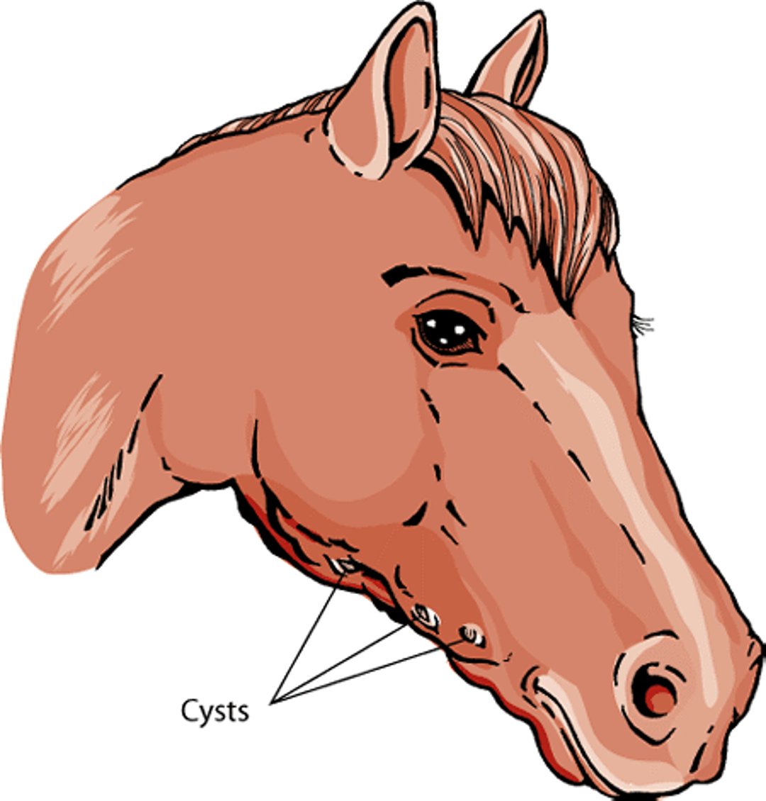
Cysts of the Head and Neck
Cysts (lumps) in the head and neck can be caused by imperfections during fetal development. These need to be distinguished by your veterinarian from abscesses or lumps caused by infection or other disease. Dentigerous cysts arise from abnormal tooth development and often contain tooth fragments. They are most often seen in association with the wolf teeth (first premolar) or canine teeth in mares. These cysts can be found in horses less than 3 years of age, and they may be difficult to distinguish from cystic sinuses, another condition which can result in face or jaw distortion. Surgical removal of the cysts is required. The veterinarian can then make a final diagnosis based on examination of the removed tissue. Other types of congenital cysts occur in horses but are very rare.
Horse head cysts

Hernias
A hernia is the protrusion of a portion of an organ or tissue through an abnormal opening. In horses, hernias can involve an abnormal opening in the wall of the diaphragm (the sheet of muscle that separates the chest from the abdomen) or the abdomen. The defect may allow abdominal organs to pass into the chest or bulge beneath the skin. Hernias may be congenital (present at birth) or result from injury. Signs of a hernia vary from none to severe and depend on the amount of herniated tissue and its effect on the organ involved.
Types of Hernias in Horses
General Area | Specific Type | Description |
|---|---|---|
Diaphragm | Retrosternal (Morgani) | Abdominal contents extend into the sac surrounding the heart |
Abdomen | Umbilical | Abdominal contents protrude at the site of the navel |
Inguinal | Abdominal contents protrude into the groin, above the scrotum of male animals | |
Scrotal | Abdominal contents protrude into the scrotum (the sac surrounding the testes) of male animals |
Hernias involving the abdominal wall are called umbilical, inguinal, or scrotal, depending on their location ( see Table: Types of Hernias in Horses). Diagnosis of umbilical hernias is usually simple, especially if the veterinarian is able to push the hernia back through the abdominal wall (called reducing the hernia). If not, the hernia must be differentiated from an umbilical abscess, which is common in horses. The veterinarian may need to use a fine needle to collect tissue or fluid for confirmation of the diagnosis. Umbilical hernias vary in size and may contain only fat, or in more severe cases, intestinal loops. These hernias are corrected by surgery. The tendency to develop hernias may be inherited.
Inguinal or scrotal hernias occur when sections of the intestine protrude into the inguinal canal located in the groin. They are common in horses, particularly draft breeds and warmbloods. It is suspected that these hernias are inherited. Signs can range from a nonpainful swelling in the groin or scrotum to sudden and severe colic, particularly if an intestinal loop is trapped. Veterinarians may be able to diagnose an inguinal hernia on examination, which may include a rectal examination. Inguinal hernias in colts often resolve spontaneously during the first year of life. Veterinarians may also treat foals by pushing the hernia back into the abdomen and bandaging the area. Fortunately, inguinal hernias rarely cause a significant problem in foals. Hernias that do not spontaneously resolve early in life should be surgically corrected to prevent later complications, which are more common in stallions. If the hernia is blocking blood flow to the intestines, it is a medical emergency and urgent treatment is necessary.
Congenital hernias in the diaphragm can also occur in horses. A retrosternal or Morgani hernia has been described in which a hernial sac protrudes into the chest. Affected horses display signs of colic, and the hernia can be repaired with surgery. When a severe injury causes a diaphragmatic hernia, signs include trouble breathing, tiring easily, lethargy, and weight loss.
Stomach
Pyloric stenosis results from muscular thickening of the “exit” of the stomach (pyloric sphincter). This obstructs the flow of digested food from the stomach to the small intestine. It can be acquired or inherited, but the inherited form is rare in horses. Signs may include loss of appetite and other signs, such as abdominal pain, that are usually associated with colic. Treatment is through dietary modification and medication. In more severely affected cases, surgery may be beneficial.
Small and Large Intestine
Maldigestion is a condition in which certain foods are not properly digested. Malabsorption occurs when nutrients are not properly absorbed into the bloodstream. Both of these conditions often cause chronic, persistent digestive system problems, including weight loss and diarrhea. There are many potential causes, which may be inherited or acquired. Malabsorption and maldigestion are often treated with a combination of dietary changes and medication; the exact treatment will depend on the condition being treated.
A defect in the colonic nerves called ileocolonic agangliosis is seen in white foals produced by matings of Overo horses. (Overo is a pinto color pattern that describes a solid color with splashes of white.) Although the foals appear normal at birth, they soon develop colic and an impaction of meconium (the first stool) due to lack of motility of the digestive tract. The condition is fatal. The affected horses are white and have blue irises. Diagnosis can be confirmed by the lack of specific nerves in the colon. Adult Overo horses can be genetically screened before breeding to reduce the likelihood of this defect.
Congenital defects of the rectum and anus generally result from arrested embryonic development. Inherited closures (atresias) of the small- and large-intestinal tracts are uncommon in large animals and are always fatal. A missing or underdeveloped colon (atresia coli) has been reported in several breeds of horses. Segmental aplasia (also called rectal agenesis) is a condition in which the rectum is closed off before reaching the anus. Surgical correction is difficult because the location of the closure varies, and damage to nerves in the area may occur during surgery.
Liver
A portosystemic shunt can occur in foals. In a healthy animal, blood coming from the intestines is processed by the liver, which removes toxins from the bloodstream before they reach the brain or other organs. In an animal with a portosystemic shunt, however, blood bypasses the liver through one or more “shortcuts” (shunts) and enters directly into the general circulatory system.
Signs of a portosystemic shunt include nervous system disturbances (staggering, wandering, blindness, circling, and seizures) and a failure to grow and thrive. Signs may be most pronounced at feeding time. A definite diagnosis is made by using an opaque dye to highlight the blood vessels, followed by x-rays. This procedure can identify the location of the shunt and determine whether it is single or multiple. In some cases, the shunt may be seen with ultrasonography. It also allows the veterinarian to assess whether surgical correction is possible. The signs of the condition may be controlled with dietary changes, medications, and supportive care. The outlook is uncertain, and animals with multiple shunts tend to do poorly.
Congenital hepatic fibrosis is an inherited condition in Swiss Freiberger horses (also called the Franches-Montagnes horses) that has been traced back to one stallion. Affected foals are 1 to 12 months old. Most show signs of liver disease. Biopsies of liver tissue show evidence of severe damage and the development of connective tissue (fibrosis) within the liver.
Other liver developmental anomalies include hepatic (liver) cysts, which are inherited in Swiss Freiberger horses. All affected horses can be genetically traced back to one stallion. These liver cysts are part of a larger syndrome that can also cause cysts in the kidneys and/or the pancreas. Affected foals usually show signs in the first year of life.
Signs of hepatic cysts include:
weight loss
jaundice
neurologic disturbances
abdominal distention
fever
colic
For More Information
See professional content regarding congenital and inherited disorders of the digestive system.


