


Cats can develop many different cardiovascular diseases. The ones discussed below are the most common.
Degenerative Valve Disease
This acquired disease is characterized by a thickening of the heart valves. Degenerative valves do not close properly and allow blood to flow backwards through the heart. The mitral valve is most commonly affected. This allows blood to return from the left ventricle to the left atrium. With time, enlargement of the left atrium and left-side heart failure can develop. If the tricuspid valve (located between the right atrium and right ventricle) is affected, right-side heart failure can develop. Degenerative valve disease is uncommon in cats.
In cats, there are no signs during the early stages of the disease, although a heart murmur can be heard. As the disease becomes severe and overwhelming, heart failure can develop, and affected cats may be unwilling to lay down or have difficulty breathing. A veterinarian can often diagnose degenerative valve disease based on physical examination findings and appropriate imaging procedures, which may include chest x-rays and echocardiography (ultrasonography). Because x-rays can be inconclusive, your veterinarian may recommend that you count the number of your cat's breaths while it sleeps or rests. If your cat's sleeping respiratory rate is elevated, it may have abnormal fluid buildup within the lungs (pulmonary edema) or within the chest cavity (pleural effusion). Regular monitoring of the sleeping respiratory rate can also help assess your cat's response to treatments. Arrhythmias may develop as the disease progresses and can be detected with electrocardiography.
Affected cats can live for years with appropriate treatment. Treatment is often not begun until signs of heart failure start to appear or when fluid in the lungs is found on chest x-rays. Optimal treatment should be planned by your veterinarian for each stage of disease. Treatment for early signs of congestive heart failure includes diuretics to control the accumulation of fluid in and around the lungs and ACE inhibitors to reduce adverse hormonal effects caused by compensatory mechanisms. Other drugs or therapy (such as those needed to control arrhythmias) may be added as needed. Survival times can vary.
Disorders of the Heart Muscle (Cardiomyopathy)
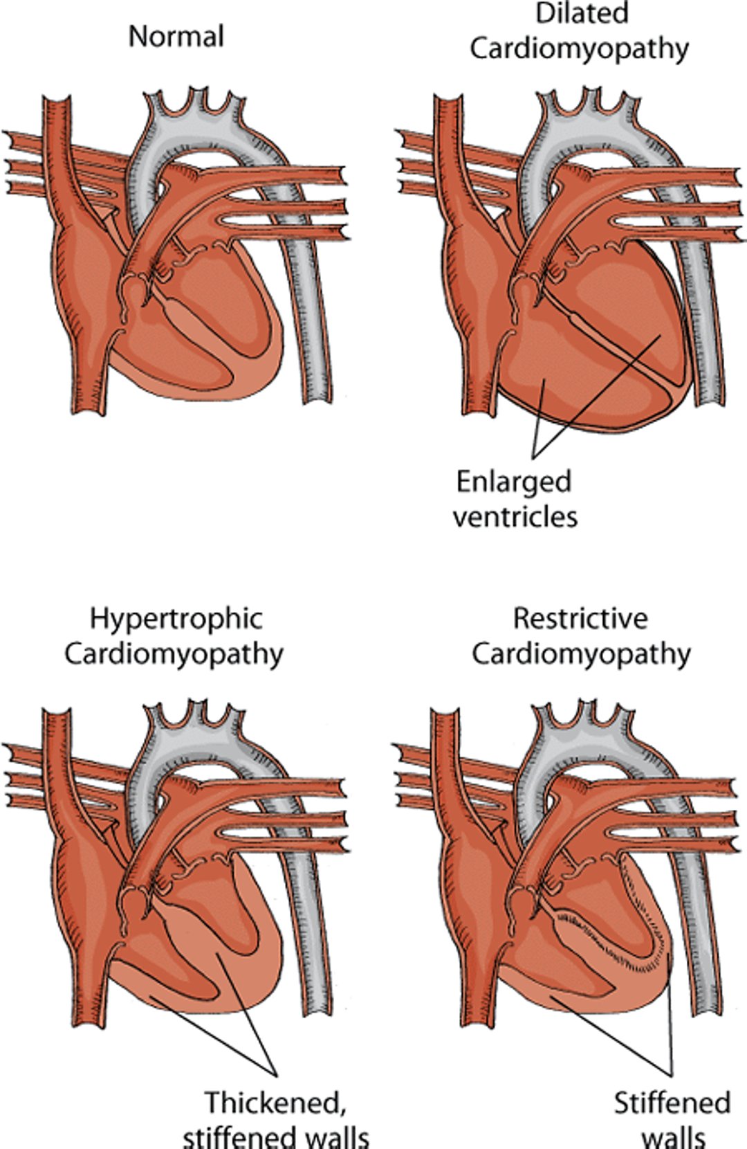
Cardiomyopathy is the name for any disease that mostly affects the heart muscle. Most cardiomyopathies of animals are diseases with no known cause that are not the result of any generalized or primary heart disease. In cats and other animals, they have been classified as dilated cardiomyopathy, hypertrophic cardiomyopathy, arrhythmogenic right ventricular cardiomyopathy, and restrictive or unclassified cardiomyopathy. Cardiomyopathy can also occur as a result of other diseases. In these cases, they are usually called secondary myocardial diseases.
Dilated Cardiomyopathy
Dilated cardiomyopathy is a disease that causes the progressive loss of the heart muscle’s ability to contract. Several forms of secondary dilated cardiomyopathy exist; for example, one form in cats is caused by a nutritional deficiency of taurine, an amino acid. The incidence in cats has decreased dramatically since the discovery in 1987 that taurine deficiency was responsible for most cases. Since then, taurine has been added to all commercial cat foods. Today, most cases are not taurine responsive and reflect primary disease of unknown cause. When cases of taurine-responsive dilated cardiomyopathy are occasionally seen, it is in cats fed noncommercial foods (such as vegetarian diets, baby food, or home-cooked meals) or commercial dog food. Be sure to speak with your veterinarian about your cat's diet, especially if you do not use a commercial cat food.
Types of cardiomyopathy in cats

Cats with heart failure caused by dilated cardiomyopathy have extreme difficulty breathing due to fluid accumulation in the lungs (pulmonary edema) and/or in the chest cavity (pleural effusion). The respiratory distress often progresses rapidly and may not respond to treatment. The outlook is grave for cats with dilated cardiomyopathy that is not taurine responsive. Half of affected cats die within 2 weeks of diagnosis. Cats that have taurine responsive cardiomyopathy also have a high risk of death. However, cats that can be kept alive long enough for taurine supplementation to become effective (2 to 3 weeks) have an excellent outlook.
Hypertrophic Cardiomyopathy
Hypertrophic cardiomyopathy is a condition in which the walls of the left ventricle thicken and become stiff as a result of a heart muscle disorder. The chamber of the left ventricle shrinks in size, leading to enlargement of the left atrium and potentially heart failure eventually. It is the most common primary heart disease diagnosed in cats. It occurs in families in certain breeds of cats, such as Persians, Sphynx, Norwegian Forest Cats, Bengals, Turkish Vans, Maine Coons, Ragdolls, and American and British Shorthairs. The disease may be seen in cats from 3 months to 17 years of age, although most patients are middle aged. Males and females are equally affected, but males tend to develop severe disease at a younger age. The disease is caused by an inherited genetic defect.
Affected cats may not have any signs or may have sudden onset of difficulty breathing, collapse, and weakness or paralysis of the hind limbs. They may also die suddenly. A veterinarian may note abnormal heart sounds, including murmurs and gallop heart sounds. However, at least one out three cats with hypertrophic cardiomyopathy do not have a murmur. Fluid may accumulate in the lungs and in the space between the lungs and the chest wall. Blood clots may form in the heart, then travel and lodge at the point where the aorta divides near the hips (called saddle thrombi), leading to muscular weakness or paralysis of the hind limbs. X-rays and echocardiography (ultrasonography) may be helpful to diagnose the condition as well as to determine the best treatment.
Treatment is directed at controlling signs of heart failure, improving cardiac function, and reducing the incidence of blood clots. Diuretics (to reduce fluid buildup), oxygen, calcium-channel blockers, beta-blockers, or ACE inhibitors may all be considered. Many mildly affected cats have a good longterm outlook; however cats with congestive heart failure have a poorer outlook.
Restrictive Cardiomyopathy
Restrictive cardiomyopathy develops when the left ventricle becomes stiff without significant thickening of the muscle fibers. The cause is not known. The stiff heart muscle fibers cause the ventricles to resist filling with blood between heartbeats, leading to left atrial enlargement and possibly heart failure. To confirm a diagnosis, echocardiography (ultrasonography) is usually required. Signs and treatment are similar to those for hypertrophic cardiomyopathy (see above); however, the outlook seems to be worse, especially in cats with congestive heart failure.
Arrhythmogenic right ventricular cardiomyopathy
Arrhythmogenic right ventricular cardiomyopathy is a rare cause of heart muscle failure in cats. It is restricted primarily to the right side of the heart, but may also involve the left ventricle. It is characterized by a fibrous and fatty muscle of the right ventricle causing progressive heart muscle failure. Signs include difficulty breathing, increased breathing rate, fainting, and nonspecific signs such as loss of appetite and lethargy. Treatment is similar to that for dilated cardiomyopathy (see above). The longterm outlook is generally poor.
Unclassified Cardiomyopathy
Unclassified cardiomyopathy is a disease of cats with obvious abnormalities of the heart muscle on echocardiography (ultrasonography) that do not clearly fit into any other category. The cause is not known. Signs and treatment are similar to hypertrophic cardiomyopathy; however the outlook is worse, especially for cats in heart failure.
Other Causes of Heart Muscle Failure
A form of cardiomyopathy called atrial standstill destroys the muscle wall of the atrium and occasionally affects the muscle wall of the ventricle. This disease has been reported in some cats with coexisting dilated cardiomyopathy. Treatment is rarely effective.
Endocardial fibroelastosis is a disease of unknown cause that leads to thickening of the lining of the left atrium, left ventricle, and mitral valve. It is a rare cause of heart muscle failure in young cats, especially Siamese and Burmese breeds. Affected cats are usually less than 6 months old when they develop left-side heart failure. Signs, treatment, and outlook are similar to dilated cardiomyopathy (see above).
Infective Endocarditis
The endocardium is the thin membrane that lines the heart cavity. Infection of the endocardium typically involves one of the heart valves, although endocarditis of the cavity’s wall may occur. Infection is caused by bacteria carried in the blood. The infection gradually destroys the valve and keeps it from working properly. The disease is rare in cats, with the aortic and mitral valves being most commonly affected.
Infected blood clots form on affected valves. Pieces of blood clots can dislodge and travel to other parts of the body, including the nervous system, digestive tract, urinary system, reproductive tract, and joints. Signs vary depending on where the infected blood clots lodge, but affected animals usually have a murmur, fever, weight loss, and fatigue. The condition can also lead to heart failure, which causes cats to have trouble breathing. Veterinarians use blood tests, x-rays, echocardiography (ultrasound), and electrocardiography to diagnose the condition.
Treatment is directed at controlling signs of congestive heart failure, resolving any significant arrhythmias, killing the bacteria that started the infection, and eliminating the spread of infection. Controlling heart failure requires the use of diuretics and ACE inhibitors. Antibiotics are also necessary.
Pericardial Disease
The pericardium is the membrane surrounding the heart. When fluid builds up in the pericardium (called pericardial effusion), pressure on the heart increases. The increased pressure gradually compresses the heart, interfering with its ability to pump blood. This condition is called cardiac tamponade. This compression of the chambers significantly affects blood circulation and causes swollen jugular veins and accumulation of fluid in the abdomen. In addition, too little oxygen reaches the body’s tissues. This condition is uncommon in cats and is most often caused by congestive heart failure. Other causes include cancer (usually lymphoma in cats), infections (for example, feline infectious peritonitis), injury, and chamber rupture.
Cats with cardiac tamponade require urgent treatment. Medical treatment may not be able to rapidly reduce fluid buildup within the membrane surrounding the heart. Pericardiocentesis (inserting a needle through the membrane to withdraw the fluid) is commonly used to remove fluid quickly. The underlying cause should also be treated, whenever possible.
High Blood Pressure (Hypertension)
Systemic hypertension is an increase in the body’s blood pressure. There are 2 major types of systemic hypertension. Essential (primary) hypertension, which is of unknown cause, is common in humans but rare in cats. Secondary hypertension results from a specific underlying disease. In cats, the most common causes are kidney disease and hyperthyroidism (overproduction of thyroid hormones). Veterinarians typically do not measure blood pressure in all cats. However, if your cat has kidney disease, hyperthyroidism, or evidence of damage caused by hypertension, blood pressure should be measured.
Cats with extremely high blood pressure may have no signs that are visible to the owner. Sudden blindness is the most common sign. Blood tests may help with diagnosis of the cause of high blood pressure. Treatment should be started in cats with sustained and severe high blood pressure, or in pets with sustained high blood pressure and an underlying cause (such as kidney failure). A drug that widens small arteries is used to treat systemic hypertension in cats.
Pulmonary hypertension is elevation of blood pressure in the lungs. Possible causes include increased thickness of blood (for example, an abnormal increase in red blood cells) and increased pulmonary blood flow (caused by, for example, a ventricular septal defect, patent ductus arteriosus, or an atrial septal defect). Other causes include abnormalities of the blood vessels in the lungs, which may be caused by heartworm disease, narrowing of the arteries within the lungs, left-side heart failure, or blood clots within the lungs. Signs are similar to those seen in right-sided congestive heart failure, such as accumulation of fluid around the lungs or in the abdomen. A swollen and pulsating jugular vein may be noted. Doppler echocardiography (ultrasonography) is the most likely method of confirming the diagnosis. Treatment is usually not effective, and the outlook is poor. The best chance for a successful outcome is the identification and treatment of the underlying disease.
For More Information
Also see professional content regarding acquired heart and blood vessel disorders.


