


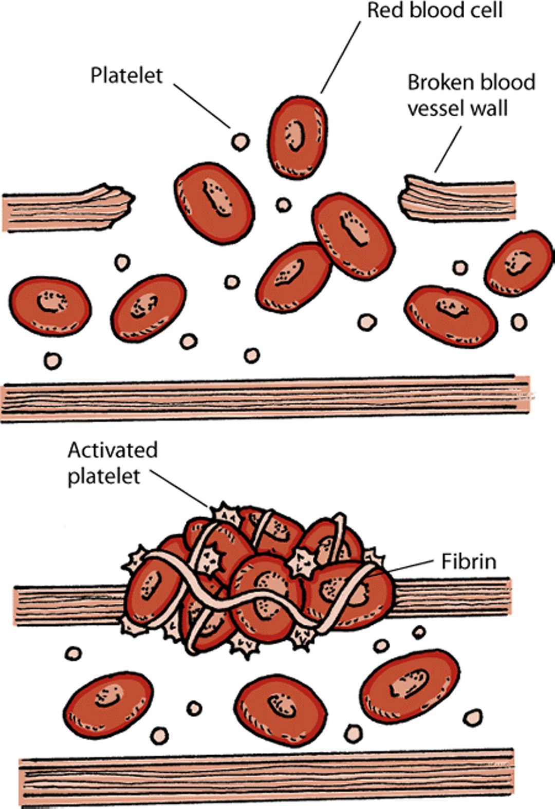
When bleeding occurs in an organ or body part, a process is set in motion to stop the bleeding. This is called hemostasis. In order to work, hemostasis requires an adequate number of platelets, the right amount of blood clotting proteins (often referred to as factors), and blood vessels that constrict properly. When an injury occurs, the wall of the blood vessel breaks. A normally responsive blood vessel will narrow so that blood flows more slowly, allowing the clotting process to begin. Platelets also rush to the broken wall where certain proteins change the platelets’ shape from round to spiny so that they can stick to blood cells, the broken vessel wall, and to each other. Other proteins form long strands called fibrin. These fibrin strands form a net that traps and helps hold together the platelets and blood cells, creating a clot that plugs the break in the vessel wall. After the clot has formed and stabilized, other proteins stop the clotting process and eventually dissolve the clot.
Bleeding disorders may be present at birth (congenital) or occur later (acquired). Defects in blood clotting proteins usually show up as delayed bleeding and bruising deep in tissues, while platelet defects usually show up as superficial small bruises, nosebleeds, black stools caused by bleeding into the bowels, or prolonged bleeding at injection and surgery sites.
Abnormal clotting (also called hypercoagulation) leading to blocked arteries may be inherited disorders of anticlotting proteins or acquired disorders. Acquired clotting diseases are more common in animals than are inherited disorders.
Blood clotting tests can help identify animals with defective clotting proteins. However, the tests are not very sensitive, so an animal must have a severe deficiency for the tests to find the problem. Newer tests may provide more information, but they are not widely available.
Hemostasis

Congenital Clotting Protein Disorders
Many different proteins are involved in the clotting process. Deficiencies of any of these proteins can cause bleeding disorders. Congenital clotting protein disorders are present at birth. In a severe deficiency or defect of clotting proteins, signs will appear at an early age. Severe defects are usually deadly. Animals may be stillborn or die shortly after birth. Lack of clotting proteins or vitamin K (which is also part of the clotting process) in a newborn animal may make a clotting defect worse. If the amount of any particular clotting protein is 5 to 10% of normal, the newborn may survive, but will usually show signs of illness before 6 months of age. It is during this time, when numerous routine procedures (for example, vaccination, dewclaw removal, and neutering or spaying) are usually done, that a bleeding tendency may be noticed by your veterinarian. Most of the clotting protein disorders present at birth in domestic animals are defects of a single protein.
Hypofibrinogenemia (an abnormal shortage of fibrinogen in the blood), accompanied by severe bleeding, has been reported in Saint Bernards and Vizslas. Dysfibrinogenemia (abnormally functioning fibrinogen) has been reported in an inbred family of Russian Wolfhounds (Borzois). Dogs with the disorder had mild bleeding problems (such as nosebleeds), but injury or surgery resulted in life-threatening bleeding. Intravenous transfusion with fresh or fresh-frozen plasma (the liquid portion of blood) is the best treatment to stop the bleeding.
Factor II (prothrombin) disorders are rare. Prothrombin is one of the proteins that plays a role in clotting of blood. Boxer dogs have been reported to have prothrombin that is present in normal amounts in the body but does not function normally. A disorder of Factor II has been reported in English Cocker Spaniels. Signs in affected puppies, such as nosebleeds and bleeding gums, decrease with age. Adults bruise easily or have inflamed skin. Treatment is by transfusion of whole blood or plasma.
Deficiency of Factor VII, another clotting protein, has been reported in Beagles, English Bulldogs, Alaskan Malamutes, Alaskan Klee Kai, Miniature Schnauzers, Boxers, and mixed-breed dogs. Usually, it is not associated with sudden, unexplained bleeding, but affected dogs may have bruising or excessive bleeding after surgery. Prolonged bleeding after giving birth has been reported. Factor VII deficiency is most often diagnosed coincidentally when clotting tests are performed.
Hemophilia A (Factor VIII deficiency) is the most common inherited bleeding disorder in dogs. Usually, females carry the gene for the disease without showing any signs, while males do show signs. In affected puppies, prolonged bleeding is seen from the umbilical cord after birth, from the gums during teething, and after surgery. Lameness due to bleeding into a joint, sudden clot formation, and oozing of blood in the body cavity also are common signs in dogs with less than 5% of normal Factor VIII activity. Animals with 5 to 10% of normal activity typically do not bleed spontaneously but bleed more than usual after an injury or surgery. Small dogs may be less likely to show signs of illness. Carrier animals have higher levels of Factor VIII (40 to 60% of normal), and the results of their clotting tests are usually normal. The diagnosis is harder to confirm in animals less than 6 months old because their livers may not yet have produced enough of the clotting proteins. Treatment requires repeated transfusions of whole blood or plasma until bleeding has been controlled.
Hemophilia B (Factor IX deficiency) is less common than hemophilia A. It has been reported in several breeds of purebred dogs and a mixed-breed dog. Females are usually carriers and rarely have signs of the disease. Signs are similar to those of hemophilia A. Animals with extremely low Factor IX activity (less than 1% of normal) usually die at birth or shortly thereafter. Animals with 5 to 10% of normal Factor IX activity may suddenly develop blood clots, bleeding in the joints, oozing of blood in the body cavity, or organ bleeding. Gums may bleed excessively during teething. Some animals have no signs until injury or surgery. Carrier animals with 40 to 60% of normal Factor IX activity usually have no signs and normal results on blood clotting tests. Treatment requires transfusion with fresh or fresh-frozen plasma. Often, internal bleeding into the abdomen, chest, central nervous system, or muscles occurs, and may not be noticed until a crisis happens.
Congenital clotting protein disorders involving deficiencies of Factor X, Factor XI, Factor XII, and prekallikrein have been reported in a few dogs but appear to be extremely rare. Finally, poor clot strength has caused bleeding after surgery in some Greyhounds.
Acquired Clotting Protein Disorders
Most clotting proteins are produced in the liver. Therefore, liver disease can lead to decreased levels of clotting proteins, particularly Factors VII, IX, X, and XI, and proteins that break up clots. Severe liver disease can also lead to a condition known as disseminated intravascular coagulation (see below). Fibrinogen, the protein in blood that is made in the liver and converted to fibrin in response to tissue damage, and von Willebrand’s factor, which is produced outside the liver and helps platelets stick to the blood vessel wall and to each other, can be increased in liver disease.
Dogs that eat rat poison may have blood clotting problems because the poison reduces the liver’s production of clotting proteins. Affected animals may have blood clots and bruising of superficial and deep tissues. Often, the animals do not bleed within the first 24 hours after eating the poison. Vitamin K1, given by injection and then by mouth, is the usual treatment, but may cause side effects, including anemia or allergic reactions. If you suspect your dog has eaten any type of rat or mouse poison, this is an emergency and an immediate trip to your veterinarian is appropriate.
Disseminated intravascular coagulation (DIC) is a condition in which small blood clots develop throughout the bloodstream, blocking small blood vessels and consuming the platelets and clotting factors needed to control bleeding. It usually develops after a triggering event, such as severe infection, heat stroke, burn, tumor, or severe injury. In many cases, the signs are uncontrolled bleeding and the inability to form a normal clot. Death is caused by extensive blood clots or collapse of circulation, leading to the failure of one or several organs. Your veterinarian will determine and attempt to correct the underlying problem causing this condition. Intravenous fluids are extremely important for maintaining normal circulation. The underlying cause must be treated promptly, and anti-clotting drugs may be necessary. DIC is a very serious disorder and is often fatal. Chances of survival are increased with an earlier diagnosis, but early diagnosis can be difficult and requires specialized testing.
Platelet Disorders
Disorders of platelets include having too few platelets (thrombocytopenia) or having platelets that do not work properly. Each type of disorder can be either congenital (present at birth) or acquired later in life. Thrombocytosis is having too many platelets. It may occur as a response to a physiologic or disease process, or, rarely, it may be a component of blood cancer.
Congenital Thrombocytopenia
Gray Collies may develop a disorder called cyclic hematopoiesis (see ), which is characterized by 12-day cycles during which all types of blood cells, including platelets, decrease. All cell types are affected, but neutrophils (the most common type of white blood cells) are most affected. Mild to severe platelet shortages can be seen, and excessive bleeding is a potential complication. This disorder is deadly; most dogs with the disease die from infections before 6 months of age. Even dogs that receive frequent antibiotic treatments usually die by 3 years of age from amyloidosis, a condition in which abnormal proteins build up in the body’s organs. Treatment with proteins that stimulate the production of neutrophils in the bone marrow may provide temporary—but not longterm—improvement.
Hereditary macrothrombocytopenia occurs in about 50% of Cavalier King Charles Spaniels. These dogs have a decreased number of platelets with the presence of giant platelets. This benign (harmless) condition is discovered on routine blood screens.
Acquired Thrombocytopenia
Acquired thrombocytopenias are common in dogs. Many causes have been identified, most involving the immune system destroying platelets.
Rickettsial diseases, caused by organisms in the genera Ehrlichia and Anaplasma, cause mild to severe loss of platelets in dogs. Infection may include short- and longterm changes in the number of platelets and other blood cells. Ticks are the usual carriers of the infection. Infected dogs may show no signs or may have nosebleeds, bleeding into the bowels (resulting in black stools), bleeding of the gums, and prolonged bleeding after vaccination or surgery.
Thrombocytopenia due to immune system dysfunction occurs when the immune system makes antibodies that destroy platelets or platelet-producing cells in the bone marrow. Signs include tiny, purplish red spots on the gums or skin (called petechiae), bruising, bleeding into the bowels resulting in black stools, or nosebleeds. An evaluation of the bone marrow may be necessary to help determine if circulating platelets or the platelet-forming cells are targeted by the antibodies. Corticosteroids are the usual treatment, although other drugs are sometimes used. A blood transfusion may be necessary for anemic dogs. Affected animals should rest to help prevent injuries and abnormal bleeding. If an animal has repeated episodes of the disease, the spleen is sometimes removed.
Thrombocytopenia caused by vaccination has been reported in dogs that have been vaccinated repeatedly with certain types of vaccines (modified live adenovirus or paramyxovirus vaccines). The platelet loss occurs 3 to 10 days after repeat vaccination, usually lasts for only a short time, and may be so mild that it is not noticed unless it happens at the same time as another clotting disorder. Studies have not shown an association between vaccination and immune-mediated destruction of platelets; however, it may rarely occur.
Thrombocytopenia caused by drugs has been reported in dogs. Some drugs and classes of drugs (including estrogen and some antibiotics) suppress the production of platelets in the bone marrow. Other drugs (including aspirin, acetaminophen, penicillin, and others) destroy platelets circulating in the bloodstream. Drug reactions are rare and unpredictable. Platelets usually return to normal shortly after the drug is discontinued. Drug-induced bone marrow suppression may last longer, however. If your dog is taking one of these drugs, your veterinarian will likely monitor the blood count to check for any serious reductions in the number of platelets.
Thrombocytopenia caused by cancer can occur in dogs. Certain types of cancers can trigger a condition called disseminated intravascular coagulation (see ,), which destroys many platelets. Some cancers can also stimulate the immune system and inflammation to consume platelets. Abnormal bleeding can also occur in cancer patients due to abnormal platelet function and inflammation of blood vessels.
Congenital Platelet Function Disorders
Several types of platelet function disorders can be present at birth (congenital). Specialized tests are usually required to detect them.
Canine thrombopathia has been reported in Basset Hounds. Affected dogs have nosebleeds, tiny purplish red spots (petechiae), and bleeding of the gums. This disorder should be suspected in Basset Hounds that have these signs, along with normal levels of platelets and von Willebrand’s factor. Specific diagnosis of this disorder requires specialized platelet function testing. There is no specific treatment, but in cases of severe bleeding, plasma or whole blood transfusions may be needed.
Glanzmann thrombasthenia, previously called thrombasthenic thrombopathia, has been diagnosed in Otterhounds and Great Pyrenees dogs. Affected dogs have prolonged bleeding times and form large bruises easily. A large number of oddly shaped, giant platelets are seen in blood tests. Platelets from dogs with this disorder do not clump together or separate as they normally should. There is no specific treatment. In cases of severe bleeding, transfusions of plasma or whole blood can be given.
Von Willebrand disease is caused by a defective or deficient von Willebrand’s factor. Von Willebrand factor is the protein that carries an important clotting factor (Factor VIII) in the blood and that regulates the first step in clot formation. It is the most common inherited bleeding disorder in dogs and occurs in nearly all breeds and in mixed breeds. The disorder is most common in Doberman Pinschers, German Shepherds, Golden Retrievers, Miniature Schnauzers, Pembroke Welsh Corgis, Shetland Sheepdogs, Basset Hounds, Scottish Terriers, Standard Poodles, and Standard Manchester Terriers.
Three types of von Willebrand disease are known. The most common, Type 1, occurs in dogs with low von Willebrand factor and results in mild to moderate signs. Dogs with Type 2 also have a low amount of the factor but have moderate to severe signs. The factor is absent in dogs with Type 3, which is most frequently seen in Shetland Sheepdogs and Scottish Terriers. Signs include bleeding of the gums, nosebleeds, and blood in the urine. Some puppies may bleed excessively only after injection or surgery. Signs of von Willebrand disease are similar to those of platelet disorders or thrombocytopenia. Laboratory tests are required to confirm the diagnosis. Active bleeding episodes may require transfusion with whole blood or plasma. Dogs with Type 1 disease may respond to treatment with desmopressin acetate.
Acquired Platelet Function Disorders
Dogs with thrombocytopenia due to immune system dysfunction (see ) also may have an acquired platelet functional defect. Dogs can have excessive bleeding tendencies without a severe drop in the number of platelets.
Several diseases have been associated with acquired platelet function disorders. A bone marrow tumor called multiple myeloma increases the amount of antibodies circulating in the blood. This can affect platelets and reduce their ability to form a blood clot. Longterm kidney disease can also decrease the ability of platelets to stick together. Many drugs can also impair platelet function; however, the impairment may not be noticed unless another clotting disorder is also present.
Blood Vessel Disorders
Certain defects present at birth or diseases can cause severe inflammation of the blood vessels and bleeding disorders.
Ehlers-Danlos Syndrome
This syndrome, also known as rubber puppy disease or cutaneous asthenia, is caused by a defect (present at birth) in protein connective tissue in the skin. This causes weak structural support of blood vessels and can lead to blood clots and easy bruising. The disorder has been reported in dogs and people but is rare. The most striking sign is loose skin that stretches to a greater than normal degree and tears easily. There is no treatment.
Rocky Mountain Spotted Fever
This disease is caused by an organism (Rickettsia rickettsii) that is transmitted by ticks. The rickettsial organisms invade and kill blood vessel cells, which leads to blood vessel swelling and bleeding. Infected dogs may have nosebleeds, bruises, blood in the urine, bleeding into the bowel, or bleeding of the retina (the membrane at the back of the eye). In severely affected dogs, disseminated intravascular coagulation may occur (see ).
Canine Herpesvirus
This virus generally affects puppies that are 7 to 21 days old. Widespread inflammation and destruction of the blood vessels is accompanied by bleeding of tissues surrounding blood vessels. The disease usually results in death within 24 hours after signs begin.
Blood Clotting Disorders
Abnormal blood clotting (known as pathologic thrombosis) is the uncontrolled clotting of blood, which causes blocked arteries. Inherited blood clotting disorders that occur in humans are not known in animals. However, there are several acquired clotting disorders that can occur. Certain diseases in animals have been associated with increased risk of blood clots. These have been seen in dogs with kidney disease, overactive adrenal glands, cancer, longterm decrease in the production of thyroid hormones, and rarely, immune-mediated hemolytic anemia (see ).
Some kidney diseases cause a decrease in the anticlotting protein called antithrombin III. Other abnormalities found in kidney disease include an increased tendency for platelets to clump together and a decrease in the enzyme that dissolves blood clots. Increased blood clotting is thought to be caused by several different factors.
Having too much cholesterol in the blood has been associated with increased risk of blood clots. Diseases that cause this include an increase in adrenal gland activity, diabetes, kidney disease, deficiency of thyroid hormones, and inflammation of the pancreas. All have been associated with increased risk of blood clot formation, often in the lungs.
The best treatment for an animal with blood clots is to diagnose and treat the underlying disease, along with providing good supportive care. Maintaining blood flow to the tissues is critical. Your veterinarian may prescribe medication to dissolve or prevent clots. In other cases, transfusions may be the most effective treatment.
For More Information
Also see professional content regarding bleeding disorders.


