


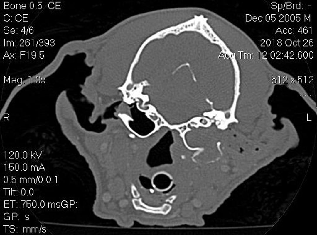
Courtesy of Dr. Michelle Woodward.

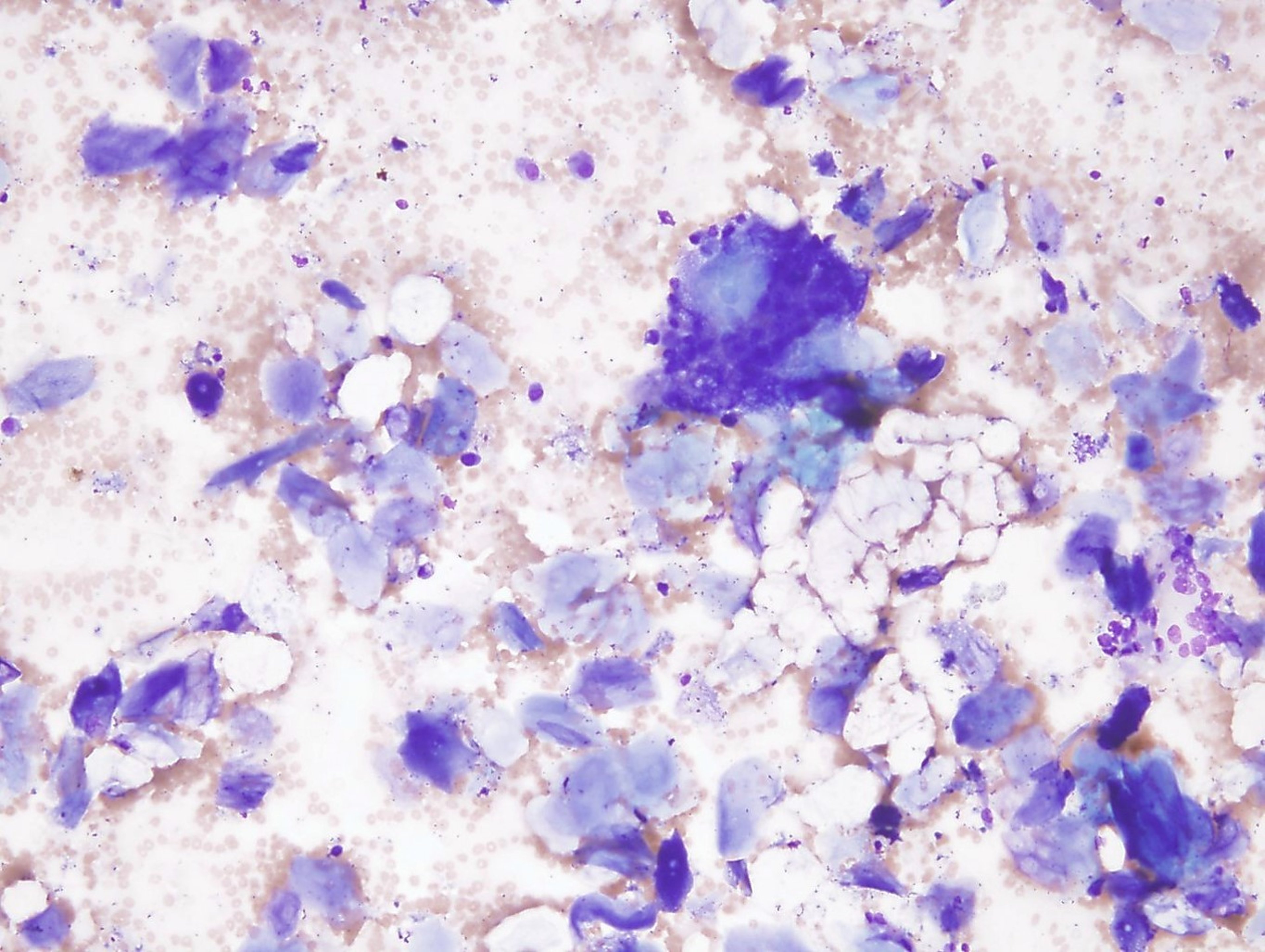
Courtesy of Dr. Shannon Dehghanpir.
Cholesteatomas are cystic tumors that can expand in the middle ear (most commonly in dogs). They can be locally destructive, lead to secondary inflammation, and may induce a secondary otitis externa/media. Although the pathogenesis of these tumors isn't completely understood, they may be associated with eustachian tube dysfunction (leading to tympanic membrane invagination), may occur secondary to chronic otitis media, or may be secondary to surgery of the ear canal and middle ear. There is no significant breed predilection, and the age of affected patients varies.
Clinical signs often include those associated with otitis externa/media (head shaking, pain on palpation of ear/bulla or opening of mouth, exudate in the external ear canal, facial nerve palsy). In chronic cases, the cholesteatoma may not be visible on otic examination due to stenosis, exudate, and chronic changes. When visible, the tumor is frequently pearly white and protrudes from the middle ear to the external ear canal, with the tympanic membrane no longer intact. A complete neurologic examination is warranted, focusing on dysfunction of the facial nerve.
Diagnosis involves advanced imaging to assess the changes to the middle ear. Bulla radiographs can be performed if CT or MRI are not available, but they do not provide as much information. Cholesteatomas will cause lysis, sclerosis, and proliferation of the bulla. The bulla may also expand and be filled with soft tissue. Otitis externa may also be evident. Samples for cytology and histology should be submitted when possible to confirm the type of lesion present. Culture of the middle or external ear should also be considered based on cytology because of the potential for secondary infection.
Surgical treatment may be curative in 50% of cases and, if possible, should be done early in the disease to limit tumor expansion. However, surgery even in later-stage disease may be palliative. Bulla osteotomy or total ear canal ablation with bulla osteotomy should be considered. As much diseased tissue as possible should be removed. Medical management may include treating secondary infections and use of corticosteroids (systemic and topical) to reduce inflammation.


