


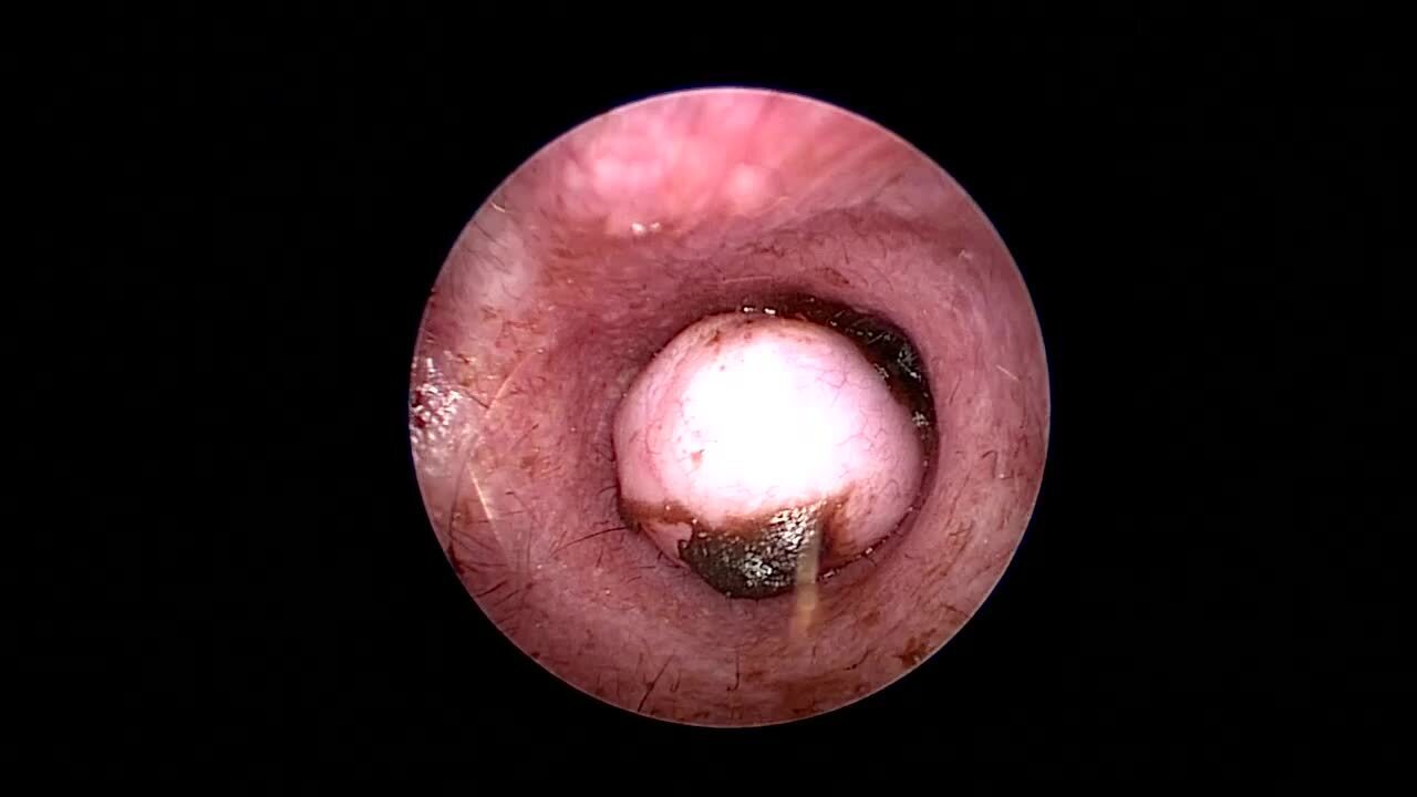
Ceruminous gland tumors are best visualized in a clean ear using a video otoscope. These tumors can be pedunculated or broad based, and they are smooth or multilobulated in appearance (see ).

In dog breeds other than the American Cocker Spaniel, ceruminous gland tumors are often primarily in the vertical ear canal. In the American Cocker Spaniel, these tumors can occur in the horizontal ear canal as well.
Biopsy of the mass can be helpful for diagnosis; however, results from superficial ear canal biopsies are often incorrectly reported as polyps with granulation tissue covered by epithelium and bacteria or inflammatory cells. Deep en bloc biopsies of the same tissue are often correctly reported as tumors.
CT or MRI can be useful in assessing the tympanic bulla more completely and in determining the extent of tumor invasion, especially with malignant tumors.
Benign ear canal tumors can be surgically removed via lateral ear canal resection for access to the tumor mass. Laser surgery, especially when used in conjunction with a video otoscope, has made removal of these tumors relatively easy, obviating the need to open the canal surgically.
More aggressive surgeries, such as total ear canal ablation and bulla osteotomy, are recommended for malignant tumors of the middle ear. Lateral ear canal resection for malignancies is associated with a > 75% recurrence rate.
Median survival time for animals with malignant ear canal tumors has been reported to be > 58 months in dogs and > 11.7 months in cats (1). Dogs with extensive tumor involvement have a less favorable prognosis.
Radiation therapy can be used to treat excised ceruminous gland adenocarcinomas in dogs and cats; a 1-year progression-free survival rate of 56% has been reported (2). No data are available on the efficacy of chemotherapy for otic tumors of dogs and cats.
The prognosis of animals with otic neoplasia can best be determined by histological examination of removed tissues.
For More Information
Abdelgalil AI, Mohammed FF. Clinical, ultrasonographic and histopathological diagnosis of ceruminous gland tumors in cats. Vet Res Forum. 2021;12(3):277-281.
Pieper JB, Noxon JO, Berger DJ. Retrospective evaluation of ceruminous gland tumors confined to the external ear canal of dogs and cats treated with biopsy and CO2 laser ablation. J Vet Int Med. 2023;37(6):2385-2390.
Also see pet owner content regarding earwax gland tumors in dogs and cats.
References
London CA, Dubilzeig RR, Vail DM, et al. Evaluation of dogs and cats with tumors of the ear canal: 145 cases (1978-1992). J Am Vet Med Assoc. 1996;208(9):1413-1418. doi:10.2460/javma.1996.208.09.1413
Théon AP, Barthez PY, Madewell BR, Griffey SM. Radiation therapy of ceruminous gland carcinomas in dogs and cats. J Am Vet Med Assoc. 1994;205(4):566-569. doi:


