

Anatomy of Ratites
All female ratites have a single left ovary and oviduct. The oviduct consists of:
the infundibulum, where fertilization occurs
the magnum, where the thick albumin is put on
the isthmus, where the inner and outer shell membranes are added
the uterus, where the shell is added
the vagina
The opening of the vagina into the cloaca is on the left side of the cloaca at the ten o’clock position.
The male ratite has two intra-abdominal testicles located near the kidneys. During the breeding season, the testicles increase 200%–300% in size. The cock does not produce sperm when not in season.
Cocks in the ratite family all have a phallus that serves to transport semen from the ejaculatory ducts in the cloaca of the male to the cloaca of the female. The phallus is shaped differently in ostriches, emus, and rheas; however, the function is similar. All ratite phalluses have a dorsal groove through which the semen travels.
Breeding of Ratites

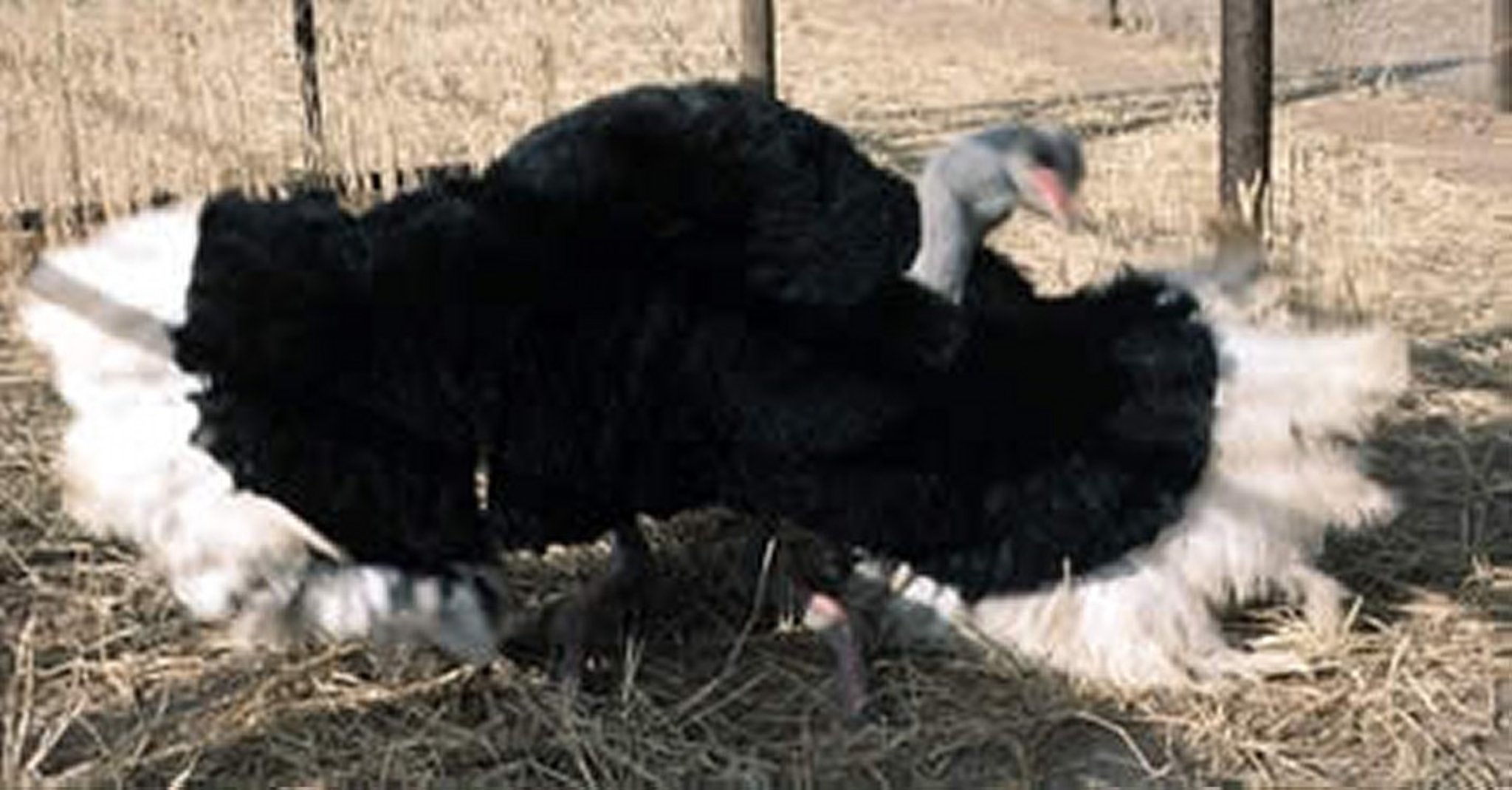
Male ostrich kanteling. Kanteling is a territorial behavior in which the bird sits on its hocks and fans both wings while swinging the head back and forth at the level of the back.
Courtesy of Dr. Karen Hicks-Alldredge.

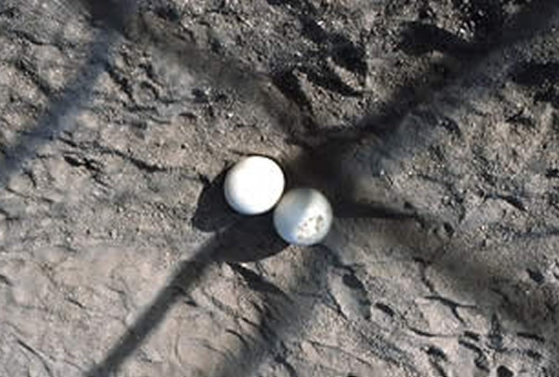
An ostrich communal dump, or scrape (nest). Using its claws, the male scrapes the nest in the ground to a depth of 30–60 cm and a diameter of 3 m. The dominant female lays the first eggs in the nest. The nest will usually hold ~20 eggs for incubation.
Courtesy of Dr. Karen Hicks-Alldredge.
Ostriches and rheas are long-day breeders. The season is controlled by the photoperiod (length of daylight) as well as the ambient temperature; in North America, the breeding season is spring and summer. The emu is a short-day breeder and lays in the late fall and winter. Once the breeding season has started, the cock and hen become reproductively active. The ostrich cock develops external "breeding" colors and begins to display; the hen begins to flutter. This behavior continues for days to weeks before egg laying begins. In captivity, it is often recommended to place one male with two female ostriches for optimal reproductive success. In general, females become reproductively active before the males. Therefore, early eggs are often infertile. This is also true for emus and rheas. Ostrich hens lay every other day during the proper season if the eggs are collected daily. In the wild, normal clutch sizes for ratites are 15 to 25 eggs, whereas 30 is average in a captive production facility. The range is from 0 to 167 consecutive eggs.
Hatching of Ratites
If eggs are gathered and stored for “batch setting,” they should be cooled, if possible, to 15.6°C (60°F). Physiologic zero—or the point at which the embryo stops developing—is 22.2°C (72°F). The egg is laid with the embryo in the 60,000 cell stage, and cooling the egg before incubation ensures that all cells develop in synchrony. In general, hatching eggs should not be washed if they are clean. If eggs are wet or dirty, they should be washed in a warm (43.3°C [110°F]) disinfectant solution (eg, chlorhexidine, sodium hypochlorite, quaternary ammonia) before cooling.

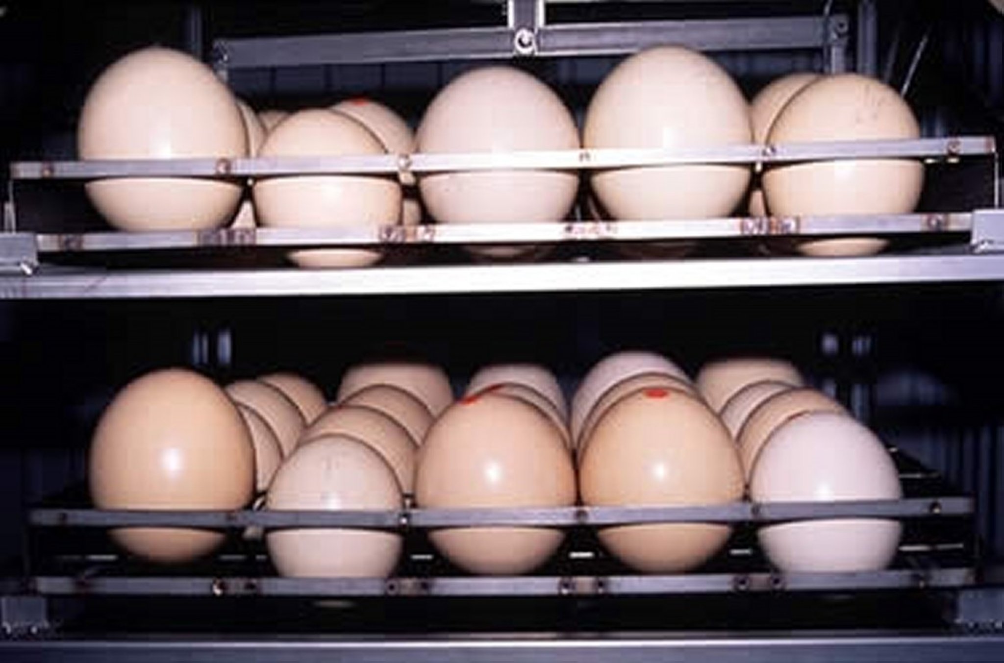
Ostrich eggs set in an incubator. Note the size variation. Artificial incubation is an art, and success is correlated to the quality of the incubator and the knowledge of incubating ostrich/ratite eggs. The temperature to incubate ostrich chicks is 36.4°C (97.6°F). To achieve the most young birds from the fertilized eggs hatched, artificial incubation should be used. The internal environment of the incubator and environment of both the building in which it sits and the environment outside affect the development of the chicks in the eggs. Consequently, the quality of the incubator, to maintain an optimal condition for embryo development, is critical for successful hatching.
Courtesy of Dr. Karen Hicks-Alldredge.
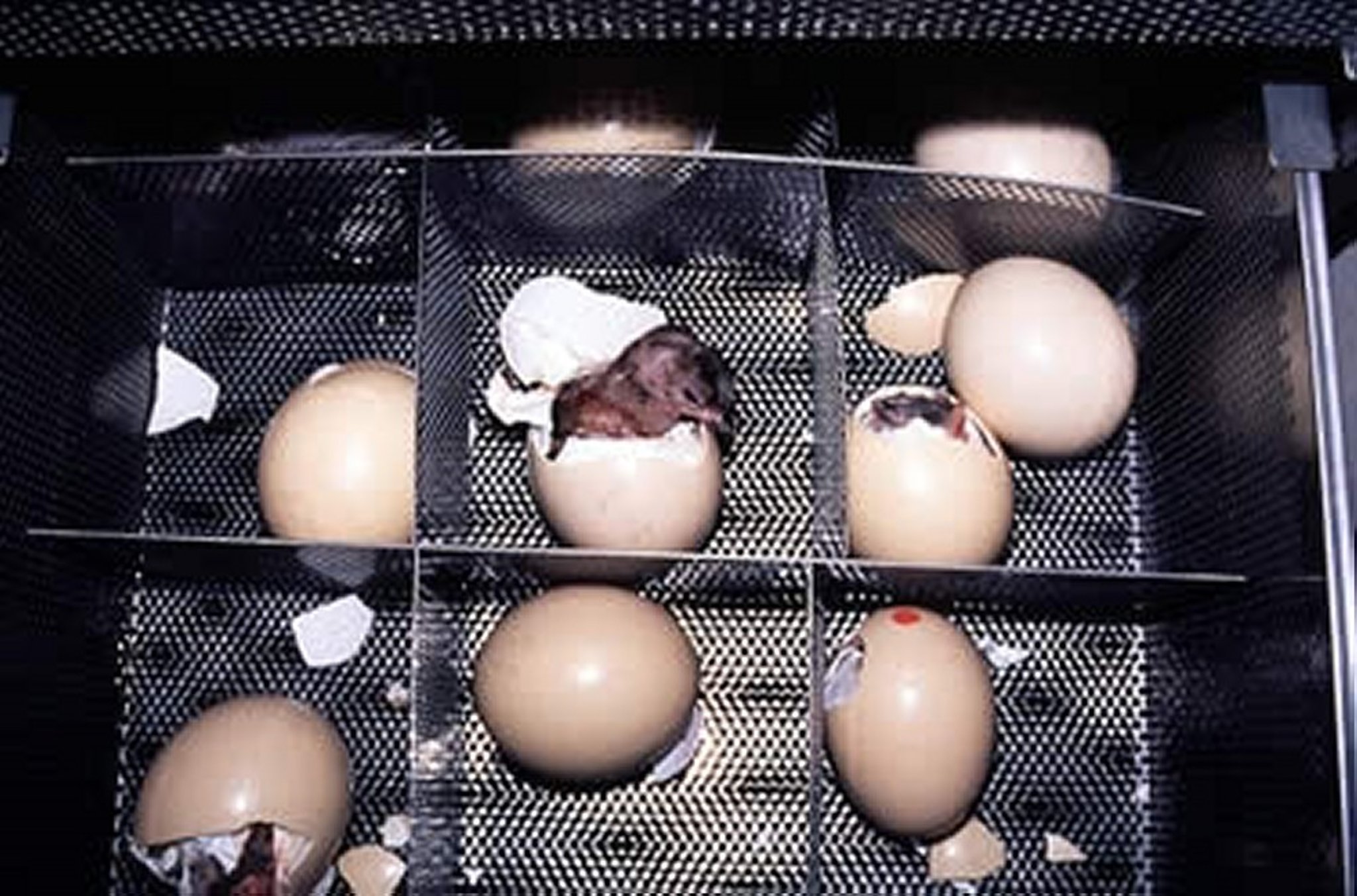
Ostrich chicks hatching. When ostrich chicks are hatching, the recommended relative humidity of the hatcher is 25%, although this may vary depending on individual characteristics and egg size. Higher humidity may lead to wet, edematous chicks. Before a chick is removed from the incubator, the area of the yolk sac on the ventral body wall should be examined to ensure that there has been proper involution; if so, a 7% iodine solution may be applied to the area to protect against infection.
Courtesy of Dr. Karen Hicks-Alldredge.
Commercial incubation of eggs is performed in forced air incubators. Ostrich eggs generally are incubated at 36.4°C (97.6°F) for 40–42 days and moved to the hatcher at 40 days or when the chick “pips” (breaks the egg). Emu eggs are incubated at 36.1°C–36.7°C (97°F–98°F) for 54–58 days and moved to the hatcher at 50–52 days or at “pipping.” Rhea eggs are incubated at 36.7°C (98°F) for 40 days and moved to the hatcher at 38 days or at “pipping.” Whereas temperature in the hatcher is the same as incubation, the humidity should be higher.
For optimal performance of hatchery operations, excellent basic management and biosecurity practices must be followed, including “all-in/all-out” management. Groups of newly hatched birds should be placed in an area of adequate space (to run and exercise their rapidly growing legs and bodies), heat, and ventilation, with fresh water and feed. Mixing groups of young birds should be avoided (to reduce stress) until chicks are at least 3 months old.
Reproductive Diseases of Ratites
Many diseases can result in reproductive failure, either through failure to produce eggs or through production of abnormal or contaminated eggs. Bacterial salpingitis or metritis is common. The etiologic agents vary, as does the severity of the infection. In mild cases, only the uterus or shell gland is affected (metritis), and clinical signs range from abnormal shells to no egg production at all. Infection may result from retrograde bacterial invasion (from breeding or uterine fatigue), extension of an airsacculitis, or perforation of the abdominal cavity by a foreign body. Affected hens generally have a history of erratic egg production, malformed or odoriferous eggs, or a sudden stop in production. On physical examination, temperature and respiration are variable, there may be a discharge below the cloaca, and hens may have a fetid odor. Affected hens have WBC counts ranging from 8,000 to >100,000/mcL. Ultrasonography and radiology are useful to assess the amount and consistency of exudates in the oviduct. If a bacterial salpingitis is diagnosed through a caudal oviductal (vaginal) culture, subsequent treatment is based on organism isolation and sensitivity.


