


Chlamydial conjunctivitis is an acute, chronic, or recurrent infection of the conjunctiva with intracellular bacteria of the family Chlamydiaceae. A variety of animals can be affected. Although often subclinical, infection can also lead to acute or chronic purulent inflammation of the conjunctiva with or without the presence of keratitis or other clinical signs. Diagnosis is confirmed by cytology in some species or by PCR assay. Treatment typically involves administration of tetracycline-class antimicrobials. Systemic administration is preferred because chlamydial conjunctivitis can result from systemic infection and/or occur simultaneously with shedding at other anatomical sites.
Chlamydial conjunctivitis is a well-recognized ocular disease that affects a wide range of domestic and wild animal species. Initially described in association with conjunctival inflammation in companion animals and livestock, the condition is caused by obligate intracellular bacteria of the family Chlamydiaceae.
Chlamydial conjunctivitis is clinically important because of its potential to present as either a localized conjunctival infection or part of a broader systemic illness, with signs and severity varying by host species, age, and immune status. The occurrence of this disease in both individual animals and population settings, particularly those involving close contact or group housing, highlights the need for timely diagnosis, appropriate therapeutic intervention, and consideration of potential zoonotic transmission.
Etiology and Epidemiology of Chlamydial Conjunctivitis
Chlamydiae are obligate intracellular bacteria that form inclusions in the cytoplasm of epithelial cells. The developmental cycle of chlamydiae involves an alternation between the intracellular reticulate body and the extracellular elementary body, which is the infectious form of the organism.
Chlamydiae infect the mucosa of a variety of anatomical sites, including the GI tract, reproductive tract, and conjunctiva. Although some infections can be localized, animals are typically infected systemically, resulting in pathological changes and chlamydial shedding at various anatomical sites.
The conjunctiva is a typical site for chlamydial infection and shedding. Several members of the family Chlamydiaceae have been associated with conjunctivitis in the host species they infect, including Chlamydia caviae (associated with guinea pigs), Chlamydia suis (pigs), Chlamydia psittaci (birds, sheep), and Chlamydia pecorum (pigs, cattle, sheep, other ruminants such as reindeer, wildlife such as koalas, crocodiles, etc).
C pecorum is a ubiquitous cause of ocular infections in livestock; however, the overall contribution of this species to infectious conjunctivitis is unclear. Infection by C pecorum as a serious cause of conjunctivitis in koalas is well documented, and it has the potential to progress toward chronic scarring.
Chlamydial conjunctivitis in cats is caused by Chlamydia felis. Chlamydia pneumoniae has also been detected in cats with conjunctivitis.
C psittaci has been occasionally isolated in dogs and also in sheep.
Trachoma and inclusion conjunctivitis in humans are caused by Chlamydia trachomatis.
Parachlamydia acanthamoebae is a chlamydia-like organism that resides and proliferates within free-living amoebas in the eyes of cats, guinea pigs, pigs, and sheep with conjunctivitis. The pathogenic role of these organisms and their amoebic hosts is unclear.
Although chlamydial conjunctivitis in cats has been referred to as feline pneumonitis, chlamydiae rarely cause pneumonia in cats. The infection always involves the eye, occasionally causing signs of rhinitis, with sneezing and nasal discharge. Although positive antibody titers to C felis are common in some cat populations, the organism is rarely isolated from clinically healthy cats.
Cats with chlamydial conjunctivitis are generally < 1 year old, and cats 2–6 months old appear to be at highest risk of infection. Cats with conjunctivitis that are > 5 years old are very unlikely to be infected, and cats < 8 weeks old might be less at risk because of the presence of maternal antibodies.
Feline chlamydial conjunctivitis is transmitted via direct, close contact between cats. Transmission via fomites is possible, but it is likely to occur only in heavily contaminated environments because chlamydiae survive poorly in the environment. Infected cats also shed chlamydiae from their rectum and vagina; however, whether venereal transmission is possible has not been confirmed.
Weak evidence suggests that chlamydiae might be capable of causing reproductive disease and lameness in cats; however, these associations have not been definitively documented.
Chlamydial infection is one of the most common causes of conjunctivitis in guinea pigs populations, in which it is also known as guinea pig inclusion conjunctivitis. As with cats, young guinea pigs, especially at 1–2 months old, are predisposed to the disease. Subclinical infection can also occur. Rhinitis, lower respiratory tract disease, and genital infections, causing salpingitis and cystitis in female guinea pigs and urethritis in males, can also occur.
Clinical Findings of Chlamydial Conjunctivitis
In cats, the incubation period after exposure to chlamydial infection in another cat ranges from 3 to 10 days. Clinical signs of chlamydial infection in cats can include serous to mucopurulent ocular discharge, nasal discharge, and sneezing. Cats with signs of rhinitis in the absence of conjunctivitis are unlikely to be infected with C felis.
Early signs of chlamydial conjunctivitis include unilateral or bilateral conjunctival hyperemia, chemosis, and serous ocular discharge, with prominent follicles on the inside of the third eyelid in more severe cases. Keratitis is rare, and if present, might be the result of coinfection with organisms such as feline herpesvirus 1.
Chlamydial conjunctival signs are most severe 9–13 days after onset and then become mild over the next 2–3 weeks. In some cats, clinical signs can last for weeks despite treatment, and recurrence of signs is not uncommon. Untreated cats can harbor the organism for months after infection.
In livestock (pigs, sheep, cattle), eye infections are often subclinical, and the GI tract is the primary site of infection. When chlamydial conjunctivitis develops, it can be accompanied by other well-recognized chlamydial diseases, including polyarthritis.
Conjunctivitis in livestock is typically characterized by the early development of bilateral epiphora, chemosis, and conjunctival hyperemia, with disease progressing to prominent conjunctival follicle formation and corneal neovascularization.
Guinea pigs can develop mild to severe conjunctivitis, with conjunctival hyperemia, chemosis, and mucopurulent ocular discharge.
Diagnosis of Chlamydial Conjunctivitis
Chlamydial PCR assay in cases of purulent conjunctivitis
Cytological examination of conjunctival secretions
In cats, chlamydial conjunctivitis should be differentiated from conjunctivitis caused by feline herpesvirus 1 and feline calicivirus, and in guinea pigs, from mycoplasmal and other bacterial infections (eg, pink eye caused by Bordetella bronchiseptica). Diagnosis is best confirmed via PCR assay for chlamydial DNA on conjunctival swabs; however, causality should be confirmed by exclusion of other well-known causes of infectious conjunctivitis in the host species affected.
Cell culture for Chlamydia spp is sensitive and specific but not widely available or practical for routine diagnostic purposes. A special chlamydial transport medium is required to transport specimens for culture. Although not ideal, dry swabs may also be used to collect specimens for chlamydial PCR assay.
A diagnosis of ocular chlamydiosis can also be made by demonstration of intracytoplasmic chlamydial inclusions in exfoliative cytological preparations (see ). Scrapings for cytological examination are prepared by lightly but firmly moving a spatula over the conjunctiva and smearing the scraped material onto a glass slide; the preparation is air-dried and stained. Chlamydial inclusions, which contain reticulate bodies, are round and generally stain purple with Romanowsky stains.
Pearls & Pitfalls
|

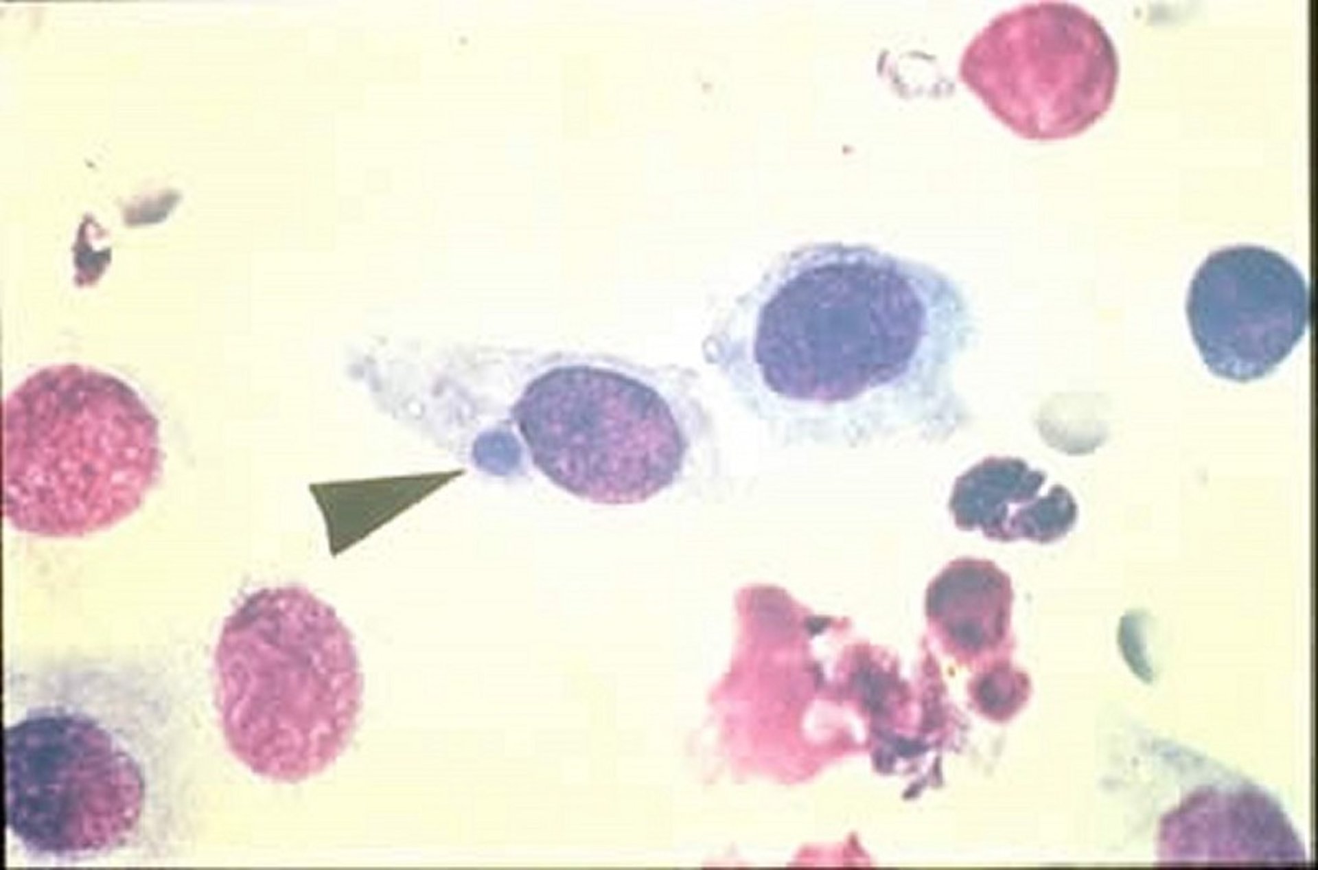
Photomicrograph of a conjunctival cytological sample from a cat, showing inclusion bodies from chlamydial keratoconjunctivitis. The arrowhead points to a stained intracellular inclusion (adjacent to the cell's nucleus). Chlamydial intracellular inclusions typically range in size from 15 to 30 mcm.
Courtesy of Dr. Glenn A. Severin.
Cytological examination of the conjunctiva from guinea pigs with chlamydial conjunctivitis generally reveals a neutrophilic inflammatory response. Inclusions are generally visible only early in the course of chlamydial infection and sometimes not at all. Melanin granules and remnants of some ophthalmic preparations can be mistaken for inclusions, leading to false-positive results, so other diagnostic tests are recommended to confirm the diagnosis.
Because of problems with the sensitivity and specificity of most commercially viable serological assays for chlamydial infections in animals, serological testing is not useful for diagnosing chlamydial conjunctivitis.
Prevention and Treatment of Chlamydial Conjunctivitis
Systemic administration of tetracycline-class antimicrobials
Vaccines only for cats
Vaccines against chlamydiosis are available for cats but not for other species. Feline chlamydial vaccines do not provide complete protection from infection; however, they can decrease disease severity and infection rates. Cat chlamydial vaccines are considered non–core vaccines, and their use should be considered only in environments, such as catteries, where chlamydiosis is endemic.
Nearly all Chlamydia isolates are susceptible to tetracyclines. Systemic therapy is superior to topical therapy and is logical, given that organisms are shed from sites other than the conjunctiva. Tetracycline resistance has been documented as a growing problem in C suis isolates from pigs.
In cats, the treatment of choice for chlamydiosis is doxycycline (10 mg/kg, PO, every 24 hours; alternatively, 5 mg/kg, PO, every 12 hours, to mitigate adverse GI effects) for at least 4 weeks (1). Treatment for up to 6 weeks has been required to eliminate infection in some cats. All cats in a given household must be treated.
In parrots as well, the treatment of choice for chlamydiosis is doxycycline (25–35 mg/kg, PO, every 24 hours, or 75–100 mg/kg, IM, every 5–7 days, for 6 weeks) (2).
Fluoroquinolones (eg, enrofloxacin, pradofloxacin) and amoxicillin-clavulanic acid also have been used successfully to treat both feline and avian chlamydiosis; however, they might be less effective than doxycycline. Azithromycin does not appear to be effective.
Zoonotic Risk of Chlamydial Conjunctivitis
Zoonosis from birds infected with Chlamydia psittaci is well recognized as a risk to human health. On rare occasions, C felis and C caviae have been isolated from humans living in close contact with infected cats and guinea pigs:
Follicular conjunctivitis was described in a single immunocompromised person found to be infected with C felis (3).
One report described detection of C caviae in a person with serous ocular discharge who worked with approximately 200 diseased guinea pigs (4).
A cluster of cases of severe atypical pneumonia in humans was linked to C caviae infection of pet guinea pigs (5).
C suis has been documented in the eyes of pig farmers and slaughterhouse workers who have come into contact with infected pigs (6).
Routine hygiene practices, such as handwashing before and after handling sick animals, might decrease the potential for transmission of these organisms from affected animals to humans.
Key Points
Chlamydial infections are a common cause of infectious conjunctivitis in a variety of domesticated and wild animal species.
When chlamydial etiology is suspected, prompt treatment with systemic administration of tetracycline-class antimicrobials is warranted.
Appropriate infection control, including handwashing after contact with infected animals, should be used to decrease the potential zoonotic risk of chlamydial conjunctivitis.
For More Information
Guideline for Chlamydia felis. European Advisory Board on Cat Diseases (ABCD).
Also see pet owner content regarding chlamydial conjunctivitis in cats.
References
European Advisory Board on Cat Diseases. Guideline for Chlamydia felis. Updated November 6, 2024. Accessed October 1, 2025.
Balsamo G, Maxted AM, Midla JW, et al. Compendium of measures to control Chlamydia psittaci infection among humans (psittacosis) and pet birds (avian chlamydiosis), 2017. J Avian Med Surg. 31(3):262-282. doi:10.1647/217-265
Wons J, Meiller R, Bergua A, Bogdan C, Geissdorfer W. Follicular conjunctivitis due to Chlamydia felis—case report, review of the literature and improved molecular diagnostics. Front Med (Lausanne). 2017;17(4):105 doi:10.3389/fmed.2017.00105
Lutz-Wohlgroth L, Becker A, Brugnera E, et al. Chlamydiales in guinea-pigs and their zoonotic potential. J Vet Med A Physiol Pathol Clin Med. 2006;53(4):185-193. doi:10.1111/j.1439-0442.2006.00819.x
Ramakers BP, Heijne M, Lie N, et al. Zoonotic Chlamydia caviae presenting as community-acquired pneumonia. N Engl J Med. 2017;377(10):992-994. doi:10.1056/NEJMc1702983
De Puysseleyr L, De Puysseleyr K, Braeckman L, Morré SA, Cox E, Vanrompay D. Assessment of Chlamydia suis infection in pig farmers. Transbound Emerg Dis. 2016;64(3):826-833. doi:10.1111/tbed.12446


