

Diaphragmatic hernia is protrusion of abdominal organs or tissue through a congenital opening or traumatic rupture in the diaphragm. This may cause respiratory signs or may be subclinical. Thoracic radiography is highly diagnostic, and treatment centers on stabilization and surgical repair.
Diaphragmatic hernia is a disruption in the continuity of the diaphragm with subsequent shift of abdominal viscera into the thorax. Causes are trauma (more common in dogs), substantial increase in abdominal pressure (eg, dystocia; more common in horses), or congenital (more common in cats). Clinical signs range from mild and vague in chronic or congenital cases to severe respiratory distress in acute, traumatic cases. Thoracic radiographs are usually diagnostic. Surgical correction is required for definitive treatment. (Also see hernias.)
Etiology of Diaphragmatic Hernia in Animals
In small animals, automobile-related trauma is a common cause of diaphragmatic hernia, although congenital defects of the diaphragm may also result in herniation (eg, peritoneopericardial diaphragmatic hernia).
In horses, diaphragmatic hernia may occur, less commonly, congenitally, or after trauma, dystocia, or recent strenuous activity.
Diaphragmatic hernias are extremely rare in cattle.
Clinical Findings of Diaphragmatic Hernia in Animals

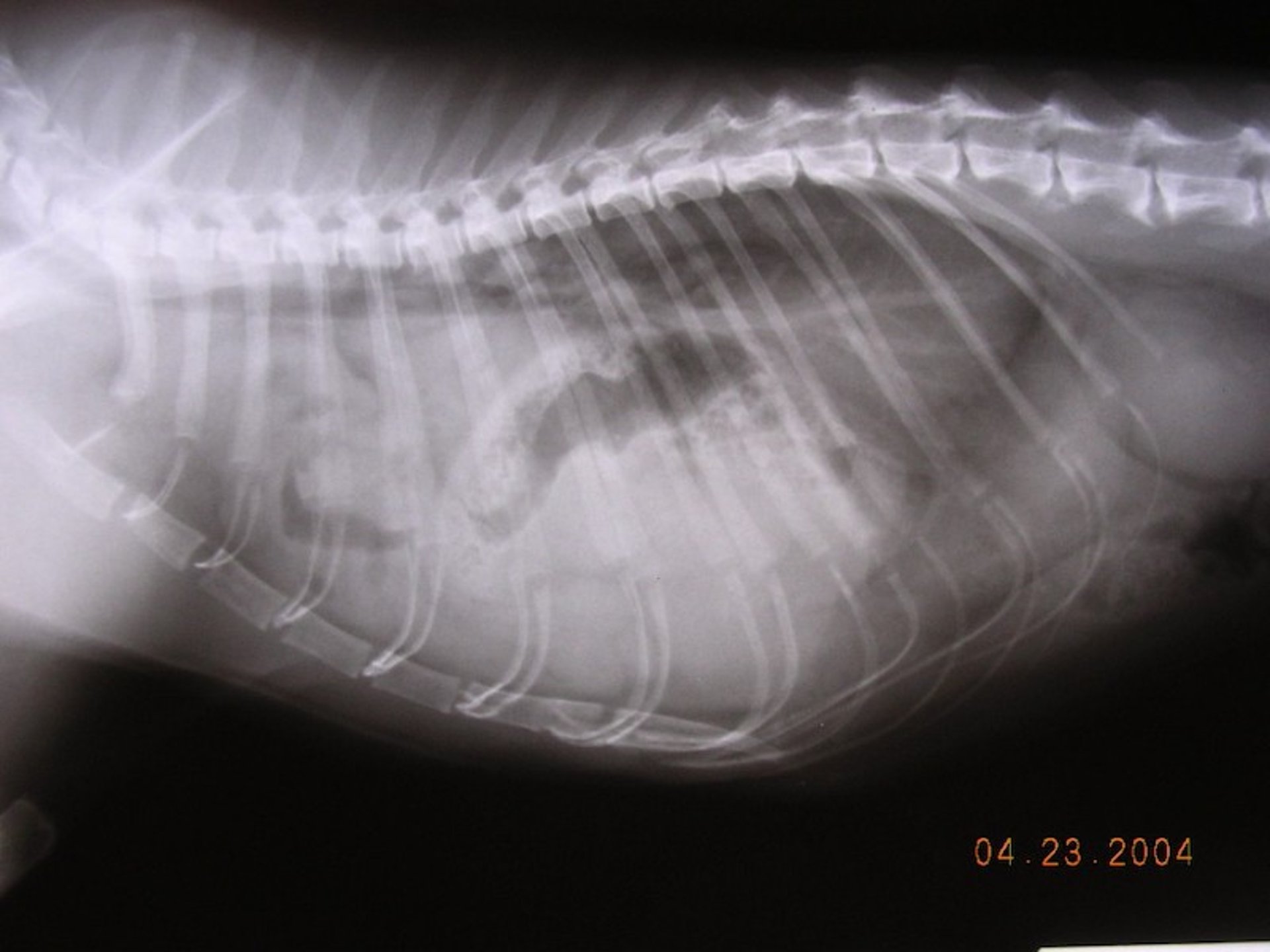
Diaphragmatic hernia. Note gas-filled loops of bowel (gas opacity) overlapping lung fields.
Courtesy of Dr. Joe Hauptman.
Clinical signs of diaphragmatic hernia vary, depending on extent, duration, precipitating events, and species affected. Dogs and cats are characteristically dyspneic in acute cases. The degree of dyspnea may vary from subclinical to life-threatening, depending on the amount of herniated viscera. If the stomach is herniated, it may bloat, and the animal's condition may deteriorate rapidly. In chronic cases, systemic clinical signs such as weight loss may be more prominent than respiratory signs.
Physical examination findings may include absence of lung sounds or presence of gastrointestinal sounds on thoracic auscultation. Congenital peritoneopericardial diaphragmatic hernia is most frequently an incidental finding, although clinical signs may be related to the respiratory or gastrointestinal systems or due to compromised venous return to the heart.
Horses most frequently are presented for evaluation with acute, severe colic secondary to displaced intestines or with respiratory signs and dyspnea.
In cattle and water buffalo, diaphragmatic hernias may be associated with traumatic reticulitis and herniation of the reticulum.
Diagnosis of Diaphragmatic Hernia in Animals

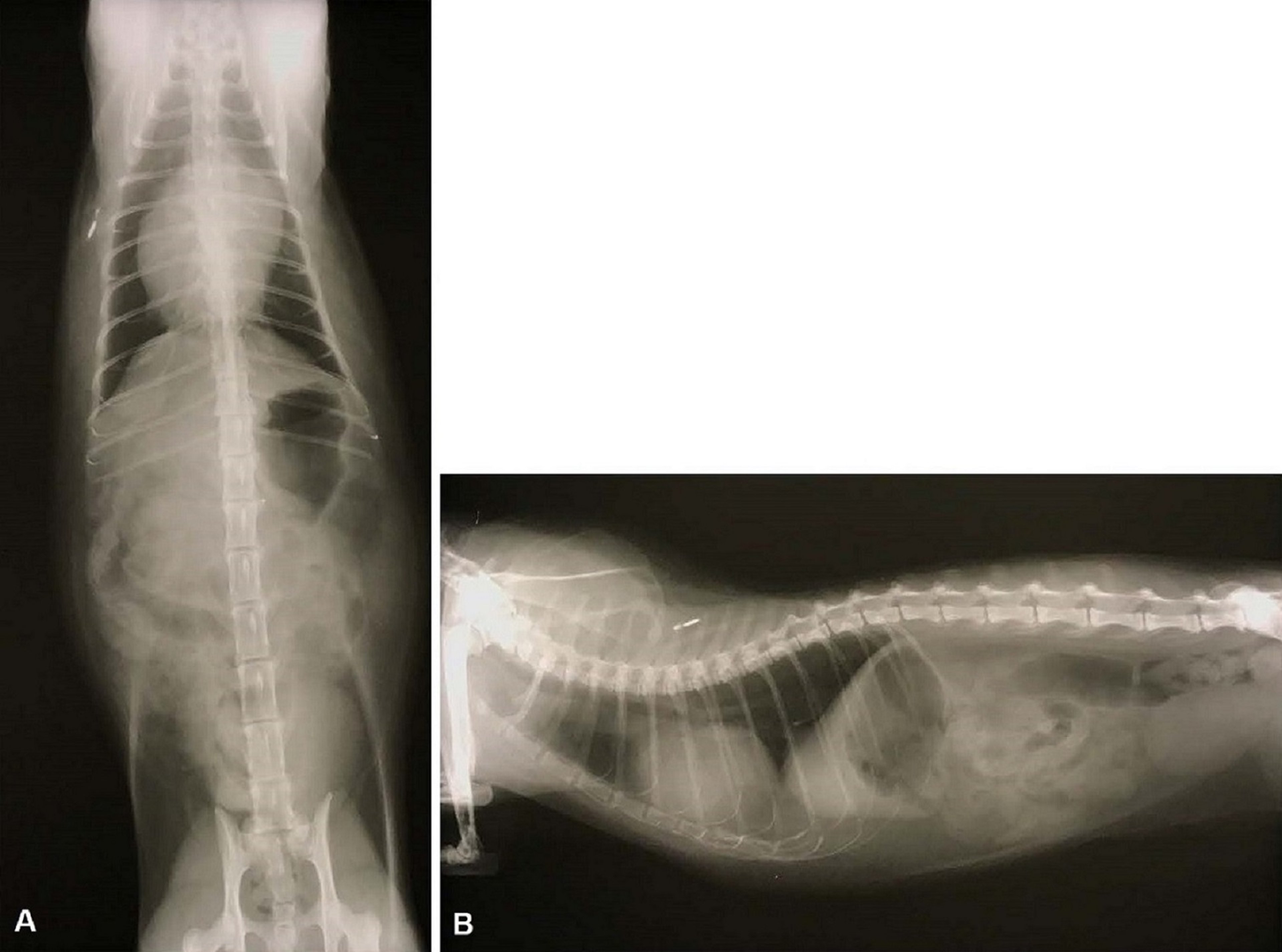
Ventrodorsal (A) and right lateral (B) survey radiographic views of a cat with incidentally diagnosed peritoneopericardial diaphragmatic hernia (PPDH). Note the enlarged cardiac silhouette, which overlaps with the diaphragm.
Courtesy of Dr. Nicholas Roman.
Clinical evaluation
Thoracic radiography
Careful physical examination, including auscultation and percussion, usually suggests the presence of thoracic disease. Tachypnea may be accompanied by signs of severe respiratory distress, such as cyanotic mucous membranes, rapid respiratory rate, or muffled heart and lung sounds. A rapidly deteriorating animal may have the stomach in the thorax with subsequent bloating or torsion.
In many cats with congenital diaphragmatic hernias (especially peritoneopericardial diaphragmatic hernia), the hernia is diagnosed as an incidental finding.
Thoracic radiographs are usually diagnostic. Loss of diaphragmatic contour, abdominal viscera in the thorax, and displacement of viscera from the abdomen may be apparent. Contrast radiography may rarely be necessary to make the diagnosis. Barium may be administered by mouth (gastrointestinal series), or water-soluble contrast may be injected intraperitoneally (celiogram). If available, ultrasonography is frequently useful. Radiographs may be difficult to interpret in patients with pleural effusion. In horses and cattle, radiographs may be more difficult to obtain.
If available, advanced imaging (eg, CT or MRI) may be indicated in some cases.
Laboratory tests (eg, CBC, serum biochemical analysis, urinalysis, and blood gas analysis) and ECG may provide information about the degree of patient compromise. Thoracocentesis may be indicated in cases of pleural effusion or pneumothorax and may yield samples for laboratory analysis. Surgical exploration of the abdominal cavity may be necessary for definitive diagnosis.
Treatment of Diaphragmatic Hernia in Animals
Stabilization
Supportive care
Surgery
Stabilization of the emergent patient, particularly if in respiratory distress, is the first priority. Oxygen supplementation, analgesia, and cardiovascular support (eg, intravenous fluids) should be provided first. Surgery to replace the abdominal contents and repair the diaphragm is the preferred treatment. If the diaphragmatic tear is chronic, it is necessary to be especially careful with anesthesia, because re-expansion pulmonary edema is more likely.
For peritoneopericardial diaphragmatic hernias, patients without clinical signs or that are poor surgical candidates may be managed medically. However, surgical treatment should not be unduly delayed when indicated.
Key Points
Diaphragmatic hernia can be acute (eg, traumatic) or chronic (usually congenital).
Clinical signs can range from nonexistent (eg, incidental finding) to severe respiratory distress.
Thoracic radiographs are usually diagnostic, and surgical repair is the definitive treatment.
Re-expansion pulmonary edema is more common after chronic diaphragmatic hernia repair.
For More Information


