


Listeriosis, caused by Listeria monocytogenes, has been reported in a wide range of avian species. Sporadic cases can occur in chickens, particularly after stressful events, and may cause encephalitis or septicemia and sudden death. Diagnosis is confirmed by isolation, real-time PCR assay, or immunohistochemical identification of the organisms in infected tissues. Antimicrobials can be effective treatment. There is a noteworthy risk of zoonotic transmission.
Listeriosis (circling disease) in birds is caused by the bacterium Listeria monocytogenes. Sporadic outbreaks as well as isolated cases of listeriosis occur in chickens. An an important zoonotic foodborne pathogen, L monocytogenes is of considerable concern to the food industry.
Etiology and Pathogenesis of Listeriosis in Poultry
The causative organism of listeriosis, Listeria monocytogenes, is a gram-positive, non–spore-forming, facultative intracellular, facultative anaerobic, rod-shaped bacterium. Several lineages, serotypes, and strains are recognized.
In chickens, listeriosis has been associated with septicemic and encephalitic forms of disease.
In ruminants, the encephalitic form of listeriosis develops after the organism enters through minor injuries in the conjunctiva or oral and nasal mucosa with subsequent migration along peripheral nerves to the brain. It is unknown if this same route of infection occurs in birds.
Epidemiology of Listeriosis in Poultry
Listeria monocytogenes is commonly present in temperate areas of the world. It is ubiquitous and commonly found in the environment, including soil, sewage, feces from animals, and surface water. In temperate zones, the primary habitats of the organism are soil and decaying vegetation. The organism is common in poorly preserved stored corn silage.
L monocytogenes has been isolated from the intestinal tract of healthy animals, including various species of mammals, birds, and fish. Although many species of birds, including chickens, turkeys, pigeons, ducks, geese, pheasants, canaries, and cockatiels, are susceptible to natural infection, clinical listeriosis in birds is rare.
Generally, young birds are more susceptible to infection and more likely to develop clinical signs than are older birds.
Transmission occurs via ingestion, inhalation, or wound contamination.
Contamination of poultry farms with fecal material from nearby food animal farms (eg, cattle or swine farms) is an important source of infection, especially after a rain or flooding. Wounds from beak trimming and vaccine injection are possible sites of entry for the organism.
The disease often follows stressful events; outbreaks have been reported after beak trimming and with either cold and damp or hot and humid conditions in poultry houses.
Pearls & Pitfalls
|
Clinical Findings of Listeriosis in Poultry
In the septicemic form of listeriosis in poultry, clinical signs are not specific and include a short period of listlessness and lethargy, followed by death. Sudden death may occur without premonitory signs.
In the encephalitic form, ataxia, lateral recumbency with leg paddling, twisting and backward retraction of the neck, and paralysis occur in affected birds.
Lesions
Myocardial necrosis and inflammation (myocarditis) is the most common lesion in birds affected with the septicemic form of listeriosis (see septicemic listeriosis image). Grossly, the affected areas of the myocardium are yellow or yellow-tan and dry, due to necrosis. Often, there are splenomegaly, necrotic foci in the liver, and pericarditis.
Other lesions reported in broilers include ascites and petechial hemorrhages in the myocardium, liver, kidneys, and spleen.

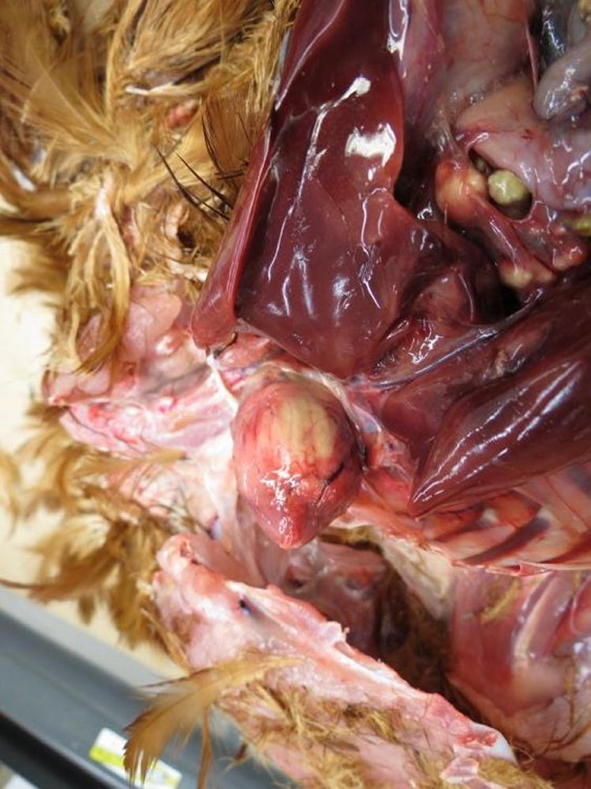
Courtesy of Dr. Tahseen Abdul-Aziz.
In the encephalitic form of listeriosis, no gross lesions are observed in the brain; however, histopathologic lesions are remarkable and include foci of malacia (necrosis), mixed inflammatory infiltrates predominantly composed of lymphocytes, marked perivascular lymphocytic cuffs, and gliosis.
Lesions are usually regionally extensive and found in the medulla oblongata, where they are generally most severe, and in the optic lobes and cerebellum. Gram stain of tissues usually reveals gram-positive bacteria within the lesions. L monocytogenes is usually abundant in myocardial lesions but not in brain lesions.
Diagnosis of Listeriosis in Poultry
Bacterial isolation
Real-time PCR assay
Immunohistochemical staining
In the septicemic form of listeriosis in poultry, gross and histopathologic lesions in the myocardium should arouse suspicion and allow preliminary diagnosis.
Diagnosis is confirmed by isolation, which does not require special media. Diagnosis is also confirmed by immunohistochemical staining to demonstrate L monocytogenes in the tissues or by isolation of the organism, usually from the liver or spleen in the septicemic form and the brain in the encephalitic form.
Samples can be cultured directly on blood agar plates or blood agar containing 0.05% potassium tellurite. Direct culture using brain tissue may not always be successful because of low numbers of organisms in the tissue. Recovery of L monocytogenes increases substantially if a portion of the specimen is refrigerated for 4–8 weeks and subcultured weekly; this is known as cold enrichment.
Alternatively, tissue may be macerated or blended with a general nutrient broth (eg, tryptic soy broth, brain heart infusion) at a ratio of 1:10. The broth medium is incubated at 35°C (95°F) for 5–7 days and examined daily for growth.
Multiple real-time PCR assay kits are commercially available for the species-specific identification of L monocytogenes.
Differential diagnoses for septicemic listeriosis include other types of bacterial septicemia, such as colibacillosis, pasteurellosis, and erysipelas.
Differential diagnoses for encephalitic listeriosis include viral encephalitides (eg, Marek's disease and virulent Newcastle disease). With virulent Newcastle disease, neurologic signs (torticollis, opisthotonos) typically follow high mortality in the flock, and lesions may be present in visceral organs with viscerotropic velogenic Newcastle disease virus infection.
Treatment and Prevention of Listeriosis in Poultry
Antimicrobial treatment
Antimicrobials may be used successfully to treat the septicemic form of listeriosis. In vitro, L monocytogenes is susceptible to penicillin, tetracycline, erythromycin, gentamicin, and trimethoprim-sulfamethoxazole. Treatment of the encephalitic form is usually unsuccessful.
Prevention should focus on identifying and eliminating potential sources of infection.
Zoonotic Risk of Listeriosis in Poultry
Listeriosis is a serious zoonotic disease. L monocytogenes is recognized as an important foodborne pathogen in humans and is of great concern to the public and poultry industry. This is particularly noteworthy because Listeria can survive cold temperatures for long periods of time; therefore, refrigeration and even freezing are not effective in preventing the transmission of the infection to humans.
Outbreaks of listeriosis usually follow exposure to raw or uncooked poultry products but have also occurred after consumption of contaminated ready-to-eat poultry meat products.
Key Points
Listeriosis is a bacterial disease caused by Listeria monocytogenes; listeriosis in birds is rare.
In birds, listeriosis occurs in septicemic or encephalitic form.
Diagnosis is confirmed by bacterial isolation, real-time PCR assay, or immunohistochemical identification of the organisms in infected tissues.
If poultry products are contaminated with Listeria, they could represent a serious zoonotic risk.
For More Information
Kariyawasam S. Listeriosis. In: Williams SM, Dufour‐Zavala L, Jackwood MW, et al, eds. A Laboratory Manual for the Isolation, Identification, and Characterization of Avian Pathogens. 6th ed. American Association of Avian Pathologists; 49-54.
Gasanov U, Hughes D, Hansbro PM. Methods for the isolation and identification of Listeria spp. and Listeria monocytogenes: a review. FEMS Microbiol Rev. 2005;29(5):851-875. doi:10.1016/j.femsre.2004.12.002


