

The canine nasal mite is a parasite of the nasal passages and paranasal sinuses of dogs, with corresponding clinical signs of sneezing, nasal discharge, and epistaxis.
The canine nasal mite, also known as Pneumonyssoides caninum or Pneumonyssus caninum, has been reported worldwide, including the US, Canada, Japan, Australia, South Africa, Italy, France, Spain, Norway, Sweden, Finland, Denmark, and Iran.
Etiology and Epidemiology of Canine Nasal Mites
The canine nasal mite has most commonly been reported in dogs and has also been reported in a silver fox. There does not seem to be a breed, age, or sex predilection, although one report suggested that dogs >3 years old were affected more often and that large-breed dogs had a higher incidence than small breed dogs.
The mites live in the nasal passages and paranasal sinuses. The complete life cycle of P caninum is not known or understood. Transmission is thought to be via direct and indirect contact between dogs. There is no evidence to suggest that P caninum presents a zoonotic risk.
Clinical Findings of Canine Nasal Mites
The most common clinical signs associated with nasal mite infestation include:
sneezing
nasal discharge
epistaxis
stridor
head shaking
facial pruritus
Diagnosis of Canine Nasal Mites
Clinical signs
Diagnostic imaging (eg, CT)
Rhinoscopy/endoscopy and nasal lavage
Differential diagnoses based on the clinical signs include:
rhinitis
nasal neoplasia
dental disease
oronasal fistula
nasal foreign body
To exclude concurrent systemic disease, a CBC, serum chemistry profile, and urinalysis (if indicated) should be performed. If epistaxis is present, a one-stage prothrombin time, partial thromboplastin time, and buccal mucosal bleeding time should be considered in addition to a platelet count.
Imaging modalities such as CT provide excellent images of the nasal cavity and paranasal sinuses. More invasive diagnostic procedures such as rhinoscopy, retroflex nasopharyngoscopy, nasal lavage, and nasal biopsy should be performed after imaging, because iatrogenic changes may be hard to distinguish from primary disease.
Rhinoscopy and nasal lavage are the most useful diagnostic tools. Flexible rhinoscopes allow observation of the nasal choanae. This area is best visualized by putting a u-bend in the rhinoscope (retroflexed view) and advancing it into the oral cavity until it can be hooked under the soft palate. Gentle traction is applied, and the endoscopist can view the nasal choanae or the caudal nasal passages as they enter the nasopharynx. Flooding the nasal chambers with anesthetic gas or oxygen to encourage the mites to migrate toward the nasopharynx and the endoscope has been described.

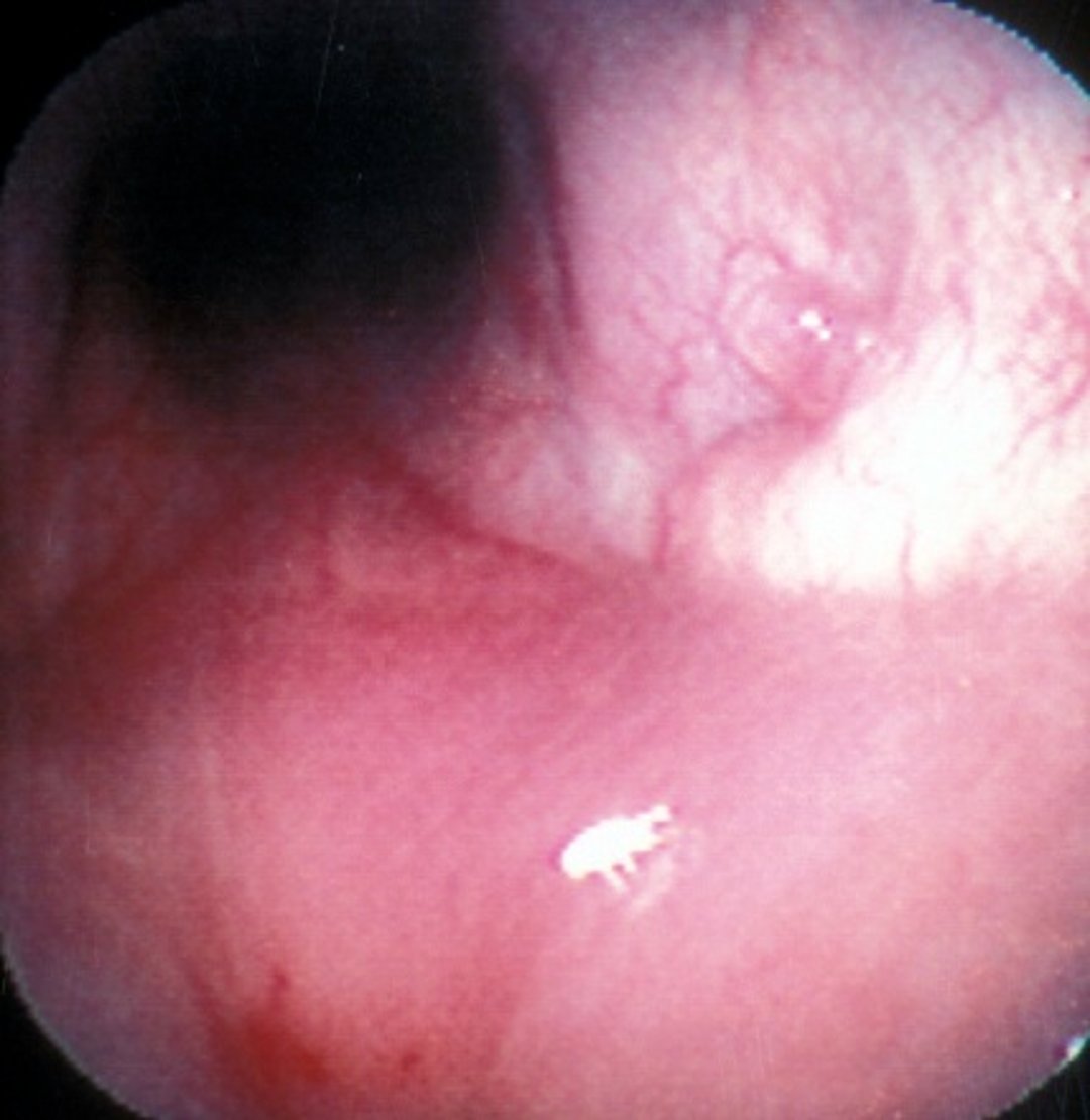
Endoscopic view of a nasal mite (P caninum) in the nasopharynx of a dog.
Courtesy of Dr. Steven L. Marks.
Nasal flushing may also help identify P caninum. This is generally performed with the dog under general anesthesia with a cuffed endotracheal tube in place. The oropharynx is packed with gauze, and saline is flushed through the external nares with a Foley catheter or a tight-fitting syringe to collect fluid from the oropharynx. Retrograde flushing can be done by placing a modified catheter behind the soft palate, occluding the nasal pharynx, and flushing with saline. This allows fluid to be collected via the external nares. In both cases, the fluid should be evaluated using an illuminated magnifying lens to look for mites.
The definitive diagnosis of nasal acariasis can be made via endoscopy or nasal flushing if the mites are identified. This does not, however, determine whether the disease is primary or secondary.
Treatment of Canine Nasal Mites
Empirical therapy
Ivermectin or milbemycin oxime (not an approved use)
No drugs are currently approved for the treatment of P caninum; however, ivermectin (200–400 mcg/kg, SC or PO), milbemycin oxime (1 mg/kg, PO, three times at 10-day intervals), and selamectin (topical) have been suggested. The optimal treatment regimen has yet to be determined. Treatment has been reported to be effective in >85% of cases, and the prognosis is excellent. However, treatment may not completely eliminate clinical signs, particularly if infection is suspected rather than demonstrated. In these cases, it is probable that the signs are the result of a concurrent upper airway disease. Treatment is based on definitive diagnosis, but empirical therapy has also been performed based on a high index of suspicion.
Key Points on Canine Nasal Mites
Nasal mites may be difficult to diagnose without endoscopy or other means of direct visualization.
No drugs are currently approved to treat nasal mites.
For More Information
Also see pet health content regarding nasal mites in dogs.


