

Over the years, colic has become a broad term for a variety of conditions that cause horses to experience abdominal pain. Because it is such a broad term, it is used to refer to conditions that vary widely in cause and severity. Your veterinarian’s understanding of your horse’s digestive system structure and function is key to making a diagnosis and providing appropriate treatment for cases of colic ( see Table: The Veterinarian’s Examination of a Horse with Colic on this page).
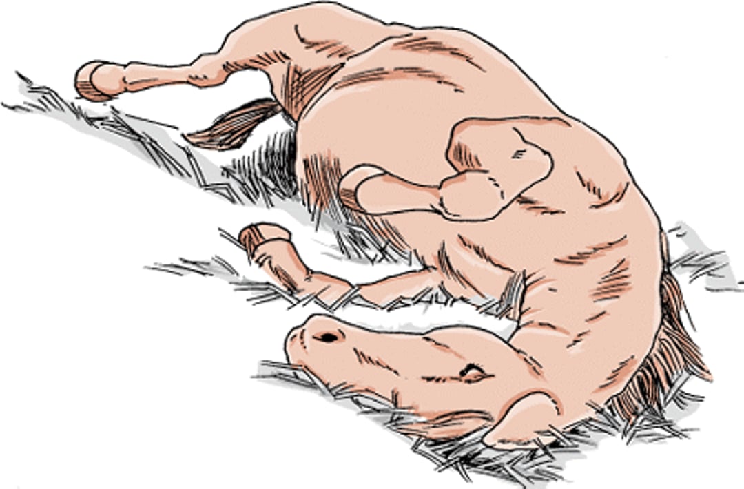
The most common signs of colic are pawing repeatedly with a front foot, looking back at the flank region, curling the upper lip and arching the neck, repeatedly raising a rear leg or kicking at the abdomen, lying down, rolling from side to side, sweating, stretching out as if to urinate, straining to defecate, distention of the abdomen, loss of appetite, depression, and a decreased number of bowel movements. It is uncommon for a horse with colic to exhibit all of these signs. Although these signs are reliable indicators of abdominal pain, they do not indicate which portion of the digestive system is affected.
Colic, horse

Diagnosis and treatment occur only after a thorough examination of the horse, including a review of its history of any previous problems or treatments. Both the location and the cause of the colic should be determined. The list of possible conditions that cause colic is long (see Common Causes of Colic in box below). For that reason, your veterinarian may begin treatment based on the most likely diagnosis and then make a more specific diagnosis later, if necessary or possible. Information you can provide includes the length and speed of progression of the colic episode, the severity of pain, whether feces have been passed, and the response to any provided treatments. It is also important to provide the horse’s deworming history (schedule, treatment dates, drugs used), when the teeth were floated last, if any changes in the type or amount of feed or water supply have occurred, any history of cribbing behavior, and whether the horse was at rest or exercising when the colic episode started. The age of the horse is also important, because a number of conditions are more likely to occur at certain ages.
The Veterinarian’s Examination of a Horse with Colic
Type of Procedure | Why It is Done |
|---|---|
Assessment of breathing and heart rate | Increased heart rate can indicate pain, dehydration, or low blood pressure. Increased breathing rate can indicate pain, fever, decrease in blood pH, or an underlying respiratory problem. |
Examination of mucous membranes (soft tissues inside of mouth) | Paleness or blueish tinge indicates poor oxygen level in blood; dryness indicates dehydration; grey discoloration indicates poor blood flow in the tissues; red discoloration can indicate shock. |
Insertion of tube through nose into stomach | Because horses cannot vomit, the tube can allow release of gas or fluid that would otherwise result in stomach rupture. For this reason, passing a stomach tube may save the horse’s life in addition to helping the veterinarian diagnose the condition causing the colic. |
Listening to various parts of the abdomen with a stethoscope | Sounds may indicate the presence of fluid, gas, impending diarrhea, and/or obstruction. Lack of sounds may indicate impaired motility or blood flow. |
Sample of abdominal (peritoneal) fluid via needle | Composition (protein and white blood cells) can reveal extent of intestinal damage. |
Rectal examination | Critical component of the exam that allows the veterinarian to feel the intestines, their position, and their content. |
Ultrasonography | Provides a view of certain abdominal organs, including the intestines. Some conditions (such as an inguinal hernia or an intussusception) may be seen. |
Treatment
Horses with colic may or may not need surgery. Almost all horses will require some form of medical treatment, but only those with certain mechanical obstructions of the intestine need surgery. The type of treatment is determined by the cause of colic and the severity of the disease. If the horse appears to have only mild pain and the heart and circulatory system are functioning normally, the horse may be treated using medication or other nonsurgical methods and the response evaluated. Ultrasonography can be used to evaluate the effectiveness of nonsurgical treatment in some cases. If necessary, surgery can be used for diagnosis as well as treatment.
If evidence of intestinal obstruction with dry, digested food is found, the main goal of treatment is to rehydrate and remove the intestinal contents. If the horse has severe pain and has signs indicating loss of fluid from the bloodstream (high heart rate and discoloration of the mucous membranes), the initial aims of treatment are to relieve pain, restore tissue blood supply, and correct any abnormalities in the composition of the blood and body fluids (such as an abnormal pH or electrolyte level).
If damage to the intestinal wall is suspected, specific medications may be administered to prevent or counteract the ill effects of bacterial toxins (called endotoxins) that leave the intestine and enter the bloodstream. If there is evidence that the colic episode is caused by parasites, one of the first goals of treatment would be to eliminate the parasites.
Pain Relief
Pain is mild in most cases of colic, and pain medication is all that is needed. This is the usual treatment if the cause of colic is believed to be a spasm of intestinal muscle or excessive gas in a portion of the intestine. If the pain is due to a more serious condition, such as an intestinal twist or displacement, some of the stronger pain medications may mask the signs that would be useful in making a diagnosis. For these reasons, whenever possible a thorough physical examination is performed before any medications are given. However, because horses with severe colic or pain may hurt themselves and become dangerous to people nearby, pain medication often must be given first. In addition, many horses with less severe problems may need pain relief until the other treatments have time to be effective.
Your veterinarian will chose a pain reliever that is least likely to cause side effects or changes in the horse's attitude. Examples include anti-inflammatory drugs, sedatives that also reduce pain, and narcotics. Although pain relief usually is provided by medications, pain can be reduced in other ways. For example, the veterinarian’s use of a stomach tube during diagnosis will also remove any fluid that has accumulated in the stomach because of an obstruction of the small intestine. The removal of this fluid not only relieves pain caused by distention of the stomach but also prevents rupture of the stomach.
Fluid Treatment
Many horses with colic benefit from fluid treatment to prevent dehydration and maintain blood supply to the kidneys and other vital organs. The fluids may be given either through a stomach tube or intravenous catheter, depending on the particular intestinal problem. Intravenous fluids may be needed for several days until intestinal function has returned, blood electrolyte (salt) concentrations are balanced, and the horse can maintain its fluid needs by drinking.
Protection against Bacterial Endotoxins
Endotoxins are a part of the outer coating of certain bacteria. Endotoxins are released when the bacteria die or multiply rapidly. Normally, endotoxins are contained within the intestines, but if the intestinal lining is damaged, they can escape into the abdominal cavity or bloodstream. Endotoxins then trigger an inflammatory response (commonly called endotoxemia) that can include fever, depression, reduced blood pressure, impaired blood circulation, blood clotting abnormalities, and eventually death. One treatment is administration of antibodies or medications designed to neutralize the endotoxin. The effectiveness of this treatment is still being studied, with some studies finding a benefit and others that do not. Other treatments include anti-inflammatory medications and certain antibiotics. In cases of colic, your veterinarian will be on the alert for damage to the intestinal lining and the possibility of complications due to endotoxins.
Intestinal Lubricants and Laxatives
A common cause of colic in horses is obstruction of the large intestine by dried digested food, sometimes mixed with sand. In most instances, lubricants or fecal-softening agents given through a stomach tube soften the impacted material, allowing it to be passed. Intravenous fluids are often given during this procedure. The horse will normally need to be muzzled to prevent further impaction of feed while the obstruction is softening. Medications include mineral oil, dioctyl sodium sulfosuccinate (a soap-like compound), and psyllium hydrophilic mucilloid (an ingredient also found in some fiber products used by humans). When mixed with water, psyllium forms a gelatin-like mass that carries ingested food along the digestive tract. Horses that live in a sandy environment or that persistently develop impactions may be given psyllium powder in their feed, as directed by a veterinarian, to help prevent impaction.
Strong laxatives that stimulate intestinal contractions are not commonly used to treat impactions and, in fact, may worsen the problem. If an impaction does not start to break down within 3 to 5 days, surgery may be necessary to remove the impacted material.
Deworming Treatments
The larvae of large bloodworms, especially Strongylus vulgaris, can cause colic by interfering with the flow of blood to the intestines. The routine use of deworming medications has reduced the occurrence of this once-common type of colic. Intestinal damage caused by the larvae of small strongyles may also cause colic, diarrhea, and loss of condition, especially in young horses. Infections with the intestinal tapeworm Anoplocephala perfoliata are also associated with intestinal impactions. Horses with parasite infections are treated with deworming medications. Complications, such as pain or intestinal obstruction, must also be treated appropriately.
However, intestinal impactions can occasionally occur after the administration of deworming medication, especially in young horses with an inadequate history of deworming. This occurs when deworming drugs that are highly effective against Parascaris equorum cause a mass of paralyzed worms to obstruct the small intestine. Some affected horses may respond to medical treatment (fluids and intestinal lubricants), whereas others may require surgery to relieve the impaction. Your veterinarian can recommend an appropriate deworming program to minimize the likelihood of this occurring in other horses on the same property.
Surgery
Usually, surgery is necessary if there is a mechanical obstruction that cannot be corrected medically or if the obstruction also interferes with the intestinal blood supply. The latter condition causes death of the horse unless surgery is performed quickly. Occasionally, surgery is needed to diagnose the problem in horses with longterm colic that have not responded to routine medical treatment.
Horses with severe pain that do not respond to pain medications usually require surgery. However, some horses with mild or moderate pain may also require surgery, a judgement your veterinarian will make based on the physical examination.
Some of the common indications for surgery in horses with colic include:
uncontrollable pain
more than 4 liters of fluid retrieved from the stomach tube
no intestinal sounds heard with a stethoscope
abnormalities within a fluid sample from the abdomen
evidence of an intestinal obstruction or displacement identified on rectal examination
If surgery is indicated, it is critical to perform it promptly to ensure the best chance of success and survival. This usually requires referral to an equine surgeon at a properly equipped facility. Depending on the cause and the extent of the damage, care after the surgery may include intravenous fluids, antibiotics, antibodies against endotoxin, anti-inflammatory drugs, or other medications. Each horse is individually treated based on its response to surgery and the development of any complications.
Outcomes for Colic
The survival rate for horses that undergo surgery to treat colic ranges from 50% to more than 80%. Your veterinarian can provide insight into the possible outcome for your horse based on physical examination findings. In general, survival rates are highest for horses with mild abdominal pain and are lowest for horses with severe pain. Horses with distended intestines, lack of abdominal sounds, red mucous membrane color, or impaired cardiovascular function (such as low blood pressure) tend to do worse than those without these signs.
Specific Causes of Colic and their Treatment
Colic can be caused by several disorders of the stomach and intestines. The most common ones are discussed here.
Distention and Rupture of the Stomach
Excessive gas or intestinal obstruction can lead to distention of the stomach. This may also be caused by overeating fermentable feeds such as grains, lush grass, or beet pulp. If untreated, this can rapidly progress to a rupture of the stomach. Signs include severe abdominal pain, increased heart rate, and retching. Once the stomach ruptures, the horse may act relieved or depressed. The outlook for survival is often excellent if the condition is recognized and treated soon enough, but stomach rupture is fatal.
Obstruction of the Small or Large Intestine
Signs of colic may occur if the small or large intestine is obstructed. The outlook depends on the location and can worsen if treatment is delayed, so rapid diagnosis and treatment are critical.
The most common condition that causes obstruction of the small intestine is impaction (blockage of the intestine by food or other materials that have been eaten). It has been linked to eating Coastal Bermuda hay and infection by the tapeworm Anoplocephala perfoliata. Young horses may be affected by impaction of the small intestine with ascarid parasites following deworming. Intussusceptions (a telescoping of a section of the intestine within an adjacent section) can also cause small intestinal obstructions, especially in horses younger than 3 years old. In the large intestine, the underlying cause of obstruction is not always known but has also been linked to coarse feed, insufficient water intake, and diseased teeth. In some areas sand may be the cause of intestinal obstruction and colic. This is especially true if there is not enough pasture and the horse is fed on the ground. The sand may accumulate in the large intestine and eventually cause a blockage. Morgan, Arabian, and Appaloosa horses may have a higher risk of impaction of the cecum compared with other breeds. Impactions also may develop because of other intestinal diseases or occur after prolonged hospitalization. Impactions within the cecum tend to occur in horses older than 8 years.
Signs in horses with impaction of the small intestine include mild to severe abdominal pain, reduced intestinal sounds, stomach reflux, and increased heart rate. Because the horse’s condition may remain stable and the pain may be mild at first, many horses with this condition are not immediately referred for surgery. The condition often requires surgery, although it may respond to treatment with fluids, pain-relieving drugs, and mineral oil if identified early. A lack of normal intestinal contractions may occur after surgery. Adhesions within the abdomen (see below) are another potential complication.
In contrast, horses with impaction of the large intestine may display more mild signs and do not usually require surgery. Almost all respond well to administration of laxatives, fluids, and pain medications. If the obstruction occurs in the descending colon, however, surgery is often necessary. The outlook for horses with impactions of the large colon is excellent, with more than 95% of horses surviving the condition. For horses with impactions of the cecum, the survival rate ranges from 60% to 80% depending on whether they are treated medically or surgically.
Adhesions
Adhesions are fibrous connections between organs within the abdomen. They generally affect the small intestine. Adhesions develop in response to abdominal injury such as surgery, longterm distention of the intestine, inflammation, or migration of larval parasites. They can squeeze or kink the intestines, leading to an obstruction. Signs range from mild, recurrent colic to severe, continual pain. Treatment involves surgery to remove the fibrous tissue and the affected portion of the intestine. Medications are also given to try to reduce the formation of new adhesions. However, adhesions often recur and the longterm outlook for horses with extensive adhesions is poor.
Inflammation of the Small Intestine
Inflammation of the first part of the small intestine is a poorly understood condition of horses. The condition has several names, including proximal enteritis–jejunitis, anterior enteritis, and duodenitis–jejunitis. It has been reported in the southeastern and northeastern US as well as in England and continental Europe. There may be fluid or bleeding within the intestinal wall, or tissue death in more severe cases. The condition was more common in the United States in the 1980s but occurs less frequently and less severely now.
Varying degrees of abdominal pain are the most common sign of the disorder. Treatment may be either medical (such as placing a stomach tube and administering fluids, pain medication, and other drugs) or surgical. About half of the cases are fatal. Laminitis, or inflammation of the hoof, is a common complication that occurs in approximately 25% of affected horses.
Lipomas
Colic caused by lipomas (benign fatty tumors) is sometimes seen in horses more than 10 years old. If the tumor is attached by a stalk to connective tissue in the abdomen, then it may wrap around a part of the intestine, shutting off its blood supply. Signs may include depression and severe abdominal pain, with rapid worsening of condition. Treatment requires removal of the tumor by surgery, along with any damaged sections of the intestine. If the problem is detected early, the outlook is good, but if surgery is not done before signs are advanced, the chances for recovery are fair to poor.
Twisting or Displacement of the Small or Large Intestine
Twisting of the intestines (volvulus, sometimes also called a torsion) occurs when the intestine rotates around its attachment to the abdominal wall. This reduces the blood supply to the intestine, leading to colic. Horses with this condition are painful and have an increased heart rate. Rapid dehydration is caused by movement of fluid into the stomach and intestine. The horse’s condition may worsen rapidly.
Portions of the small intestine can become trapped within mesenteric rents (defects within the connective tissue that connects the intestine to the body wall) or the epiploic foramen (a natural opening between the liver and major abdominal blood vessels). Signs can be vague and can vary from mild to severe pain. These entrapments require surgery to correct. If surgery is performed quickly, the outlook is good. However, the vagueness of signs can delay surgery and worsen the outlook.
Displacement, without twisting, of the large intestine may occur and also leads to obstruction. A left dorsal displacement occurs when part or all of the left colon gets displaced over the renosplenic ligament (an anatomical attachment between the kidney and the spleen). A right dorsal displacement occurs when the left colon becomes trapped between the cecum and the body wall. A portion of the colon may also be twisted in some of these horses. Horses with displacements of the large intestine do not usually worsen as rapidly as those with twisting.
Displacements of the large intestine can often be treated with medical management. Depending on the colon's location, treatment may include withholding food, administering fluids and pain medications, jogging or rolling the horse, or other medications. Surgery may also be necessary to relocate the colon, especially if pain is severe or if twisting is present.
Twisting of the intestines requires surgery to correct the positioning of the intestine. Removal of part of the intestine may also be required if it has been compromised by a lack of blood supply for too long. The outlook for recovery is good if the condition is detected and treated soon after it occurs. Adhesions (see above) may be a complication, especially if the illness is prolonged. Volvulus of the large colon can recur in as many as 20% of cases, but performing a colopexy (a surgical procedure that attaches the colon to the abdominal wall) may reduce the recurrence.
Inguinal Hernia
Inguinal hernias (commonly referred to as scrotal hernias) occur when the intestine passes from the abdomen into the inguinal canal that connects the testes to the abdomen. They occur in male horses, generally after breeding, trauma, or a hard workout. If the inguinal opening is large enough, part of the intestine may become trapped, causing colic. Hernias appear to be most common in Tennessee Walking Horses, American Saddlebreds, and Standardbreds. If the condition has been present for more than a few hours, the horse’s condition worsens rapidly. A veterinarian may be able to push the hernia back through the abdominal wall (called reducing the hernia) if it has occurred in the last few hours. Otherwise, surgery is the usual treatment and may require removal of the testicle on the affected side, along with a portion of the intestine if it has become too damaged. The chances for survival appear to be breed-dependent, with Standardbred horses having a good outlook and Tennessee Walking Horses a fair to poor outlook. Presumably, this is because Tennessee Walking Horse stallions with inguinal hernias show few signs of pain, which may delay recognition of the problem and treatment.
Enteroliths (Intestinal Stones)
Enteroliths are hard masses composed of magnesium ammonium phosphate crystals that form around a foreign object (such as a piece of wire, stone, or nail) in the large intestine of horses. Enteroliths may be seen singly or in groups and are commonly found in horses in certain parts of the United States, including California, the southwest, Indiana, and Florida. Most horses with enteroliths are about 10 years old; horses younger than 4 years old are rarely affected. Enteroliths are commonly seen in Arabian horses. A common factor associated with formation of enteroliths may be the consumption of alfalfa hay, which results in a higher pH and increased concentrations of calcium, magnesium, and sulfur in the large colon.
Many horses with this condition have a history of recurring colic, which may indicate that the enterolith(s) had caused partial or temporary blockage of the large intestine. Depending on the location of the enterolith, the horse may be in severe pain. Heart and respiratory rates increase, and the mucous membranes may be pale or pink. Generally, intestinal distention is evident to the veterinarian on rectal examination, but the mass cannot usually be felt. In areas where the problem is common, x-rays may be used to identify the enteroliths.
Treatment involves surgery to decompress the intestine and remove the stone(s). The outlook is excellent. Veterinary practices in areas where this condition commonly occurs report survival rates of 95%.
For More Information
See professional content regarding colic in horses.


