

Nasal dermatoses of dogs may be caused by many diseases, including autoimmune, infectious, environmental, neoplastic, genetic, systemic, and neurological conditions. Some conditions are pathognomonic, and some require histological examination of the skin for diagnosis. History, physical examination, and surface cytological examination can help determine if skin biopsy, treatment trial, or benign neglect is indicated.
Nasal dermatosis lesions in dogs may affect the haired bridge of the muzzle, the planum nasale (the nonhaired, or glabrous, portion of the nose), or both.
In pyoderma, dermatophytosis, and demodicosis, the haired portions of the muzzle are affected.
In systemic lupus erythematosus or pemphigus, the whole muzzle is often crusted (with occasional exudation of serum) or ulcerated.
In systemic and discoid lupus, and occasionally in pemphigus and cutaneous lymphoma, the planum nasale is depigmented, erythematous, and eventually may ulcerate. The normal cobblestone appearance of the nasal planum is effaced (see ).

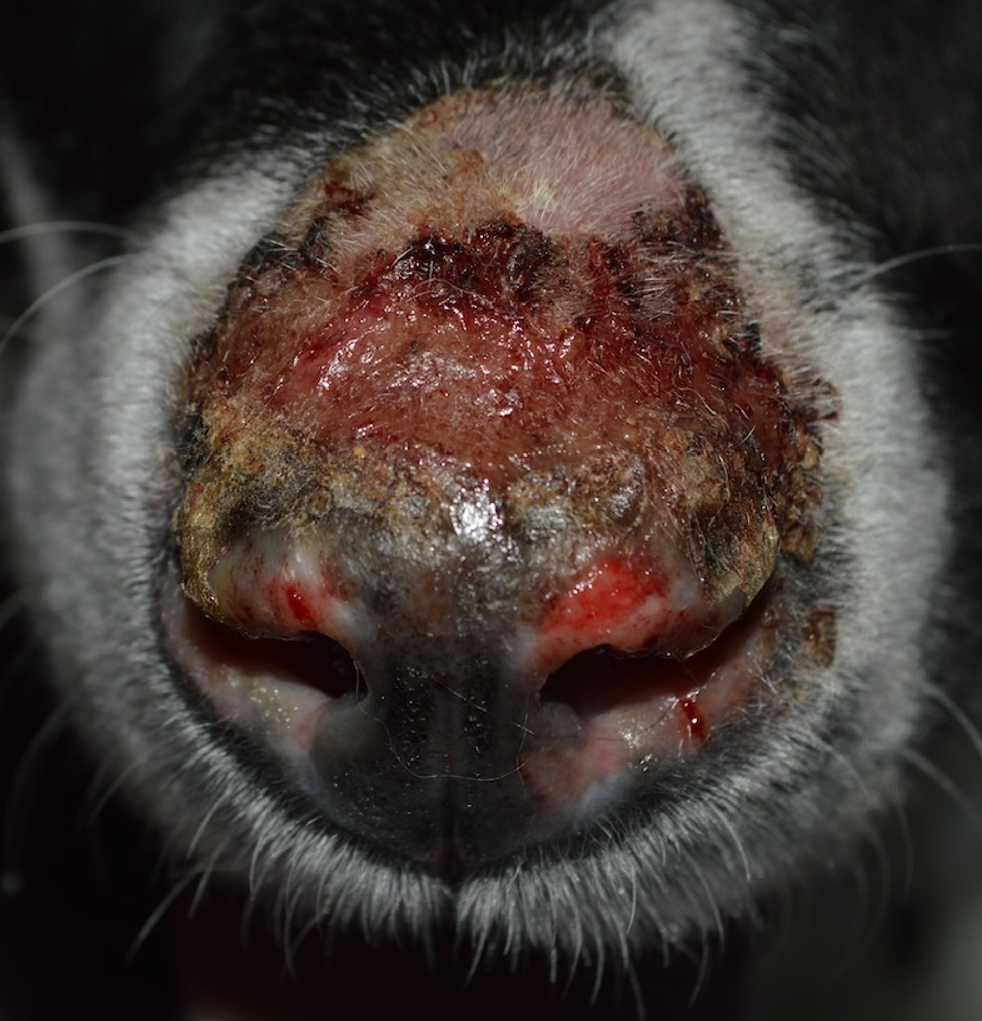
Severe erosion and crusting affecting the haired portion of the muzzle and the planum nasale in a dog with discoid lupus erythematosus.
Courtesy of Dr. Nicole A. Heinrich.
Any of these nasal dermatoses may affect the periocular areas.
Nasal dermatosis due to solar radiation probably is not as common as previously thought and may often be a misdiagnosis for the lupus variants. In true nasal solar dermatitis, the nonpigmented areas of the planum nasale are affected first, and occasionally the bridge of the nose may become inflamed and sometimes ulcerated. The lesions are worse in the summer, although lupus and pemphigus may also show this seasonal variation.
A sudden onset of nasal swelling, erythema, and exudation is often eosinophilic furunculosis; this is thought to be caused by an arthropod sting or bite.
The protozoal disease leishmaniasis may cause depigmentation of the nasal planum.
Diagnostic tests include skin scrapings, bacterial and fungal cultures, and biopsies for both histological and immunological testing. Some veterinary dermatopathologists may be able to make the diagnosis based on histopathological findings alone. If systemic lupus erythematosus is considered, blood for an antinuclear antibody test should be obtained.
Treatment depends on the cause.
For nasal solar dermatitis, a topical corticosteroid lotion (betamethasone valerate, 0.1%, topically, every 12 hours for 5–7 days, then tapered) may help relieve inflammation. Exposure to sunlight must be severely curtailed. Topical sunscreens may be effective but need to be applied at least twice daily.
Treatment for eosinophilic furunculosis is with systemic corticosteroids (prednisone or prednisolone at 1 mg/kg, PO, every 12 hours, for 1 week, after which the dosage should be gradually decreased over the course of 1 month).
The following are examples of autoimmune diseases that can affect the nasal planum:
mucocutaneous lupus
vitiligo (see )
arteritis of the nasal philtrum


Mild multifocal depigmentation of the planum nasale in a dog with vitiligo.
Courtesy of Dr. Nicole A. Heinrich.
Autoimmune diseases often cause crusting, erosion, and bleeding of the nasal planum. Depigmentation may also be present. If autoimmune disease is suspected, then a biopsy of the nasal planum should be pursued. Immunosuppressive therapy is indicated if the diagnosis is confirmed.
The most serious of the autoimmune diseases listed is uveodermatologic syndrome; this condition also affects the eyes and can quickly lead to blindness. If uveodermatologic syndrome is suspected, a biopsy should be procured and treatment with systemic corticosteroids (prednisone at 1–2 mg/kg, PO, every 24 hours, or divided every 12 hours, for a few weeks until clinical response, then tapered; maintenance dosage, 0.5 mg/kg, PO, every 24–48 hours indefinitely) should be instituted immediately.
Arteritis of the nasal philtrum is also potentially an emergency. This condition is characterized by bleeding, which can be profuse, from a lesion on the nasal philtrum.
Examples of infectious causes of nasal planum lesions include Staphylococcus spp; fungal organisms such as Cryptococcus, Sporothrix, Blastomyces, and Aspergillus; and Leishmania. Antimicrobial therapy is selected based on culture results when possible, or on other diagnostic results when culture is not rewarding.
Staphylococcus spp are the most common organisms to cause mucocutaneous pyoderma. This condition is typically characterized by mild to moderate crusting with exudate of the mucocutaneous junction of the nostrils or alar fold. Mild bleeding may also occur.
An impression smear from the exudate will reveal a large number of neutrophils and cocci.
Fungal infections are a much more serious type of infection than mucocutaneous pyoderma. They are typically characterized by swelling or severe ulceration of the nose. The nasal mucosa may also be affected, and nasal discharge may be copious. Diagnosis may be made with histological examination and fungal-specific antigen or antibody testing.
Leishmaniasis is uncommon outside of endemic areas. It is typically characterized by ulceration and hemorrhage of the nasal planum and nasal mucosa. The nasal lesions are accompanied by other cutaneous and systemic clinical signs.
Diagnosis of leishmaniasis may be made with CBC, serum chemistry, serum protein electrophoresis, urinalysis, and demonstration of the presence of Leishmania spp on cytological or histological examination or PCR assay.
Examples of environmental causes of dermatitis of the nasal planum include solar dermatitis and insect bite dermatitis. Solar dermatitis typically affects lightly pigmented skin and is characterized initially by waxing and waning erythema. The condition can progress to include crusting, erosion, and progression to other diseases, such as discoid lupus erythematosus or squamous cell carcinoma.
Solar dermatitis can be diagnosed through history, physical examination, and response to sun avoidance measures. A biopsy may be necessary in more complex cases.
Examples of neoplastic conditions that can affect the nasal planum include cutaneous lymphoma and squamous cell carcinoma (see ).
Cutaneous lymphoma is typically characterized by progressive swelling and depigmentation of the nasal planum. Typically, lesions are multifocal, affecting mucocutaneous junctions and haired skin.
Squamous cell carcinoma is typically characterized by sneezing, bleeding, and ulceration (nonhealing wounds) of the nasal planum. Histological examination is critical for diagnosis if neoplasia is suspected.

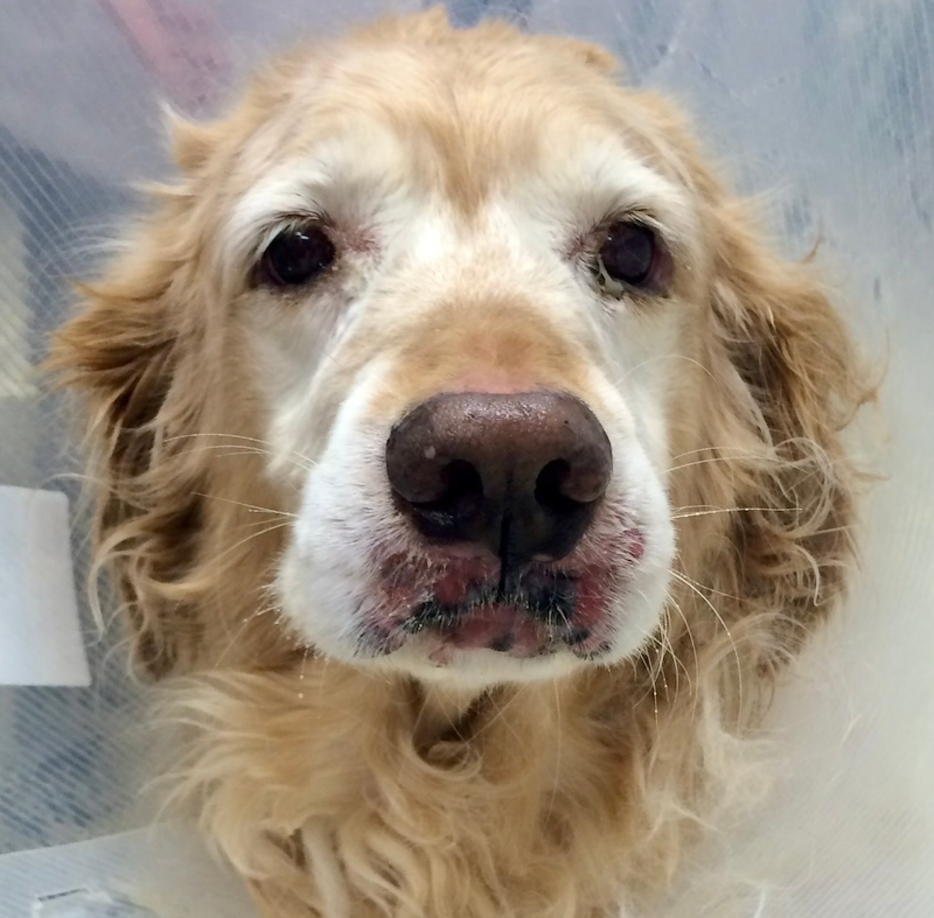
Cutaneous lymphoma causing swelling, depigmentation, and erosion of the haired skin adjacent to the nasal planum in a Golden Retriever.
Courtesy of Dr. Nicole A. Heinrich.
Genetic conditions that may affect the nasal planum include zinc-responsive dermatosis, which can also be a nutritional disease; nasal hyperkeratosis; and hereditary nasal parakeratosis of the Labrador Retriever.
Hereditary nasal parakeratosis is an autosomal recessive disorder caused by a mutation in the SUV39H2 gene that leads to crusts and fissures in the nasal planum. Diagnosis is made primarily via signalment and histological examination. Genetic testing is also available for this condition.
Zinc-responsive dermatosis is characterized by crusts and depigmentation of the nasal planum. Northern breeds are heavily predisposed, and affected animals are typically young adults (see ).

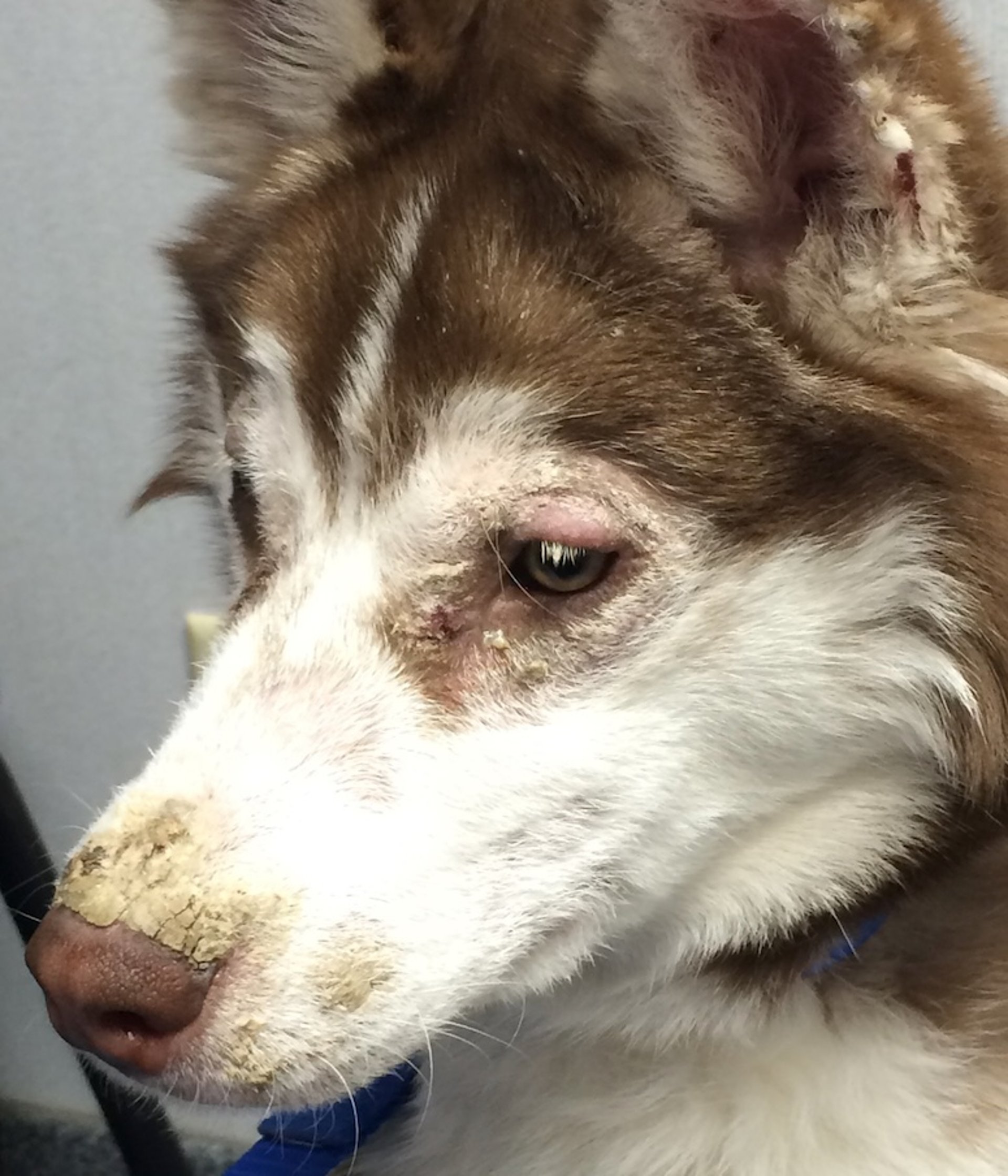
Crusting of the muzzle and periocular region in a dog with zinc-responsive dermatosis.
Courtesy of Dr. Nicole A. Heinrich.
Diagnosis is made primarily on the basis of signalment, histological examination, and response to zinc supplementation. Complete response to zinc supplementation may take weeks to months, and sometimes the type of zinc or the dose of the zinc may need to be adjusted. For this reason, histological evaluation should not be overlooked when this condition is suspected.
Nasal hyperkeratosis is characterized by fronding extensions of the dorsal aspect of the nasal planum. The condition is not painful, and inflammatory lesions are absent. This condition occurs in older adult dogs, and treatment is generally not necessary.
An example of systemic conditions that can affect the nasal planum include hepatocutaneous syndrome, a condition in which a hepatopathy or glucagonoma causes dermatitis. The nasal planum is often crusted, and lesions are always multifocal, affecting the paw pads and haired skin. In addition, dogs usually also have clinical signs of systemic illness.
Diagnosis of hepatocutaneous syndrome is made via histological examination of the skin. Additional diagnostics that should be pursued include abdominal ultrasonography, CBC, serum biochemical analysis, and urinalysis. Glucagon concentration should be measured in cases of suspected glucagonoma.
Neurological conditions that can affect the nasal planum include parasympathetic neuropathy. The facial nerve (cranial nerve VII) is a mixed nerve that contains a branch of the parasympathetic nervous system. This nerve is intimately associated with the ear as the nerve travels from the brain to various parts of the face. Damage to the parasympathetic component of the facial nerve can cause xerosis (excessive dryness) of the nasal planum because parasympathetic nerves are involved in the secretory production of the nasal mucosa. Otitis is a common cause of neuropathy. A dry nasal planum should prompt the clinician to examine the dog for otitis.
For More Information
Also see pet health content regarding nasal dermatoses in dogs.


