


Saddle sores are partial-thickness or full-thickness wounds resulting from focal pressure exerted by poorly fitted or poorly padded tack. The sores are evident as swelling, moisture, hair loss, and grossly apparent wound formation when necrotic skin sloughs. Diagnosis is based on history and physical examination. Treatment requires wound care, elimination of the ill-fitting tack, and no contact on the skin until wound healing is complete.
Saddle sores are pressure sores or ulcers that develop in horses over areas of wear from tack (especially if it is ill-fitting). Moisture from sweat or wet tack may contribute to maceration of the skin. The areas of riding horses that are under the saddle, as well as the shoulder areas of horses driven in a harness, are frequently the site of injuries to the skin and deeper soft and bony tissues.
Prolonged focal pressure can lead to decreased blood flow, tissue damage, and even necrosis. Saddle sores are frequently complicated by secondary bacterial infections. Emaciated horses are at increased risk. Prevention includes properly fitted tack as well as grooming after riding, to clean the coat of debris and remove any potentially abrasive particulates from the skin before tack is replaced for the next ride.
Clinical Findings of Saddle Sores in Horses

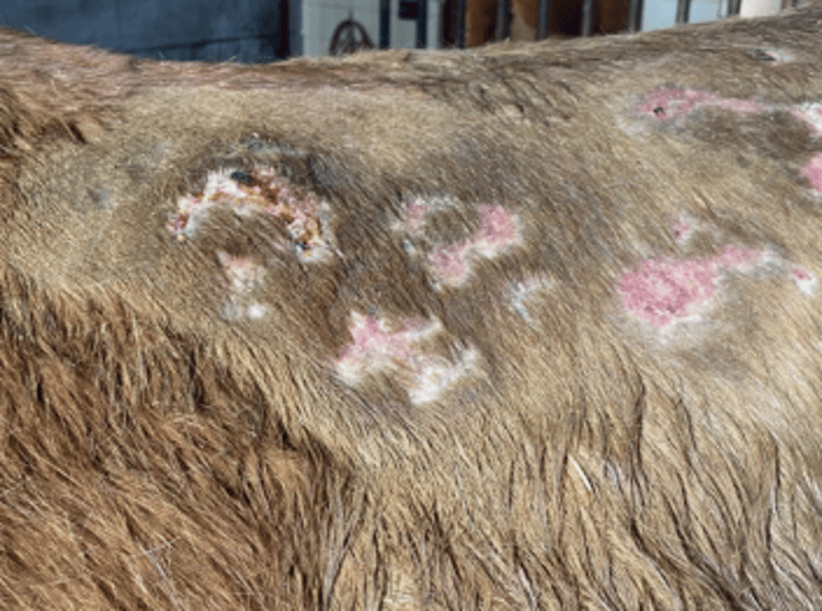
Multifocal areas of scarring alopecia and focal area of hemorrhagic crusting in the saddle region of a 13-year-old Quarter horse mare used for riding instruction.
Courtesy of Dr. Ashley Boyle.
Initial superficial clinical signs of saddle sores may be only erythema, moist exudate, and signs of folliculitis such as papules, crusts, or alopecia (see ). However, lesions can progress to include erosions, ulcers, and necrosis. Affected areas may become swollen, warm, and painful with secondary cellulitis. Advanced lesions are termed “galls.”
When the skin and underlying tissues are more severely damaged, abscesses may develop. These abscesses are warm, fluctuating, painful swellings from which purulent and serosanguineous fluid can be aspirated or that form draining tracts.
Severe devitalization of the skin and subcutis or deeper tissues results in dry or moist necrosis. Tissue may become undermined with inflammation or infection.
Chronic saddle sores are characterized by a deep folliculitis or furunculosis with fibrosis and scarring. Leukotrichia (white hair) is commonly observed in healed areas.
Diagnosis of Saddle Sores in Horses
History and physical examination
Diagnosis of saddle sores in horses is based on history and physical examination.
Recurrence of hematomas, seromas, and/or sloughing skin upon initial saddling of a young Quarter horse, Paint horse, or Appaloosa suggests the possibility of the genetic disease hereditary equine regional dermal asthenia (HERDA). A simple DNA test, performed on the hair bulbs of the tail, will confirm this diagnosis.
Treatment of Saddle Sores in Horses
Elimination of the cause
Rest
Astringent packs for drying in the acute stage
Warm compresses and antimicrobials for chronic lesions
Surgical removal of necrotic lesions
The treatment of saddle sores should be aimed at eliminating the causative factor: changing tack, increasing cushioning, and ensuring that both tack and skin beneath tack are kept as clean and dry as possible. Absolute rest of the affected parts is necessary until healing is complete.
During the early or acute stages of saddle sores, astringent packs (aluminum acetate topical solution) are indicated for their drying effects. Chronic lesions and superficially infected lesions may be treated by warm compresses and topical or systemic antimicrobials. Selection of a systemic antimicrobial should be based on cytological evaluation and bacterial culture results.
Necrotic tissue should be removed surgically.
Key Points
Saddle sores are due to pressure or friction from tack; risk factors include ill-fitting tack, moisture, and emaciated body condition.
Early clinical signs may be only erythema, crusts, alopecia, and moist exudate; however, they can progress to erosions or ulcers, cutaneous necrosis, and abscesses or cellulitis.
Management includes rest; ensuring that tack fits well and has good padding; keeping skin beneath tack clean and dry; identifying and treating secondary infections; and debriding necrotic tissue.
For More Information
American Quarter Horse Association (AQHA): Hereditary Equine Regional Dermal Asthenia (HERDA)
Veterinary Genetics Laboratory (UC Davis): Hereditary Equine Regional Dermal Asthenia (HERDA) tests
Also see pet health content regarding saddle sores in horses.


