

Primary seborrhea is a very rare keratinization disorder characterized by scaling skin with or without greasy hair. It is not pruritic. Most seborrheic animals have secondary seborrhea, in which a primary underlying disease predisposes to excessive scaling. Primary seborrhea is diagnosed by ruling out other underlying causes. Primary seborrhea is treated with frequent bathing and administration of vitamin A or a retinoid.
Seborrhea is characterized by a defect in keratinization that results in increased excessive cutaneous scaling (ie, dandruff or follicular casts).
Etiology of Seborrhea in Animals
Primary idiopathic seborrhea is an inherited skin disorder characterized by faulty keratinization of the epidermis, hair follicle epithelium, or claws.
Most seborrheic animals have secondary seborrhea, in which a primary underlying disease predisposes to excessive scaling, crusting, or oiliness, often accompanied by superficial pyoderma, Malassezia (yeast) infection, and alopecia.
The most common underlying causes of secondary seborrhea are endocrinopathies and allergies. Underlying diseases may present with seborrhea as the primary clinical problem.
Seborrhea in horses is usually secondary to either pemphigus foliaceus or equine sarcoidosis (chronic granulomatous disease).
Epidemiology of Seborrhea in Animals
Seborrhea is more common in dogs than in cats. In dogs, secondary seborrhea is more common than primary seborrhea.
Primary seborrhea occurs more frequently in the following dog breeds:
American Cocker Spaniels
English Springer Spaniels
Basset Hounds
West Highland White Terriers
Dachshunds
Labrador and Golden Retrievers
German Shepherd Dogs
A familial history of seborrhea is typical, suggesting genetic factors. The disease begins at a young age (usually < 18–24 months) and typically progresses throughout the animal’s life.
Clinical Signs of Seborrhea in Animals
Primary seborrhea is characterized by a defect in keratinization that results in increased scale formation, occasionally excessive greasiness of the skin and coat, and often secondary inflammation and infection. Primary seborrhea is not pruritic.
In secondary seborrhea, a primary underlying disease causes clinical signs similar to those of primary seborrhea. Secondary seborrhea may or may not be pruritic.
Diagnosis of Seborrhea in Animals
Ruling out underlying causes of secondary seborrhea
Clinical testing
Skin biopsy
A diagnosis of generalized primary seborrhea should be reserved for cases in which all possible underlying causes have been excluded.
To diagnose primary seborrhea, the condition must be confirmed as not pruritic and the following tests performed:
skin scrapings to rule out demodicosis
superficial skin cytologic evaluation to rule out bacterial and yeast dermatitis
blood testing to rule out endocrinopathies
If all of these tests are normal, a skin biopsy must be performed to confirm the diagnosis of primary seborrhea.
Palliative treatments that do not compromise the diagnostic evaluation should be instituted concurrently to provide as much immediate relief as possible.
The signalment (age, breed, sex) and history may provide clues in diagnosing the underlying cause. Environmental allergies (atopic dermatitis) are more likely to be the underlying cause if age at onset is < 5 years, whereas an endocrinopathy or neoplasia (especially cutaneous lymphoma) is more likely if the seborrhea begins in middle-aged or older animals.
The extent of pruritus should also be noted.
If pruritus is minimal, endocrinopathies, other internal diseases, or certain diseases limited to the skin (eg, demodicosis or sebaceous adenitis) are more likely.
If pruritus is substantial, allergies and pruritic ectoparasitic diseases (eg, scabies, fleas) should be considered instead.
Pruritus does not exclude nonpruritic disease as the underlying cause, considering that pyoderma, Malassezia infection, or inflammation from the excess scale can cause intense pruritus. However, a lack of pruritus helps to exclude allergies, scabies, and other pruritic diseases as the underlying cause.
Other important information to be obtained from the history includes the following:
polyuria, polydipsia, or polyphagia
heat-seeking behavior
abnormal estrous cycles
pyoderma
seasonal variation in clinical signs
responses to changes in diet
responses to previous medications (including corticosteroids, antibiotics, antifungals, antihistamines, and topical treatments)
risk of zoonosis or contagion
environmental influences
The duration and severity of disease as well as level of owner frustration are important factors in determining the diagnostic plan.
A thorough physical examination, including internal organ systems and dermatological examination, is the first step in identifying the underlying cause.
The dermatological examination should document the type and distribution of the lesions; the presence of alopecia; and the amounts of odor, scale, oiliness, and texture of the skin and coat (see ). Additional considerations include the following:
The presence of follicular papules, pustules, crusts, and epidermal collarettes usually indicates the existence of a superficial pyoderma.
Hyperpigmentation indicates a chronic skin irritation (such as pruritus, infection, or inflammation), and lichenification indicates chronic pruritus.
Yeast (Malassezia spp) infection should always be considered when evaluating a seborrheic patient.

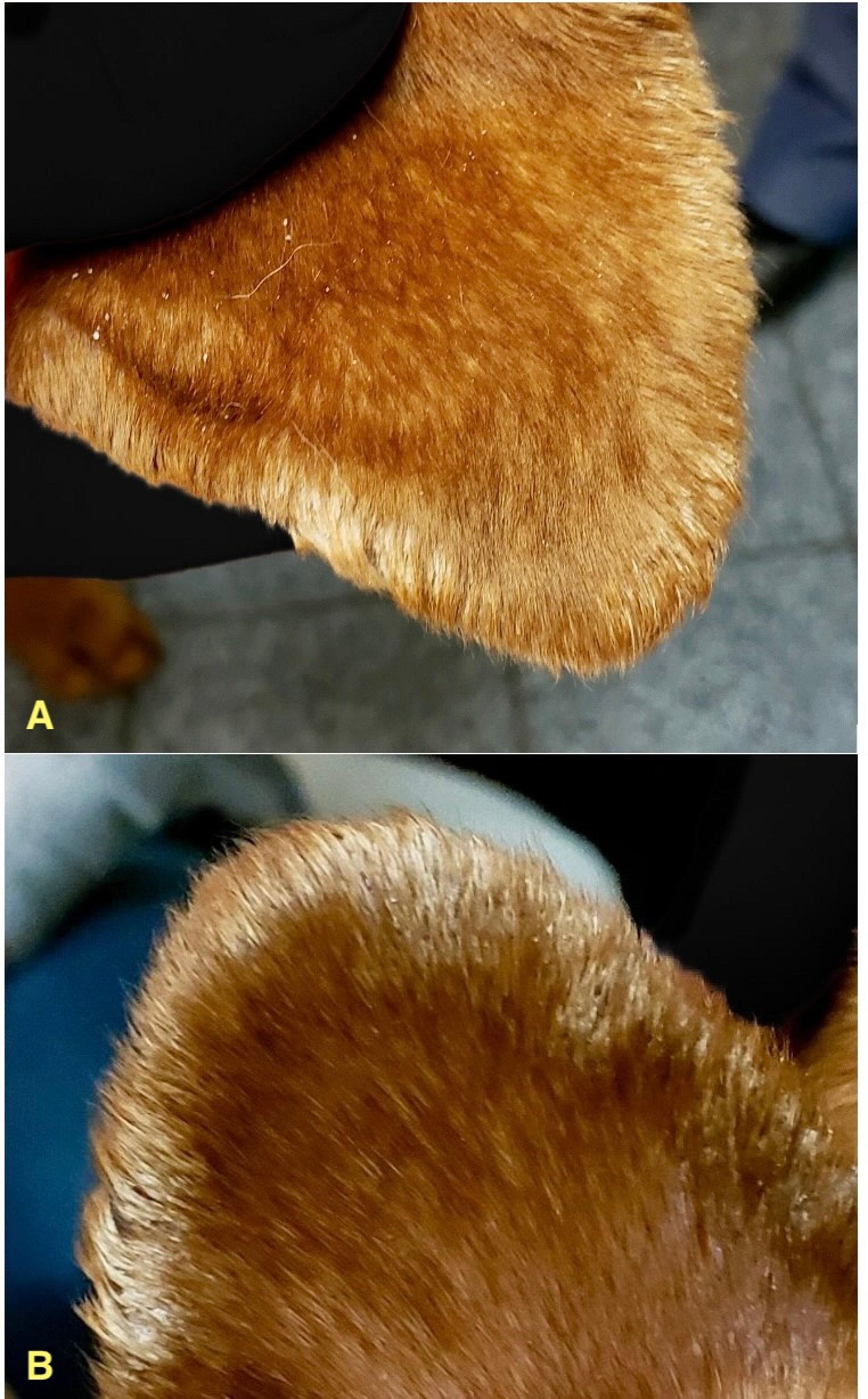
Moderately adhered, thick, fronding scale distributed along the pinnal margin of a dog. Mild thickening of the pinnal margin is also present. Images show the convex aspect of the pinna (A) and the concave aspect of the pinna (B).
Courtesy of Dr. Nicole Heinrich.
Secondary infection plays a major role in most cases of seborrhea. The sebum and keratinization abnormalities that are common in seborrhea frequently provide ideal conditions for bacterial and yeast infections. The self-trauma that occurs in pruritic animals increases the likelihood of a secondary infection.
Often, coagulase-positiveStaphylococcus spp or Malassezia spp are present. The infections add to the pruritus and are usually responsible for a substantial amount of the inflammation, papules, crusts, alopecia, and scales.
One of the first diagnostic steps is cytologic evaluation of superficial skin over affected areas to identify the quantity and type of bacteria or yeast present. If numerous cocci and neutrophils are present, pyoderma is likely. Topical treatment should be instituted for treatment of pyoderma, and systemic antimicrobials should only be used if topical treatment is not sufficient.
In a seborrheic dog with pruritus, the infection may cause all or most of the pruritus. Instead of considering allergies as the underlying disease in these dogs, nonpruritic diseases (eg, endocrinopathies) may be uncovered by addressing the infections.
After the infections have been addressed, these other diagnostic tests should be considered:
multiple deep skin scrapings
dermatophyte culture
impression smears
trichogram
flea combing
If these are negative or normal, a skin biopsy, CBC, serum biochemical profile, and complete urinalysis will complete the minimum database.
Examples of diagnostic clues include the following:
increased serum alkaline phosphatase activity (which may suggest hyperadrenocorticism or previous treatment with steroids)
increased cholesterol concentration (which may suggest hypothyroidism)
increased blood glucose concentration (which suggests diabetes mellitus)
increased BUN or creatinine concentration (which may suggest renal disease)
Treatment of Seborrhea in Animals
For primary seborrhea, frequent bathing, along with vitamin A or a retinoid
For secondary seborrhea, treatment of the underlying cause
Antimicrobial treatment
Treatment for primary idiopathic seborrhea involves bathing the dog 2–3 times per week until the desired effect has been achieved. Bathing 1–2 times per week is generally sufficient for maintenance.
A shampoo regimen can resolve pyoderma, decrease the amount of scale and sebum present, decrease pruritus, and help normalize the epidermal turnover rate. Shampoos containing ceramides, ophytrium, or phytosphingosine are often effective; however, if greasiness persists, a benzoyl peroxide or selenium sulfide shampoo can be tried.
Systemic treatment with vitamin A (8,000–10,000 units/dog, PO, every 12 hours, maximum 30,000 units/dog, PO, every 12 hours) or synthetic retinoids (eg, isotretinoin or acitretin [1 mg/kg, PO, every 12–24 hours, decreasing to 0.5 mg/kg, PO, every 48 hours, for maintenance]) can be instituted if topical treatment is not sufficient. Synthetic retinoids are known teratogens and are classified as hazardous drugs in the US. In the US, isotretinoin and acitretin are subject to a risk evaluation and mitigation strategy (REMS) and may be difficult to obtain for nonhuman patients.
Treatment for secondary seborrhea is much more complex. For treatment of seborrhea with concurrent pyoderma, a shampoo with chlorhexidine, salicylic acid, or benzoyl peroxide should be instituted. An antiseptic mousse, spray, or wipe may be useful as well.
If topical treatment is not sufficient to resolve the pyoderma, then a systemic antimicrobial should be selected based on an aerobic skin culture and the most recent antimicrobial stewardship guidelines from the International Society for Companion Animal Infectious Diseases.
Because most staphylococcal infections in seborrhea cases are superficial pyodermas, they should be treated for 3 to 4 weeks.
Epidermal collarettes may be cultured using a dry sterile culturette rolled across the collarettes. Although methicillin-resistant S pseudintermedius infections are more difficult to treat, they are not more virulent or visually striking than those due to methicillin-susceptible S pseudintermedius. Hospitalization within the past year, surgery, and previous antibiotic treatment are all possible risk factors for development of methicillin-resistant S pseudintermedius infections.
Seborrhea with concurrent Malassezia dermatitis should be treated topically with an antifungal shampoo, mousse, wipe, or spray. A systemic antifungal such as fluconazole (5 mg/kg, PO, every 24 hours for 3–4 weeks) or itraconazole (5–10 mg/kg, PO, every 24 hours) may be used if topical treatment is not sufficient.
Most active ingredients in shampoos can be classified based on their effects as keratolytic, keratoplastic, emollient, antipruritic, or antimicrobial.
Topical keratolytic products include sulfur, salicylic acid, tar, selenium sulfide, propylene glycol, fatty acids, and benzoyl peroxide. They remove stratum corneum cells by causing cellular damage that results in ballooning and sloughing of the surface keratinocytes. This decreases the scale and makes the skin feel softer.
Shampoos containing keratolytic products frequently exacerbate scaling during the first 14 days of treatment because the sloughed scales get caught in the hair coat. The scales will be removed by continued bathing; however, owners should be warned that the scaling often worsens initially.
Topical keratoplastic products help normalize keratinization and decrease scale formation by slowing down epidermal basal cell mitosis. Tar, sulfur, salicylic acid, and selenium sulfide are examples of keratoplastic agents.
Topical emollients (eg, lactic acid, sodium lactate, lanolin, and numerous oils, such as corn, coconut, peanut, and cottonseed) are indicated for any scaling dermatosis, because they decrease transepidermal water loss. They are most effective after the skin has been rehydrated and are excellent adjunct products after shampooing.
Topical antibacterial agents include benzoyl peroxide, chlorhexidine, ethyl lactate, salicylic acid, and triclosan.
Topical antifungal agents include chlorhexidine, sulfur, ketoconazole, and miconazole. Boric and acetic acids are also used as topical antimicrobials.
Given that most shampoos are a combination of products, it is important to know how individual shampoo ingredients act, as well as any additive or synergistic effects they have. The selection of an appropriate antiseborrheic shampoo regimen is based on coat and skin scaling and oiliness:
mild scaling and no oiliness
moderate to marked scaling and mild oiliness (the most common)
moderate to marked scaling and moderate oiliness
mild scaling and marked oiliness
These categories are intended to guide the type of shampoo treatment necessary; however, all factors for each individual animal should be considered.
Patients with mild scaling and no oiliness need mild shampoos that are gentle, cleansing, hypoallergenic, or moisturizing. These shampoos are indicated for animals that have mild seborrheic changes, are irritated by medicated shampoos, or have been bathed too often. These products often contain emollient oils, lanolin, lactic acid, urea, glycerin, or fatty acids. Emollient sprays or rinses are often used in conjunction with these shampoos.
Patients with moderate to marked scaling and mild to marked oiliness should be bathed with shampoos that contain sulfur and salicylic acid. Both agents are keratolytic, keratoplastic, antibacterial, and antipruritic. In addition, sulfur is antiparasitic and antifungal. Some of these shampoos also contain ingredients that are antibacterial, antifungal, and moisturizing, which can help control secondary pyoderma, Malassezia spp, and excessive scaling. Shampoos that contain ethyl lactate lower the cutaneous pH (which has a bacteriostatic or bactericidal action by inhibiting bacterial lipases), normalize keratinization, solubilize fats, and decrease sebaceous secretions. These actions also result in potent antibacterial activity.
In the past, dogs with moderate to severe scaling and moderate oiliness were often treated with tar-containing shampoos. However, because tar shampoos usually have an unpleasant odor and can be irritating, along with poor owner compliance, they usually are no longer recommended.
In patients with severe oiliness and minimal scaling, profound odor, erythema, inflammation, and a secondary generalized pyoderma or Malassezia dermatitis are often present. Shampoos that contain benzoyl peroxide provide strong degreasing actions along with potent antibacterial and follicular flushing activities. Because benzoyl peroxide shampoos are such strong degreasing agents, they can be irritating and drying. Other antibacterial shampoos are better suited in animals that have superficial pyoderma without substantial oiliness. These shampoos usually contain 2%–4% chlorhexidine (often in association with tris-EDTA) or ethyl lactate.
The follicular flushing action of benzoyl peroxide makes it helpful for patients with numerous comedones or with demodicosis. Benzoyl peroxide gels (5%) are good choices when antibacterial, degreasing, or follicular flushing actions are desired for focal areas, such as in localized demodicosis, canine acne, or Schnauzer comedone syndrome. However, these gels also may be irritating.
Key Points
Primary seborrhea is rare, but secondary seborrhea is common.
Primary seborrhea is not pruritic; however, secondary infection or inflammation can occur and create pruritus.
Primary seborrhea cannot be not cured but can be controlled with continuous treatment.
For More Information
Also see pet health content regarding seborrhea in dogs.


