


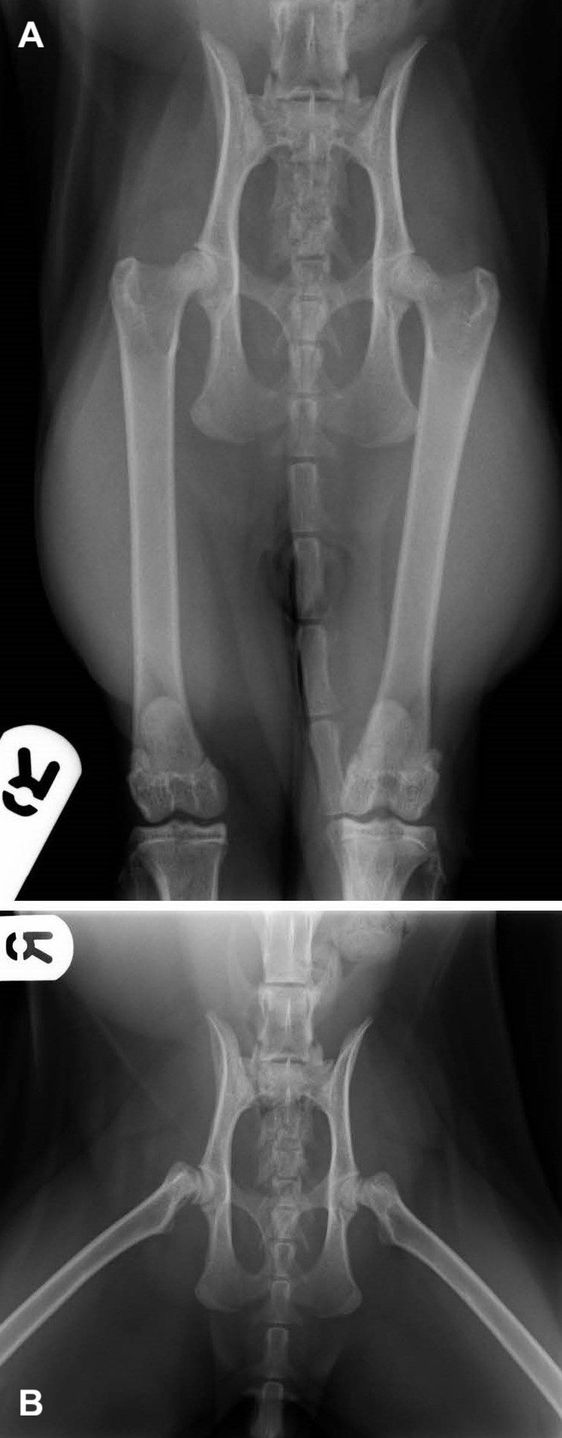
Capital physeal fracture, radiographs, cat
Ventrodorsal pelvic radiographs of a cat with a spontaneous slipped capital physeal fracture. (A) In this straight-limb view, the limb positioning (straight hindlimbs and reduction of the femoral head epiphysis) obscures the femoral epiphysis separation, so the fracture diagnosis cannot be confirmed. (B) This frog-leg (flexed) view reveals bilateral slipped capital physeal fracture of the femoral heads.
Courtesy of Dr. Pilar Lafuente.
In these topics