


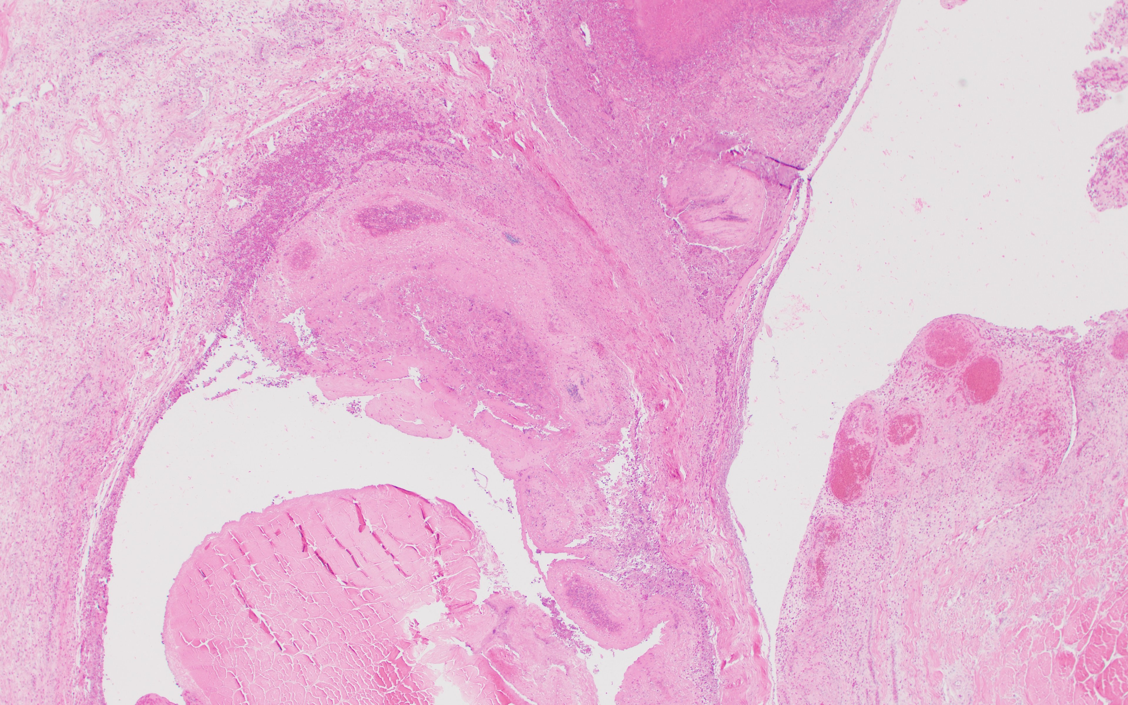
Caseous tenosynovitis, photomicrograph, poultry
Photomicrograph of a histological section of digital flexor tendon sheath from a bird with caseous tenosynovitis. Note the edema (black arrow), congestion, hemorrhage (blue arrow), necrosis, and visible bacterial colonies (red arrow). H&E stain. Original magnification, 40X.
Courtesy of Dr. Mohamed M. El-Gazzar.
In these topics