


Rhodococcal pneumonia, foal
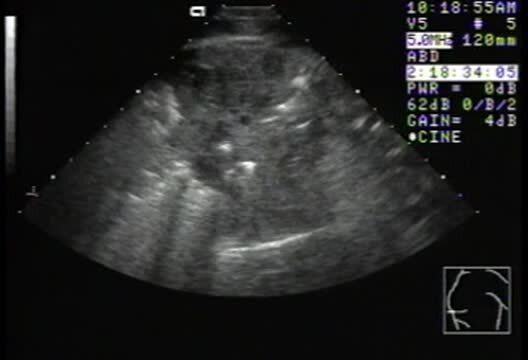
Thoracic ultrasonographic examination of a 3-month-old foal with rhodococcal pneumonia. Note pulmonary consolidation with air-filled lung visible in the periphery of the image. Small, fluid-filled abscesses can be identified in the pulmonary parenchyma.
Courtesy of Dr. Bonnie R. Rush.
In these topics