

The distal limb extends from the pedal bone to the carpus (front foot) or the tarsus (hind foot). The hoof is composed of functionally different types of horn in its different anatomic regions, and its load is supported by several structures, including the suspensory apparatus and digital cushion.

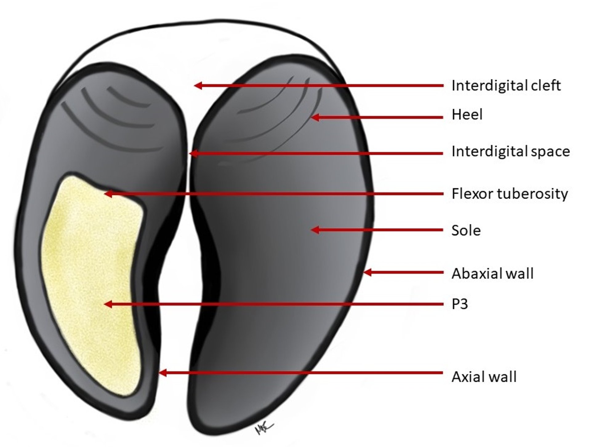
Palmar/plantar view of the bovine hoof
Courtesy of Madison Ellis-Cramer.

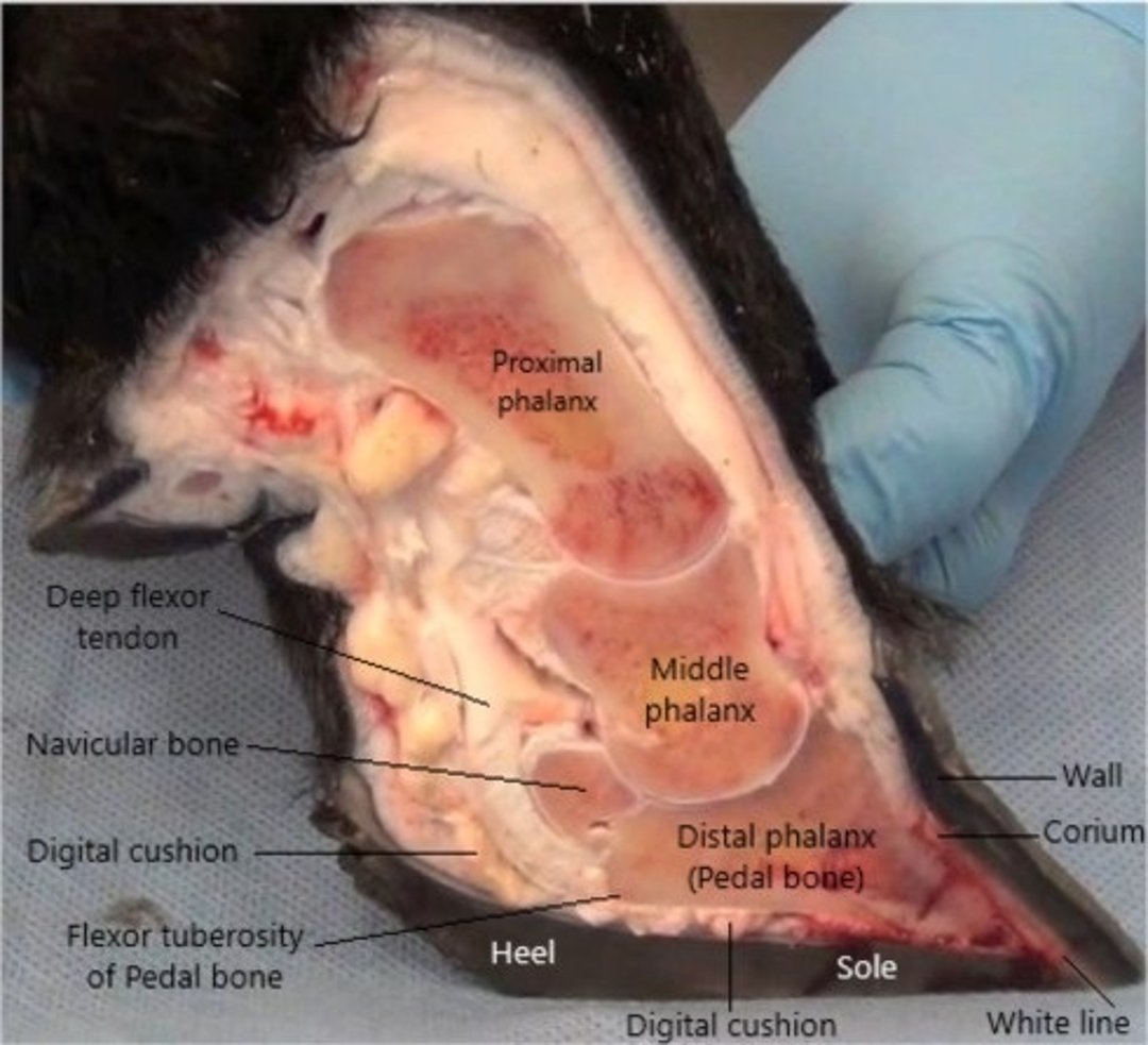
Illustration of internal anatomical structures
Courtesy of Dr. Gordon Atkins.

The distal limb is the structure distal to the carpus (in the forelimb) and tarsus (in the hind limb). Distal to the carpus and tarsus are the metacarpus and metatarsus, respectively, formed by the fused third and fourth metacarpal or metatarsal bones.
The bovine foot is the structure distal to the fetlock joint (metacarpo- or metatarsophalangeal joint). The foot consists of two digits and extends from the accessory digits (known as dewclaws) to the distal end of the third phalanx (known as the pedal bone, P3, or coffin bone). The proximal interphalangeal joint (or PIP joint) is the articulation between the proximal (first phalanx, or P1) and the middle (second phalanx, or P2) phalanges. The distal interphalangeal joint (or DIP joint) is the articulation between the middle and distal phalanges. The ventral aspect of the distal phalanx has the flexor tuberosity, where the deep flexor tendon attaches. Continuous pressure by the flexor tuberosity to the corium may result in hemorrhage and ulceration. Because of biomechanics and changes in bone length between the lateral and medial sides, the size and weight bearing between medial and lateral hooves differ between the front and hind feet. In the hind feet, the lateral hoof is larger and bears more weight than the medial hoof. In the front feet the situation is reversed: the medial hoof is larger and bears more weight than the lateral hoof.
Although the individual hooves of cows are sometimes referred to colloquially as "claws", according to the English-language naming standards of the Nomina Anatomica Veterinaria (developed by the International Committee on Veterinary Gross Anatomical Nomenclature), use of the word "claw" should be reserved to describe the last digit of a carnivore’s foot. In ruminants, pigs, and horses, the proper words used to describe the distal limb within the horn capsule are "hoof" and "hooves." Therefore, the term hoof is used throughout this chapter.
The end of each digit (hoof) is covered with modified hairless skin and has five unique segments: periople, coronary, wall, sole, and bulbar. The periople, coronary, and bulbar segments have the typical skin layers (epidermis, dermis, and subcutis); the wall and the sole, however, do not have a subcutis. The horn within and between segments is structurally different, depending on function. The horn-producing epidermal capsule, commonly known as the hoof capsule, comprises the outer surface of the hoof. Typically, the horn produced by the periople, coronary, and wall segments, collectively known as the wall, is the hardest; the horn in the proximal bulb, the softest. The horn of the white line is solely produced by the wall segment and consists mainly of cap horn. The wall and sole surfaces contain mainly tubular horn.
The dermis, more commonly called the corium, supports the function of the epidermis and contains vessels and nerves. The dermis also contains dermal lamellae, but in the wall segment, these do not feature the secondary lamellae found in horses' hooves.
The subcutis typically contains connective tissue and fat and acts as a cushion in the periople, coronary, and bulbar segments. In the bulbar segment this cushion is called the digital cushion and is part of the mechanism that protects the corium (dermis) and absorbs and dissipates forces during locomotion. Age, body condition, and metabolic changes can affect the integrity of the digital cushion.
The suspensory apparatus of the digit consists of a collagen fiber system that supports and assists in holding the third phalanx within the hoof capsule. These collagen fibers run from their point of insertion on the surface of the third phalanx through the dermis and dermal lamellae and are anchored to the basement membrane. The suspensory apparatus transfers the force applied to P3 by the animal’s body weight onto the hoof wall. The suspensory apparatus works in conjunction with the digital cushion to transfer force away from P3 and toward the hoof wall. Loosening, stretching, or any other insult to the collagen fibers may result in displacement of P3 and/or internal damage to the hoof.
Key Points
Each of the five segments of the hoof produces a structurally and functionally different type of horn.
Size and weight bearing differ between the medial and lateral hooves and between the front and hind feet.
The suspensory apparatus and digital cushion help support, absorb, and distribute forces created during locomotion.


