


Disorders of joints are among the most common causes of lameness in animals. Primary injuries to ligaments, direct trauma, and inflammatory conditions all cause pain and discomfort. Secondary changes resulting from primary disorders often manifest as chronic changes to joint tissue in the form of osteoarthritis (OA), which is diagnosed frequently and usually persists as a lifelong problem.
Degenerative changes in ligaments lead to ligament breakdown and joint instability. In dogs, partial or complete rupture of the cranial cruciate ligament is the most common joint-related injury (see ), and its incidence increases as dogs age.


Radiograph of a stifle in a dog, showing marked effusion with osteophyte formation secondary to a chronic cranial cruciate ligament tear.
Courtesy of Dr. Michael Jaffe.
Damage to a ligament is associated with chronic degeneration of the ligament fibers until the ligament breaks. Surgery to repair this injury is necessary to restore joint stability and normal function. Suspensory ligament injuries in horses can also be due to chronic degeneration, chronic repetitive trauma, or acute, excessive stress exerted on the ligament. Medical and sometimes surgical management is indicated for treating this injury.
OA is an inflammatory, degenerative, progressive disease of joints, and it is the most common joint disorder in animals. This disease causes pain, morbidity, and decreased performance in all species: it is estimated that 80–90% of dogs and cats will suffer from arthritis in middle age (1, 2), and arthritis is a major clinical problem for aged and athletic horses as well (3, 4).
OA has a multifactorial etiology; aging, trauma, abnormal or excessive mechanical forces, poor conformation, endocrine factors, genetic factors, and sepsis contribute to the development of OA. However, OA most commonly occurs as the result of a primary injury to the joint from joint instability, direct trauma, or a soft tissue injury.
Joint effusion, thickening of the synovial membrane, decreased viscosity of the synovial fluid, and damage to articular cartilage are the hallmarks of OA. Clinical examination might also reveal new bone formation (see ), loss of normal range of motion, and pain upon flexion of the affected joint. Radiographic changes include narrowing and/or unevenness of the joint space, subchondral bone lysis, subchondral bone sclerosis, osteophyte formation, joint remodeling, and periarticular new bone formation (see ).
Pearls & Pitfalls
|

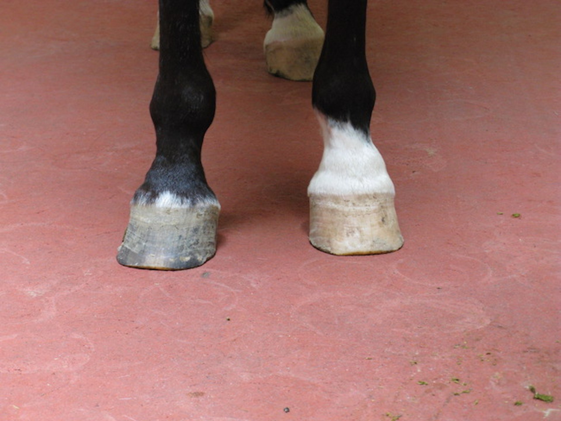
Periarticular new bone formation has caused a hard swelling (ringbone) around the proximal interphalangeal joint in the white (left) foot of this horse.
Courtesy of Dr. Stephen Adams.


Radiograph showing severe osteoarthritis of the proximal interphalangeal joint in a horse. Note the loss of joint space, new bone formation (ringbone), and subchondral lysis.
Courtesy of Dr. Stephen Adams.
OA is progressive because it creates a vicious cycle of cartilage injury: as damage accumulates, cartilage becomes less resilient to normal physiological stresses because of the loss of proteoglycan, decreased cell viability, and advancement of the tidemark of calcified cartilage. Proteoglycan loss is accompanied by increased cartilage matrix water content and decreased stiffness, and this biomechanically compromised cartilage is more susceptible to further damage.
Inflammatory joint diseases arise secondary to a variety of causes. Immune-mediated etiologies result in joint effusion and pain, and some disorders lead to erosion of cartilage and subchondral bone. Erosive polyarthritis disorders are considerably less common than nonerosive polyarthritis in small animals and include rheumatoid arthritis, feline progressive polyarthritis, and greyhound polyarthritis.
Immune-mediated polyarthritis (IMPA) is the most common nonerosive form of inflammatory joint disease in dogs and is often idiopathic. Chinese Shar-Pei, Akita, and other breeds are known to develop IMPA as a result of immune-mediated etiologies. Immune-mediated polyarthritis can develop secondary to GI infections, non-GI infections, and neoplasia. IMPA can also arise from drug reactions, as with sulfonamide use in Doberman Pinschers and with cephalosporins and penicillin derivatives. These drugs produce a type IV hypersensitivity reaction that often resolves when the medication is discontinued.
Septic arthritis is an infection of a joint. Bacteria are the most common underlying organisms; however, mycoplasmas, protozoa, rickettsiae, and mycobacteria can also cause joint infections. Of the bacterial organisms, Staphylococcus spp are the most frequently cultured in small animals. Joint infections can develop after surgery, penetrating trauma, bite wounds, and hematogenous spread from distant infections. Usually, only one joint is affected at a time.
Hereditary and developmental joint disorders include conditions such as osteochondrosis or osteochondritis dissecans (OCD), patellar luxation, hip dysplasia and elbow dysplasia in dogs (see ), equine ataxia secondary to cervical vertebrae malformation/malarticulation, angular limb deformities, and scapulohumeral dysplasia in American Miniature horses.

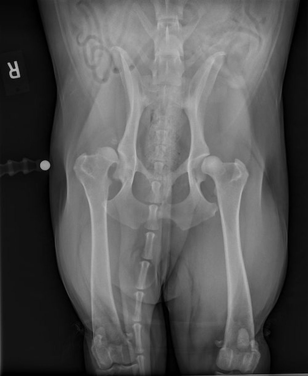
Radiographic image of hip dysplasia in a dog, showing marked subluxation and remodeling of the right coxofemoral joint with less severe changes in the left joint.
Courtesy of Dr. Mark Rochat.
Hip dysplasia in dogs results from the combined effect of many genes that influence skeletal development, connective tissue, and biomechanics. This polygenic foundation, along with external factors such as nutrition, growth rate, exercise, and body weight, determines the severity of the condition.
Extension of septic physitis into adjacent joints and damage due to continuous abnormal weight bearing in animals with angular limb deformities are other possible causes of joint disease.
Trauma to joints can occur in any species at any age. Traumatic articular fractures, ligament injuries, and joint capsule injuries can result from a direct blow to the joint or from a soft tissue strain due to injury. Severe trauma frequently results in luxation, subluxation, fracture, or instability of a joint.
Neoplasia of the joint is relatively uncommon. Bone tumors do not extend directly across the joint surface from one bone to another; however, they might metastasize. Tumors of the joint capsule, such as synovial histiocytic sarcoma, can indirectly involve the joint but are generally limited to the synovial membrane.
Pearls & Pitfalls
|
Once the underlying nature of a joint injury is diagnosed, an appropriate treatment regimen can begin. Nonetheless, it is important to note that any joint injury will result in some degree of OA developing over time.
For More Information
McIlwraith CW, Frisbie DD, Kawcak CE, van Weeren R, eds. Joint Disease in the Horse. 2nd ed. Elsevier; 2016.
Allan G, Davies S. Radiographic signs of joint disease in dogs and cats. In: Thrall DE, ed. Textbook of Veterinary Diagnostic Radiology. 7th ed. Saunders; 2018:403-433.
Also see pet owner content regarding bone, joint, and muscle disorders of dogs, joint disorders in cats, and joint disorders in horses.
References
Anderson KL, O’Neill DG, Brodbelt DC, et al. Prevalence, duration and risk factors for appendicular osteoarthritis in a UK dog population under primary veterinary care. Sci Rep. 2018;8(1):5641. doi:10.1038/s41598-018-23940-z
Hardie EM, Roe SC, Martin FR. Radiographic evidence of degenerative joint disease in geriatric cats: 100 cases (1994–1997). J Am Vet Med Assoc. 2002;220(5):628-632. doi:10.2460/javma.2002.220.628
van Weeren PR, Back W. Musculoskeletal disease in aged horses and its management. Vet Clin North Am Equine Pract. 2016;32(2):229-247. doi:10.1016/j.cveq.2016.04.003
Neundorf RH, Lowerison MB, Cruz AM, Thomason JJ, McEwen BJ, Hurtig MB. Determination of the prevalence and severity of metacarpophalangeal joint osteoarthritis in Thoroughbred racehorses via quantitative macroscopic evaluation. Am J Vet Res. 2010;71(11):1284-1293. doi:10.2460/ajvr.71.11.1284


