


A tendon is the terminal portion of a muscle that attaches at the muscle's origin and insertion on bone. Tendons transfer forces generated by muscle contractions exerted on the bone, resulting in joint movement. Several injuries of tendons are possible; they can be classified as lacerations, strains, ruptures, or avulsions.
Injuries to tendons are typically slow to heal because of the limited vascular supply to tendons. In general, tendons receive their blood supply from the musculotendinous junction and from their insertion on bone. A small percentage of blood is provided externally to the body of the tendon, and the tendon also receives nutrients via synovial fluid if it passes through a tendon sheath. However, because of this relatively poor blood supply, tendon healing can be prolonged, and the resultant repair tissue is usually of inferior mechanical strength compared to the original, uninjured tendon. This weakening is due to the rearrangement of collagen fibers in a less organized fashion as scar tissue forms.
Inflammatory conditions of the tendon are caused by acute injury or chronic overuse, such as repetitive motion or exercise-induced stress. One example is tendinitis of the superficial flexor tendon in horses, which is frequently injured by partial tearing that leads to tendinitis (see ). Another acquired injury of tendons involves traumatic disruptions by internal forces or external trauma.

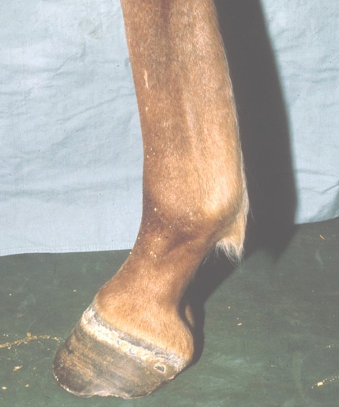
Tendinitis of the superficial digital flexor tendon in the forelimb of a horse. The tendon has a typical bowed appearance.
Courtesy of Dr. Stephen Adams.
Newer treatment modalities (stem cell injection, platelet-rich plasma injection, extracorporeal shock wave therapy), combined with specific rehabilitation programs, have improved the prognosis for recovery from tendon and ligament injuries.
Tenosynovitis
Tenosynovitis is an inflammatory condition of the synovial membrane surrounding a tendon; it is characterized by distention of the tendon sheath due to synovial effusion. The extent of synovial distention of the tendon sheath and of any resulting lameness depends on the severity of inflammation.
In horses, chronic tenosynovitis is common in the tarsal sheath of the hock (thoroughpin) and in the digital sheath (windpuffs); see . Synovial effusion at these two sites must be differentiated from synovial effusion of the tarsocrural and fetlock joints, respectively.

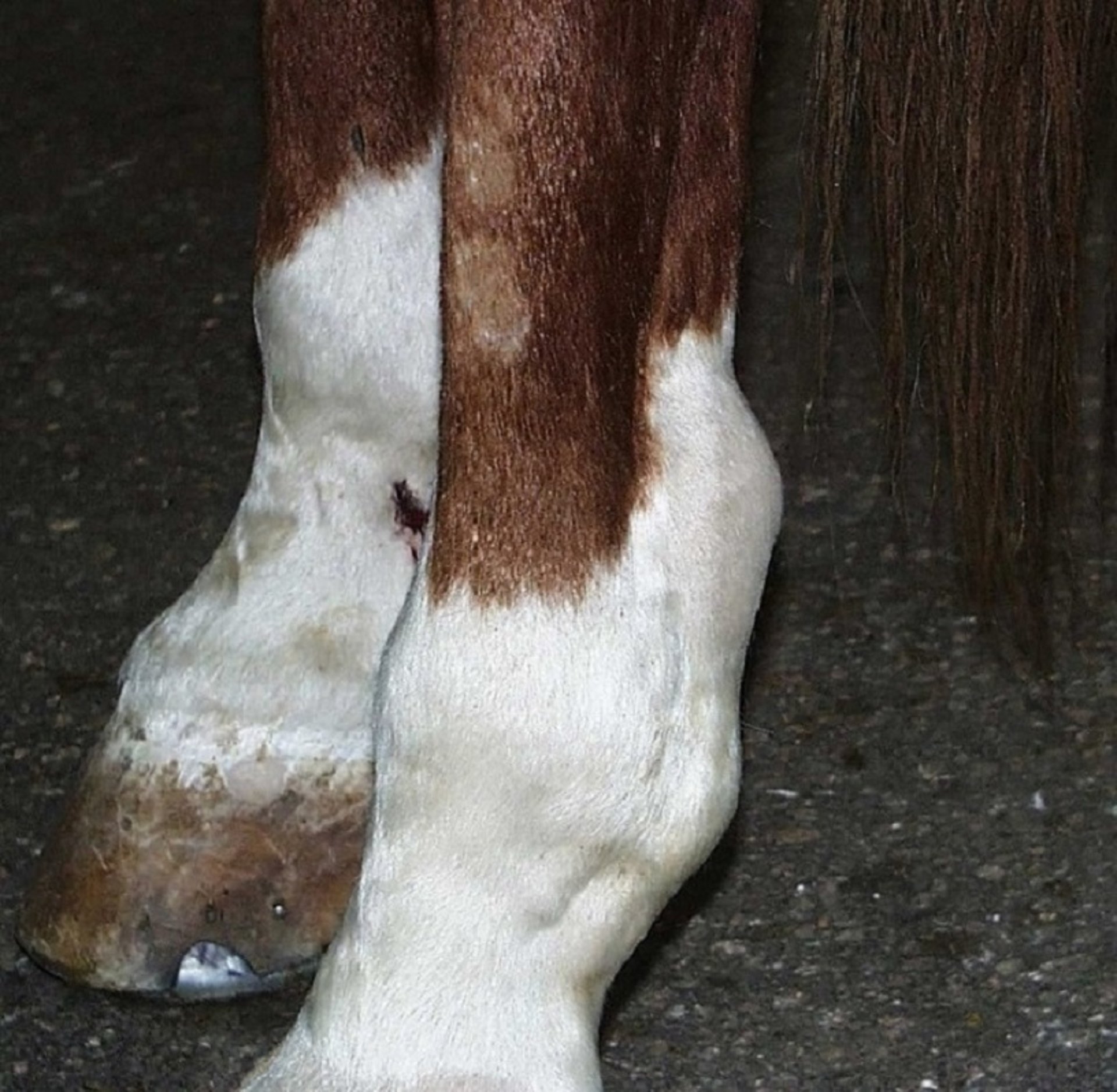
Effusion in the digital tendon sheath of this horse has caused marked swelling of the tendon sheath, which extends more proximally and distally than does the metatarsophalangeal (fetlock) joint pouch.
Courtesy of Dr. Stephen Adams.
Evaluation of horses with signs of tenosynovitis should include a lameness examination, isolation of pain to the tendon sheath using diagnostic local anesthesia, radiography (contrast radiographs can provide helpful information), and ultrasonography of the tendons enclosed within the sheath.
Bicipital tenosynovitis is an inflammatory condition of the tendon of origin of the biceps brachii muscle in dogs. This painful inflammatory condition is caused by chronic repetitive injury, acute trauma, or degenerative conditions of the shoulder joint. Diagnosis is based on a finding of pain on palpation of the tendon, radiographic signs of mineralization of the tendon in the bicipital groove of the humerus, and abnormalities noted on ultrasonography of the tendon. Direct evaluation of the tendon by shoulder arthroscopy and advanced imaging with MRI also help in diagnosing this condition.
In poultry, viral arthritis is caused by reovirus infection and leads to inflammation affecting leg joints and/or tendons. Affected birds typically are lame and have ruptured tendons. Definitive diagnosis relies on reovirus isolation and/or identification from the affected tissue via virus isolation or RT-PCR.
Treatment of Tenosynovitis
Conservative management, including rest and controlled exercise, is initially recommended for idiopathic cases of tenosynovitis in horses. Mild cases can be treated with supportive care, including bandages, cold compresses, NSAIDs, rest, and controlled exercise rehabilitation.
More severe cases of tenosynovitis, in which lameness is observed, might respond to platelet-rich plasma (PRP) injections into the tendon sheath. Extracorporeal shock wave therapy is a noninvasive treatment that can stimulate tendon healing by delivering high-energy pressure waves.
Surgery is typically reserved for chronic and severe cases of tenosynovitis. Septic tenosynovitis requires local and systemic administration of antimicrobials, lavage, drainage, and tenoscopy to debride infected tissues.
Tenosynovitis in small animals is similarly treated initially with rest, NSAIDs, and rehabilitation therapy. Intra-articular injections of corticosteroids and hyaluronic acid, or of PRP, may also be considered. NSAIDs should not be administered concurrently with corticosteroid use.
Physical rehabilitation using photobiomodulation (laser therapy), extracorporeal shock wave therapy, and manual therapy exercises help decrease inflammation and improve limb use in tenosynovitis cases. Surgery may be considered in chronic and severe cases.
For More Information
Konoplev V, Elizarkova M, Bokarev A, Kovalev S. Diagnosis of tendinites in sport horses. KnE Life Sci. 2019 (issue International Scientific and Practical Conference “AgroSMART – Smart Solutions for Agriculture”):653-658.
Canapp SO, Dycus D, Shaw KK. Disorders of the canine thoracic limb: diagnosis and treatment. In: Zink C, Van Dyke JB, eds. Canine Sports Medicine and Rehabilitation. 2nd ed. John Wiley & Sons; 2018:294-332.
Canapp SJ. 2007. Injuries in sporting and working dogs. In: Proceedings of the NAVC Conference: January 13–17, 2007, Orlando, Florida. North American Veterinary Conference; 2007.


