Nutritional myopathies develop because of dietary nutrient deficiencies or excesses. Selenium deficiency is the most common cause of nutritional myopathy in animals, and its occurrence correlates with geographical areas with low selenium status in soil and plants. Concurrent vitamin E deficiency is associated with more severe muscle damage. Clinical signs range from stiffness to lethargy and apparent dyspnea. Diagnoses can be made after postmortem examination and assessment of trace minerals, including selenium, in tissues such as liver. Dietary supplementation and parenteral administration of selenium-containing injectable products, as well as vitamin E, can prevent and reverse clinical signs.
Nutritional Myodegeneration in Ruminants and Pigs
Nutritional myodegeneration (NMD) is an acute degenerative disease of cardiac and skeletal muscle caused by a dietary deficiency of selenium or vitamin E in young, rapidly growing calves, lambs, and kids. Dams have usually consumed selenium-deficient diets during gestation.
Selenium deficiency appears to be more important than vitamin E deficiency in causing NMD. NMD occurs worldwide in areas where the soil (and therefore the derived grains and forage) is deficient in selenium, and storage conditions do not preserve vitamin E in forages. Soil in the northeastern and eastern coastal areas and northwestern regions of the US are particularly deficient in selenium. Vitamin E deficiency occurs most commonly when animals are fed poor-quality hay, straw, or root crops.
Both vitamin E and selenium have an important antioxidant function and protect cell membranes against damage from free radicals. Selenium is an essential component of five antioxidant selenoproteins, including glutathione peroxidase, and vitamin E acts as an antioxidant within lipid bilayers. Muscle degeneration is the result of oxidant damage to cell membranes and proteins, leading to a loss of cellular integrity. Young, rapidly growing animals usually are affected; however, the disease has also been reported in yearling and adult cattle.
When cardiac muscle is primarily affected, animals can be found in respiratory distress, have cardiac arrhythmias, or be found dead. In such cases, the clinical course is frequently short, with death occurring commonly in < 24 hours despite medical therapy.
When skeletal muscle is primarily affected, clinical signs of muscle weakness, stiffness, and difficulty rising are observed. Most affected animals are able to remain standing only for short periods, and locomotor muscles can be firm and painful on palpation. Animals with skeletal NMD often respond favorably to treatment and rest. Improvement is evident after a few days; within 3–5 days, animals can often stand and walk (see ).

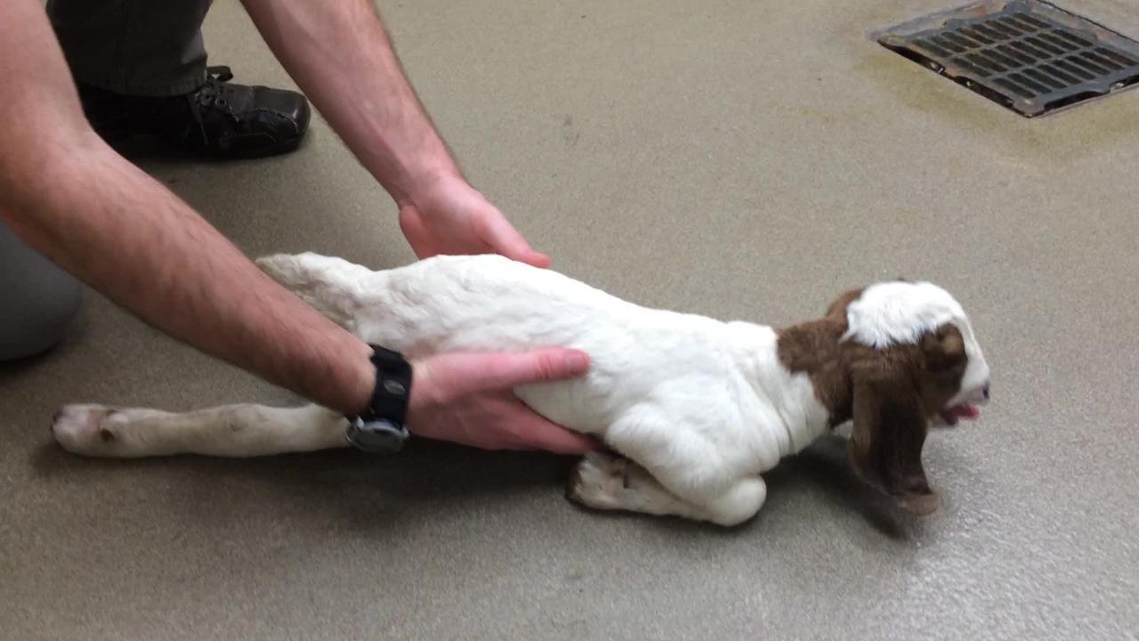
If the respiratory muscles are affected, the animal might show respiratory distress and evidence of increased abdominal effort when breathing. The muscles of the tongue can be involved, resulting in dysphagia.
Differential diagnoses include the following:
infectious diseases resulting in septicemia, pneumonia, and toxemia
cardiac anomalies caused by ionophore antimicrobials and cardiotoxic agents found in plants (eg, oleander, senna, yew, white snakeroot, and gossypol toxicity from cottonseed)
diseases causing stiffness of gait, weakness, and recumbency with no change in mental status, including:
spinal cord compression
cerebellar disease
suppurative and nonsuppurative meningitis/myelitis
polyarthritis
neurotoxins such as organophosphates and tetanus
pelvic fractures
parasitic myositis
clostridial myositis
traumatic injuries
Supportive evidence of NMD includes increased concentrations of CK, AST, and LDH. Definitive diagnosis is based on demonstrating low whole blood and liver tissue selenium concentrations. Commonly reported reference values for cattle and sheep are as follows:
Cattle 0–30 days old: whole blood 100–250 ng/mL; liver 1.5–3.5 mcg/g dry matter
Cattle > 30 days old: whole blood 120–300 ng/mL; liver 0.7–2.5 mcg/g dry matter
Sheep: whole blood 120–350 ng/mL; liver 0.8–3.0 mcg/g dry matter (1)
The critical concentration of vitamin E (alpha-tocopherol) in plasma is 1.1–2 ppm in large animals (2). Vitamin E deteriorates rapidly in plasma samples. Therefore, plasma samples for alpha-tocopherol analysis should be put on ice immediately, protected from light by wrapping them in foil, and stored at –29°C (–20°F) if analysis is to be delayed.
Pearls & Pitfalls
|
Bilaterally symmetric myodegeneration is a consistent finding in NMD. Skeletal muscle degeneration is characterized by pale discoloration and a dry appearance of affected muscle, white streaks in muscle bundles, calcification, and intramuscular edema. The white streaks in cardiac and skeletal muscle bundles represent bands of coagulation necrosis or, in chronic cases, fibrosis and calcification. In calves, the left ventricle and septum are most frequently involved, but both ventricles are usually involved in lambs. Histologically, affected muscle fibers can be hypercontracted and fragmented, with some mineralized and others infiltrated by macrophages.
The cardiac form of NMD is often acutely fatal, whereas the skeletal form can respond to injectable selenium products. The label dosage for selenium in the BO-SE formulation (2.9 mg/mL) is 0.12–0.18 mg/kg, IM or SC, for cattle, and 0.12 mg/kg for sheep (3). Dosage of these injectable products should not be greatly increased above that on the label to prevent an inadvertent selenium toxicosis.
When using vitamin E/selenium combinations, the amount of vitamin E is insufficient for supplementation; it is present only as a preservative for the solution. Injectable products that contain 300 and 500 IU/mL vitamin E as d-alpha-tocopherol are available; however, oral supplementation is the general approach to provide additional dietary levels of vitamin E. Recommended levels of supplementation for calves range from 15 to 60 mg of dl-alpha-tocopherol acetate per kg of dry feed (4).
Antimicrobials might be indicated to combat secondary pneumonia. Provision of adequate energy intake and attention to the fluid and electrolyte balance are critical if recovery is to be successful.
Under current federal regulations in the US, selenium can be incorporated into the total ration of ruminants and other species to a level of 0.3 ppm. In salt/mineral mixtures formulated for free-choice feeding, selenium can be incorporated at 90 ppm for sheep and 120 ppm for cattle. In certain areas or in herds, levels as high as 200 ppm selenium in salt/mineral mixtures might be necessary to maintain adequate selenium levels. Federal regulations limit the intake of supplemental selenium to 0.7 mg/head/day in sheep and 3 mg/head/day in beef cattle (5).
The use of rumenoreticular boluses in ruminants, which release a precise amount of selenium daily, is common in many countries. These slow-release boluses can replace supplementation by salt mixtures or by injections and are valuable in extensive grazing systems. Alternatively, individual animals can be supplemented by periodic (30- to 60-day intervals) injections of selenium/vitamin E preparations to help maintain body concentrations and assist in transplacental transfer of selenium.
Regardless of the method of supplementation, periodic blood (or tissue) sampling of animals at risk is recommended to ensure desired levels of selenium. Feeding animals properly prepared and stored hay and grain or allowing access to high-quality, green forage should ensure adequate vitamin E intake.
Hypokalemic Myopathy in Ruminants and Pigs
Hypokalemic myopathy in dairy cattle occurs when serum potassium concentrations are < 2.0 mmol/L, producing severe clinical signs of muscle weakness. Hypokalemia is often associated with another primary disorder, such as ketosis. Anorexia and enhanced potassium excretion due to the administration of one or more doses of isoflupredone acetate to ketotic cows are common causes of hypokalemia. Isoflupredone acetate has both glucocorticoid and mineralocorticoid activity, resulting in a decrease in mean plasma potassium concentration by 25% in cows 2 days after a single injection (20 mg) and 46% in cows 3 days after two injections (6).
Clinical signs of hypokalemic myopathy include severe weakness, recumbency, abnormal position of the head and neck, rumen hypomotility or atony, abnormal feces, anorexia, and tachycardia. Cardiac arrhythmias are also common. Diagnosis is based on clinical signs combined with serum potassium of < 2.0 mmol/L. Other common clinical chemistry abnormalities include ketosis, metabolic alkalosis, and increased serum CK and AST activities. Muscle biopsies reveal a vacuolar myopathy.
Restoration of whole-body potassium balance can be difficult, and serum potassium concentrations do not necessarily reflect muscle potassium concentrations. Recommended supplementation includes potassium chloride administered at an average daily dose of 42 g/100 kg (oral dose of 26 g/100 kg combined with 16 g intravenously) in moderate cases (7), or in severe cases, IV at rates not exceeding 0.5 mEq/kg/h (8). Treatment should also be directed at resolving the primary problem , such as ketosis and other causes of severe anorexia, as well as providing supportive care. Survival was reported to be 22% in one study and 79% in a case report (9).
Nutritional Myopathy of Pigs
There are several specific diseases of pigs in which muscle degeneration can be extensive (eg, mulberry heart disease) and others in which the degeneration is frequently less conspicuous (eg, hepatosis dietetica). Yellow fat disease might occur with accompanying myopathy.
Etiology of Nutritional Myopathy of Pigs
Mulberry heart disease and hepatosis dietetica are associated with diets associated with diets low in selenium or vitamin E. Administration of iron dextran to piglets having low vitamin E status can precipitate a severe myopathy (see also Iron Toxicosis in Newborn Pigs) with lesions identical to those of selenium or vitamin E deficiency. Other factors that can increase the selenium requirement include diets with low concentrations of protein (especially sulfur-containing amino acids), diets with an excess of selenium antagonistic compounds, and possibly genetic influences on selenium metabolism. Vitamin E might be less available in diets with high concentrations of polyunsaturated fatty acids, vitamin A, or mycotoxins.
Clinical Findings in Nutritional Myopathy of Pigs
Nutritional myopathy conditions have certain characteristics in common. Losses tend to occur sporadically, and rapidly growing pigs 2–16 weeks old are affected. Death almost invariably occurs suddenly and is often precipitated by exercise.
Lesions
In mulberry heart disease, the characteristic lesions are a pericardial sac grossly distended with straw-colored fluid that contains fibrin strands, and extensive hemorrhage throughout the epicardium and myocardium (see ). Microscopically, lesions are present in blood vessels and myocytes; in addition to interstitial hemorrhage, there is usually extensive myocardial necrosis together with fibrin thrombi in capillaries. If pigs survive for a few days, neurological signs can result from focal encephalomalacia.


Necropsy images from a 6-week-old pig after sudden death at 3 weeks post weaning: (A) cross-sections of the heart through the left and right ventricles, with severely pale myocardium in the center and dark red peripherally towards the epicardium and (B) diffusely red and wet lungs, demonstrating associated pulmonary edema.
Courtesy of Dr. Marta Mainenti and Dr. Rachel Derscheid.
In hepatosis dietetica, subcutaneous edema and varying amounts of transudate in serous cavities are often present. Fibrin strands adhere to the liver, which has a characteristic mottled appearance caused by irregular foci of parenchymal necrosis and hemorrhage. Acute lesions can appear as scattered, red, swollen lobules and edema of the gallbladder wall. Focal lesions of myocardial necrosis and, less frequently, skeletal myonecrosis is apparent.
Many pigs that die with selenium or vitamin E deficiency also have esophagogastric ulceration.
Diagnosis of Nutritional Myopathy of Pigs
Diagnosis of nutritional myopathy is typically made via history, clinical signs and necropsy findings. The history and gross necropsy findings might be distinctive; however, histological evaluation to demonstrate specific cardiac and skeletal muscle lesions is necessary.
Differential diagnoses for mulberry heart disease include the following:
acute septicemic diseases (eg, salmonellosis, erysipelas, and streptococcosis)
pericarditis
polyserositis
edema disease
Differential diagnoses for hepatosis dietetica include the following:
pitch poisoning
porcine stress syndrome, for pigs with prominent skeletal muscle lesions
Cases of selenium or vitamin E deficiency in pigs can be identified, as in other species, by decreased concentrations of selenium, vitamin E, and glutathione peroxidase in whole blood and tissues and by increased serum CK and AST.
Prevention and Treatment of Nutritional Myopathy of Pigs
Treatment for nutritional myopathy is primarily by supplementing rations with additional selenium or vitamin E, or both (as for ruminants). Additionally, affected pigs and their herdmates might be given injections of selenium/vitamin E to increase tissue levels rapidly. Injection of sows in late gestation increases tissue levels in newborn piglets. Care should be taken to avoid accidental overdose when injectable formulations of selenium are used for selenium-responsive conditions. The minimum reported lethal injectable selenium dose in pigs is 0.84 mg/kg, which is lower than the previously reported 0.9 mg/kg body weight (10).
Key Points
In pigs with selenium or vitamin E deficiency, the most common disease forms are mulberry heart disease or hepatosis dietetica.
Supplementation of vitamin E and selenium is critical, especially during gestation.
Other dietary risk factors, such as deficiencies in vitamin A or mycotoxins, should also be addressed to prevent selenium or vitamin E deficiency in pigs.
For More Information
Zimmerman JJ, Karriker LA, Ramirez A, Schwartz KJ, Stevenson GW, Zhang J, eds. Diseases of Swine. 11th ed. Wiley-Blackwell; 2019.
Cooper B, Valentine B. Muscle and tendon, In: Maxie MG, ed. Jubb, Kennedy, and Palmer's Pathology of Domestic Animals. 6th ed. Saunders Ltd; 2015.
References
Filley S, White H, Pirelli G, Hall JA. Selenium supplementation strategies for livestock in Oregon. EM 9094. Oregon State University Extension Service. Published 2014. Accessed August 10, 2026. https://extension.oregonstate.edu/catalog/em-9094-selenium-supplementation-strategies-livestock-oregon
Valberg SJ, Spier SJ, Parish SM, Murphy MJ, Carlson GP. Diseases of muscle. In: Smith BP, Van Metre DC, Pusterla N, eds. Large Animal Internal Medicine. 6th ed. Mosby; 2021:1421-1455.e5. doi:10.1016/B978-0-323-55445-9.00042-2
US Food and Drug Administration. Animal Drugs @ FDA. [Sodium selenite and vitamin E liquid sheep swine cattle]. NADA 012-635. https://animaldrugsatfda.fda.gov/adafda/views\#/home/previewsearch/012-635
Chauhan SS, Celi P, Ponnampalam EN, Leury BJ, Liu F, Dunshea FR. Antioxidant dynamics in the live animal and implications for ruminant health and product (meat/milk) quality: role of vitamin E and selenium. Anim Prod Sci. 2014;54:1525-1536. doi:10.1071/AN14334
US Food and Drug Administration. Selenium. 21 CFR Sec 573.920. https://www.ecfr.gov/current/title-21/part-573/section-573.920
Coffer NJ, Frank N, Elliott SB, Young CD, van Amstel SR. Effects of dexamethasone and isoflupredone acetate on plasma potassium concentrations and other biochemical measurements in dairy cows in early lactation. Am J Vet Res. 67(7):1244-1251. doi:10.2460/ajvr.67.7.1244
Sattler N, Fecteau G, Girard C, Couture Y. Description of 14 cases of bovine hypokalaemia syndrome. Vet Rec. 1998;143(18):503-507. doi:10.1136/vr.143.18.503.
Stämpfli H, Fecteau G, Oliver-Espinosa O. Hypokalemia. In: Smith BP, Van Metre DC, Pusterla N, eds. Large Animal Internal Medicine. 6th ed. Mosby; 2021:403-406. doi:10.1016/B978-0-323-55445-9.00022-7
Sattler N, Fecteau G. Hypokalemia syndrome in cattle. Vet Clin North Am Food Anim Pract. 2014;30(2):351-357. doi:10.1016/j.cvfa.2014.04.004
Peixoto PD, Oliveira KD, França TN, et al. Experimental and iatrogenic poisoning by sodium selenite in pigs. Pesqui Vet Bras. 2017;37(6):561-569. doi:10.1590/S0100-736X2017000600005