

Also see Diseases of the Peripheral Nerves and Neuromuscular Junction.
Congenital Neuromuscular Disorders in Dogs and Cats
Congenital Laryngeal Paralysis in Dogs
Congenital laryngeal paralysis occurs in Bouvier des Flandres (autosomal dominant) and in Siberian Huskies, Rottweilers, and Bull Terriers < 1 year old. It results in exercise intolerance and inspiratory dyspnea.
Diagnosis is confirmed by visualization on laryngoscopy.
Congenital laryngeal paralysis with diffuse peripheral neuropathy occurs in several breeds, including Dalmatians, Rottweilers, and Pyrenean mountain dogs (Great Pyrenees). An autosomal recessive mode of inheritance is suspected.
The prognosis for dogs with untreated congenital laryngeal paralysis is guarded to poor. With surgical unilateral cricoarytenoid lateralization, some dogs have been able to resume activity.
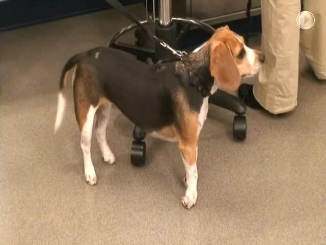
Congenital Myasthenia Gravis in Dogs and Cats
Congenital myasthenia gravis (autosomal recessive) has been described in a number of dog and cat breeds, including Parson Russell Terrier, Smooth Fox Terrier, smooth miniature Dachshund, English Springer Spaniel, Labrador Retriever, Golden Retriever, Sphynx cat, and Devon Rex cat.
The disorder is due to either a deficiency or a dysfunction of the acetylcholine receptor, and no circulating antireceptor antibody is evident in the more common acquired form of the disease.
Several mutations have been associated with congenital myasthenic syndromes: CHRNE (Jack Russell Terrier), CHAT (Old Danish Pointing Dogs), and several mutations in COLQ (Labrador Retriever, Golden Retriever, Sphynx cat, and Devon Rex cat).
Clinical signs of congenital myasthenia gravis usually start at the age of 5–10 weeks in dogs and 10–16 weeks in cats. The characteristic finding is exercise-induced weakness, often associated with megaesophagus.
The prognosis is more guarded than in acquired myasthenia gravis.
Congenital myasthenia gravis has also been described in cats, in which weakness, ventroflexion of the head and neck, and difficulty swallowing also occur.
A presynaptic form of congenital myasthenia gravis (autosomal recessive) occurs in 12- to 16-week-old Old Danish Pointing Dogs. Postsynaptic congenital myasthenic syndromes (such as mutations of the CHRNE gene) can respond to acetylcholinesterase inhibitors, whereas presynaptic and synaptic forms do not show a response.
Treatment with acetylcholinesterase inhibitorscan be effective (pyridostigmine at 0.25–3 mg/kg, PO, every 12 hours as needed, indefinitely) for some breeds (1); however, anticholinergic medications have been shown to worsen clinical signs in the Labrador Retriever variant (2).
Pearls & Pitfalls
|
Congenital Myoclonus of Labrador Retrievers
Congenital myoclonus of Labrador Retrievers (familial reflex myoclonus) causes stimulus-induced muscle spasms/hypertonicity from an early age. Puppies might be unable to walk or even maintain a sternal position because of extensor rigidity that is triggered with movement. At rest, muscles are relaxed.
The neurological examination is normal, but stimulus from performing parts of the examination (eg, reflexes) results in generalized stiffness.
Treatment with diazepam and clonazepam has not been effective.
The prognosis is very poor.
Congenital Megaesophagus in Dogs and Cats
Congenital megaesophagus is inherited in Wire Fox Terriers and Miniature Schnauzers and possibly also in German Shepherd Dogs, Great Danes, Irish Setters, Newfoundlands, Chinese Shar-Pei, Greyhounds, and Siamese cats.
Clinical signs include regurgitation and aspiration pneumonia.
Treatment with sildenafil oral suspension (1 mg/kg, PO, every 12 hours, as needed) can decrease the number of regurgitation episodes and increase weight gain (3).
The prognosis is guarded.
Inherited Neuromuscular Disorders in Dogs and Cats
Inherited Polyneuropathy of Leonberger Dogs
Inherited polyneuropathy of Leonberger dogs is a distal neuropathy, with an age at onset of 1–9 years.
Clinical signs include weakness, exercise intolerance, change in bark, and dyspnea. A high-stepping gait, representative of sensory dysfunction, is often present.
No treatment for this disease is currently available.
Pedigree analysis suggests X-linked inheritance. Genetic testing is available through UC San Diego's Comparative Neuromuscular Laboratory.
Hypertrophic Neuropathy of Tibetan Mastiffs
Hypertrophic neuropathy of Tibetan Mastiffs is an autosomal recessive disease that has been recognized in the US, Switzerland, and Australia.
The age of onset is 7.5–10 weeks.
Clinical signs include rapidly progressive paresis and muscle hypotonia. Hyporeflexia is marked, but sensory function is preserved.
Demyelination and remyelination are evident on nerve biopsy.
The disease can stabilize, and some puppies might regain the ability to walk, but they remain weak. There is no treatment, and the prognosis is guarded.
Neuropathy of Hereditary Hyperchylomicronemia in Cats
Neuropathy of hereditary hyperchylomicronemia (hyperlipidemia) is a suspected autosomal recessive disorder that causes generalized peripheral neuropathy in cats.
Affected cats show decreased lipoprotein lipase activity. Serum cholesterol and triglyceride concentrations are typically increased.
Clinical signs do not develop until the age of at least 8 months. Hyperlipidemia results in the deposition of lipid granules within nerves. Evidence suggests that the clinical signs can be controlled by a low-fat diet. Blood samples from affected cats have the appearance of “cream of tomato soup.”
Musladin-Lueke Syndrome in Beagles
Musladin-Lueke syndrome (MLS) is an autosomal recessive connective tissue disorder, recognized in Beagles, that affects muscles, bone, heart, and skin. It is an autosomal recessive disorder caused by a mutation in the ADAMTSL2 gene.
Clinical signs of MLS most prominently reflect muscle fibrosis and contractures. The resulting posture is that of the animal walking on tiptoes (see ). The abnormal posture is present shortly after birth. Other clinical signs of MLS include thickened cartilage of the ear and wide-set eyes.

Seizures can occur concurrently. It is not yet known whether these seizures are related to the primary disorder or are due to a concurrent disorder. There is no treatment.
The human counterpart of MLS (geleophysic dysplasia) is progressive and frequently fatal; in dogs, however, the disease appears to stabilize.
Alaskan Malamute Polyneuropathy
Alaskan Malamute polyneuropathy affects 10- to 18-month-old dogs of this breed and is caused by a mutation in the NDRG1 gene.
Clinical signs include laryngeal paralysis, exercise intolerance, decreased postural reactions, paraparesis progressing to tetraparesis, hyporeflexia, and muscle atrophy.
Electromyography shows diffuse fibrillation potentials and positive sharp waves. On nerve biopsy, there is axonal necrosis with demyelination.
There is no effective treatment, but clinical signs stabilize in some dogs. In most affected dogs, however, progressive disability leads to euthanasia.
Primary Hyperoxaluria in Dogs and Cats
Primary hyperoxaluria (l-glyceric aciduria) is a rare, inherited (autosomal recessive) neurofilament disorder of domestic shorthair cats and Coton de Tulear and Tibetan Spaniel dogs.
Neurological signs have not been reported in dogs; in cats, however, clinical signs include renal disease and weakness due to peripheral neuropathy.
Signs develop at the age of 5–9 months in cats and 3–4 weeks in dogs.
A plantigrade stance is the most prominent sign of primary hyperoxaluria, and spinal reflexes are sometimes decreased. Urine contains increased oxalate and l-glycerate concentrations.
Genetic mutations in the AGXT and GRHPR genes have been identified in the Coton de Tulear.
There is no treatment.
Sensory Neuropathy in Longhaired Dachshunds
Sensory neuropathy in longhaired Dachshunds (probably autosomal recessive) causes pelvic limb ataxia at the age of 8–12 weeks. Urinary and GI function might also be disturbed. Proprioception, spinal reflexes, and pain sensation are depressed, and self-mutilation can occur. There is a loss of myelinated fibers in sensory nerves and in selected areas of the spinal cord.
There is no treatment; however, affected dogs can have a relatively normal quality of life, provided that self-mutilation does not occur.
Sensory Neuropathy in Border Collies
Sensory neuropathy in Border Collies is an autosomal recessive disease that manifests as ataxia, lack of proprioception, and abnormal sensory testing.
The age of onset is usually 5–7 months, and the disorder progresses relentlessly. Euthanasia is the common end point.
Disruption of the FAM134B gene has been identified in association with this disease.
Sensory Neuropathy in Pointers
Sensory neuropathy in Pointers occurs in English Pointers (autosomal recessive) in the US and in Shorthaired Pointers in Europe. Self-mutilation of the digits is the main clinical sign, and the age of onset is < 6 months. Pain perception is absent in the pelvic limbs and depressed in the thoracic limbs. There is neuronal loss in dorsal root ganglia.
There is no treatment, and the prognosis is poor.
Polyneuropathy of Bengal Cats
Polyneuropathy of Bengal cats is a chronic, relapsing polyneuropathy that results in weakness from demyelination. The age of onset ranges from 3 to 44 months.
Clinical signs include paresis, plantigrade stance, exercise intolerance, and stiff or stilted gait.
Signs affect predominantly the pelvic limbs but can progress to tetraparesis. Muscle atrophy, weight loss, and stunted growth might occur.
There is no specific treatment. Remyelination and recovery can occur; however, residual deficits often remain, and relapses are possible.
Key Points
Congenital and inherited neuromuscular conditions in dogs and cats most commonly have a guarded to grave prognosis, with minimal to no available treatment options.
Signalment and clinical signs, alongside muscle/nerve biopsy and electrodiagnostic testing (electromyography, nerve conduction testing), provide a diagnosis.
Whereas most conditions are isolated to the muscle, nerve, or neuromuscular junction, a few neuromuscular conditions can affect multiple systems, including the heart, bone, and skin.
For More Information
Coates JR, O'Brien DP. Inherited peripheral neuropathies in dogs and catsVet Clin North Am Small Anim Pract. 2004;34(6):1361-1401.
Vite CH. Myotonia and disorders of altered muscle cell membrane excitability. Vet Clin North Am Small Anim Pract. 2002;32(1):169-187.
Shelton GD, Engvall E. Muscular dystrophies and other inherited myopathies. Vet Clin North Am Small Anim Pract. 2002;32(1):103-124.
Also see pet owner content regarding brain, spinal cord, and nerve disorders of dogs and cats.
References
Gaschen F, Jaggy A, Jones B. Congenital diseases of feline muscle and neuromuscular junction. J Feline Med Surg. 2004;6(6):355-366. doi:10.1016/j.jfms.2004.02.003
Mignan T, Targett M, Lowrie M. Classification of myasthenia gravis and congenital myasthenic syndromes in dogs and cats. J Vet Intern Med. 2020;34(5):1707-1717. doi:10.1111/jvim.15855
Quintavalla F, Menozzi A, Pozzoli C, et al. Sildenafil improves clinical signs and radiographic features in dogs with congenital idiopathic megaoesophagus: a randomised controlled trial. Vet Rec. 2017;180(16):404. doi:10.1136/vr.103832


