

Physical Examination of Reptiles
The successful diagnosis and treatment of reptile diseases requires proper restraint and performance of a variety of clinical techniques. Although the principles are similar to those used for domestic animals, a number of reptile-specific peculiarities exist. It may be possible to observe calm specimens unrestrained, permitting assessment of demeanor, locomotion, and obvious neurological disorders (eg, lameness, paralysis, paresis, and head tilt). Observation of reptiles within their usual environment is particularly valuable and should be done whenever possible.
How reptiles are restrained for physical examination depends a great deal on their temperament. Nervous or aggressive species are best restrained at all times using towels, snake hooks, clear plastic containers, and restraint tubes. Gauntlets severely decrease the clinician’s tactile sensation but might be required when dealing with large lizards or small to medium-sized aggressive crocodilians. When dealing with large or otherwise potentially dangerous reptiles, veterinary staff, zookeepers, and private owners should give careful consideration to their own safety.
In many cases, chemical agents can expedite procedures and considerably decrease risks to both the reptile and its human handlers. Given the improvements in reptile sedation and anesthesia, even manageable reptiles may be preferentially sedated or anesthetized for procedures that would otherwise take longer to accomplish and cause unnecessary patient stress or discomfort. However, sedatives and anesthetics may affect clinical results, especially hematologic test results, and should be kept in mind when results are evaluated.
The decision to examine a potentially dangerous reptile should be made with due regard to both legislative and safety requirements. No Testudines (turtles and tortoises) are legally classified as dangerous; however, several species (eg, snapping turtles, Chelydridae) have a ferocious bite that makes them formidable opponents. In addition, the Convention on International Trade in Endangered Species of Wild Fauna and Flora (CITES) may also have implications for which reptiles may be kept as pets (see Appendices 1 and 2). Even some common pet species (eg, corn snakes) might be illegal in some endemic areas, whereas native venomous snakes might be freely collected because they are considered vermin or pests. (See .)


Corn or rat snakes are common pet species, but a license may be required for ownership where they are endemic. They are nonvenomous and docile, with relatively simply husbandry and nutritional requirements.
Courtesy of Dr. Jessica Comolli.
The risks of reptile-borne zoonoses are probably no greater than for other animal groups, and basic personal hygiene, such as thorough handwashing, after handling reptile patients will minimize these risks. The major reptile-related zoonoses include infection with Salmonella, Pseudomonas, Mycobacterium, Cryptosporidium, and Rickettsia spp and pentastomids (arachnid lung parasites). Major public concern centers on the commensal reptile Salmonella spp, and veterinary clinicians are advised to obtain a copy of the policy statement and client brochure on this subject produced by the Association of Reptilian and Amphibian Veterinarians.
Every reptile must be accurately weighed; an accurate weight is important to avoid deaths associated with overdoses of drugs, particularly anesthetics and aminoglycosides. In addition, serial weight measurements permit an appraisal of growth and captive management, response to treatment, and disease progression or resolution. Relating body weight to length and conformation gives an assessment of body condition. The snout-vent length of lizards, and especially snakes, is worth noting, because organ position and growth can be calculated as a result. Chelonian body condition relies on relating total weight to straight carapace length or body volume.
Transillumination of the coelom using a cold light source can be used to visualize the internal structures of small lizards and snakes and is particularly useful to confirm suspected GI tract impactions and foreign bodies. Care must be exercised if a hot light source (eg, incandescent spotlight) is used because of the possibility of burns.
Auscultation of reptiles is difficult and often unrewarding. Electronic stethoscopes with moistened gauze between the shell or scales and the stethoscope diaphragm can be helpful. Doppler ultrasonography is particularly useful to determine heart rates in reptiles.
Physical Examination of Snakes
The head of an aggressive snake or a snake of unknown disposition should be identified and restrained before opening the transportation bag to remove the animal (see ). In general, the head of the snake is held behind the occiput (back of the skull), using the thumb and middle finger to support the lateral aspects of the cranium. The index finger is placed on top of the head. The other hand is used to support the body.

Aggressive, nonvenomous snakes (or those of unknown disposition) should have the head supported and restrained with one hand and the body weight supported by another hand (or additional people if snakes are large).
Courtesy of Dr. Jessica Comolli.
Restraining the snake’s head in this manner supports the cranial-cervical junction, which, having only a single occiput, may be prone to dislocation. When dealing with large boids, a second, third, or even fourth handler is required to support the body during the examination. Sedating a large, pugnacious snake is usually safer and more convenient than risking injury to the snake, owner, or staff.
Nonvenomous species should be supported using one, two, or more hands, depending on size. Nervous or aggressive snakes can be restrained using plexiglass tubes or sedated before examination. The clinician should attempt to gauge muscle tone, proprioception, and mobility. Systemically ill serpents will often be limp, lack strength, and be less mobile. Head carriage, body posture, cloacal tone, proprioception, skin pinch, withdrawal, and papillary and righting reflexes can be used to assess neurological function.
The entire integument, particularly the head and ventral scales, should be thoroughly examined for evidence of dysecdysis (poor shedding), trauma, parasitism (especially from the common snake mite [Ophionyssus natricis] and ticks), and microbial infection. Any recently shed skin should also be examined, if available, for evidence of retained spectacles. Skin tenting and ridges may indicate cachexia (“poverty lines”) or dehydration; ticks and mites may congregate in skin folds, infraorbital pits, nostrils, and corneal rims. The infraorbital pits (where present) and the nostrils should be free from discharges or retained skin.
The eyes should be clear, unless ecdysis is imminent. The spectacle, formed from the transparent fused eyelids covering the cornea, should be smooth and should be shed when the skin is shed; any wrinkles in the spectacle usually indicate the presence of a retained spectacle. The subspectacular fluid drains through a duct to the cranial roof of the maxilla. When blocked, the buildup of fluid causes a subspectacular swelling that often becomes infected. Damage to the underlying cornea can result in panophthalmitis and ocular swelling. Retrobulbar abscessation results in protrusion of a normal-sized globe. Other ocular pathologies can include uveitis, corneal lipidosis, and spectacular foreign bodies, including slivers of wood or other foreign bodies.
Working from cranial to caudal, the veterinarian examining a snake palpates the head and body for swellings, wounds, and other abnormalities. The position of any internal anomalies, noted as a distance from the snout and interpreted as a percentage of snout-vent length, enables an assessment of possible organ involvement. Recently fed snakes have a midbody swelling associated with the prey within the stomach; handling such individuals may lead to regurgitation. Preovulatory follicles, eggs, feces, enlarged organs, and masses may be palpable. The cloaca can be examined using a dedicated otoscope or by digital palpation.
Examination of the oral cavity is often left until last, because many snakes object to such manipulation. However, even before the mouth is opened, the tongue should be observed flicking in and out of the labial notch (small groove in the upper lip) with regularity. The mouth can be gently opened using a plastic or wooden spatula to permit an assessment of mucous membrane color and of the buccal cavity for evidence of mucosal edema, ptyalism, hemorrhage, necrosis, or inspissated exudates. White deposits may indicate uric acid deposition due to visceral gout. The buccal cavity, including the larynx, should be examined for hemorrhage, foreign bodies, parasites, and discharges. Open-mouth breathing is often an indicator of severe respiratory compromise. The patency of the internal nares and the state of the polyphyodont (continually replaced) teeth should be noted.
Physical Examination of Lizards
Lizards vary considerably in size, strength, and temperament; therefore, a variety of handling techniques are required. Tegus and monitors are renowned for their powerful bites, whereas other species, particularly the green iguana, are much more likely to use their claws and tails defensively. The main problem when handling small lizards is restraining them before they flee.
Lizards should be transported in a securely tied cloth bag, so that their position can be identified and the animal secured before the bag is opened. If possible, lizards should be observed unrestrained to obtain an initial respiratory rate and to check for neurological problems. Calm lizards may be permitted to walk around the examination table or on the floor. However, if there is any concern about the pet's or handler's safety or about the possibility of the pet escaping, the lizard should be placed in a large plastic enclosure during observation.
Large lizards are best restrained with the forelimbs held caudally and laterally against their coelom and the hindlimbs held caudally and laterally against the tail base. The limbs should never be held over the spine because fractures and dislocations can occur. Nervous lizards can be wrapped in a towel to aid restraint. Smaller lizards can be restrained around the pectoral girdle, holding the forelimbs against the coelom, although care is required not to impair respiratory movements.
A lizard should never be grasped by the tail, because many species can drop the tail (autotomy) in an attempt to evade capture (see ).

Tail autotomy is an adaptation used by many, but not all, lizards to aid escape from a predator. Consequently, special care is required during the physical examination, and sedation or anesthesia may be required to avoid lizards dropping their tails.
Courtesy of Dr Jessica Comolli.
Pearls & Pitfalls
|
Restricting the vision of a lizard (eg, placing a towel over the head) is often the simplest way to facilitate handling and examination. A useful restraint technique for iguanid or monitor lizards uses the vasovagal response: gentle digital pressure applied to both orbits causes many lizards to enter a state of stupor for up to 45 minutes (or until a painful or noisy stimulus is applied). This technique enables the mouth to be gently opened without the need for excessive force.
The integument should be examined for parasites (mites and ticks) and trauma due to fighting, mating, and burns. Unlike snakes that shed their skin in a single piece, lizards tend to shed their skin in several pieces. Classically, dysecdysis and skin retention occurs around the digits and tail, causing ischemic necrosis. Extensive skin folding and tenting may indicate cachexia and dehydration.
The head should be examined for abnormal conformation. The mouth can be opened using a blunt, preferably pliable, spatula or by gently applying pressure to the dewlap. The buccal cavity, including larynx, should be examined thoroughly for evidence of trauma, infection, neoplasia, and edema, especially pharyngeal edema. The internal extent of any rostral abrasions should be evaluated. The nostrils, eyes, and tympanic scales should be clean and free of discharges. The presence of dry, white material around the nostrils of some iguanid lizards is normal, because they excrete salt through specialized nasal glands. The rostrum should be examined for trauma, often caused by repeated attempts to escape from a poorly designed vivarium or to evade dominant cagemates. The head, body, and limbs should be palpated for masses or swellings, which can be abscesses or metabolic bone disorders.
Lizards suffering from severe hypocalcemia and hyperphosphatemia may exhibit periodic tremors and muscle fasciculations. The coelomic body cavity of most lizards can be gently palpated. Food and fecal material within the GI tract, fat bodies, liver, ova, and eggs are usually appreciable. Cystic calculi, fecoliths, enlarged kidneys, impactions, retained eggs or follicles, and unusual coelomic masses may also be noted.
The cloaca should be examined visually and digitally and should be free from fecal staining. In large iguanids, renomegaly can be appreciated by digital cloacal palpation. The high incidence of dystocia in lizards necessitates sex identification during examination. Many species of lizards are sexually dimorphic, although sexing juveniles can be difficult.
Physical Examination of Tortoises, Turtles, and Terrapins
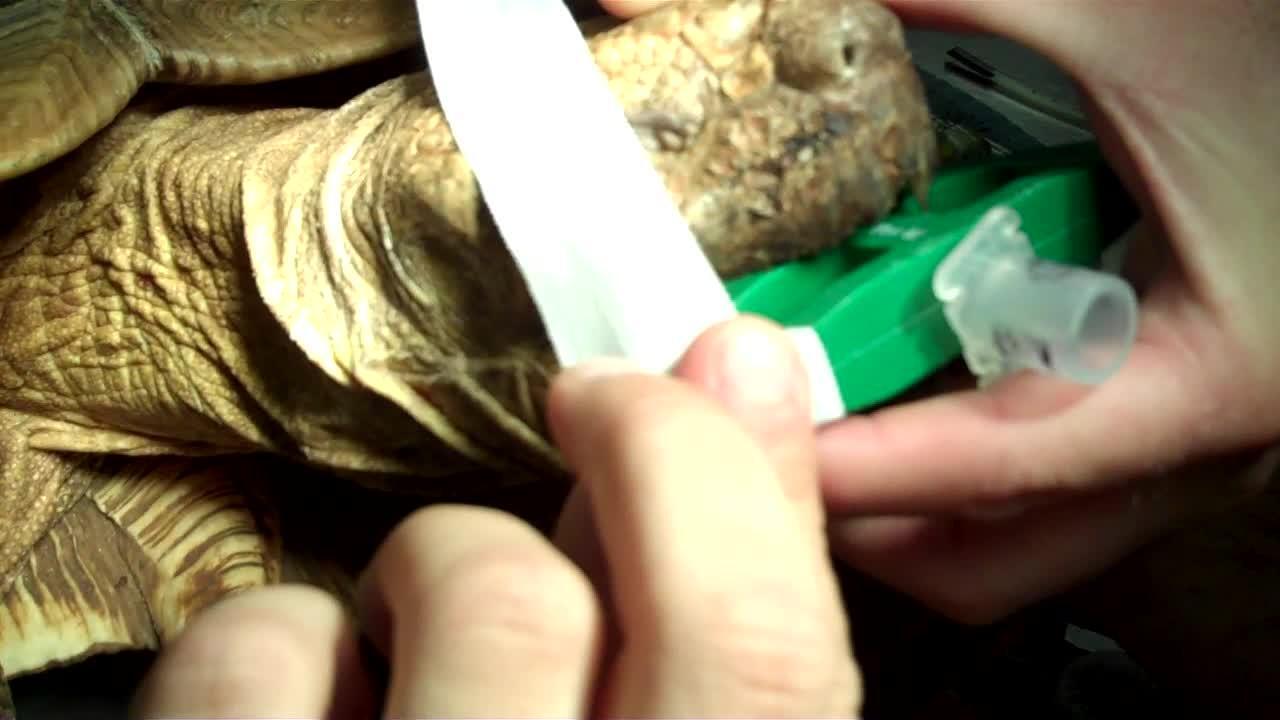
Small to medium-sized tortoises are not difficult to handle, although their strength and uncooperative nature can hinder examination (see image of ). Patiently holding the tortoise with its head down will often persuade a shy individual to protrude the head from the shell. Placing the thumb and middle finger behind the occipital condyles prevents retraction of the head. However, with larger species, it may be physically impossible to prevent a strong individual from pulling free. In such cases, sedation or anesthesia may be necessary for examination.

Small, nonaggressive tortoises can be restrained as shown, using one hand to support the shell vertically and the other around the head. For some species (eg, Testudo spp), it is possible to hold the mouth open using a finger at the oral commissure, but for those with a stronger bite, a rubber or soft plastic mouth gag should be used.
Courtesy of Dr. Stephen Divers.
The more aggressive aquatic species should be held at the rear of the carapace. Some species (eg, snapping turtles) have long necks and an extremely powerful bite, necessitating great care. Certain species also have functional hinges at the front and/or back of the plastron, and caution should be exercised not to trap a finger when the hinge closes.
Examination of the head should include the nostrils for any discharges and the beak for damage and overgrowth. The eyelids should be open and not obviously swollen or inflamed, and the eyes should be clear and bright. Conjunctivitis, corneal ulceration, and opacities are frequent presentations. The tympanic scales should be examined for clinical signs of swelling associated with aural abscessation.
Applying steady pressure to the lateral aspects of the maxilla and mandible, while simultaneously extending the neck, can open the mouth, and a mouth gag can be inserted to prevent closure. Aggressive chelonians, generally aquatic species, often threaten by open-mouth displays, which provide a good opportunity to examine the buccal cavity with minimal handling. Mucus membrane coloration is normally pale; hyperemia may be associated with septicemia or toxemia. Icterus is rare but may occur with biliverdinemia due to severe liver disease. Pale deposits within the oral membranes may represent infection or urate tophi associated with visceral gout. The larynx is positioned at the back of the fleshy tongue and may be difficult to see; however, it is important to check it for any inflammation and glottal discharges consistent with respiratory disease.
The integument should be free of damage. Aquatic species appear more susceptible to superficial and deep mycotic dermatitis, especially around the head, neck, and limbs. The withdrawn limbs can be extended from the shell of small to medium-sized chelonians by applying steady traction. Because the coelomic space within the shell is restricted, gently forcing the hindlimbs into the shell will often lead to partial protrusion of the forelimbs and head, and vice versa. A wedge or mouth gag can be used to prevent complete closure of a hinge. No chelonian will close a hinge on its own extended limb.
The integument should be examined for parasites (particularly ticks and maggots), dysecdysis, trauma, and infection that may arise due to predator attacks. Aggressive conflicts and courting trauma must also be considered in the communal environment. Limb fractures are less common in chelonians than in other reptiles; however, when they do occur they are often associated with rough handling, falls, and secondary nutritional hyperparathyroidism. Focal subcutaneous swellings are usually abscesses, but grossly swollen joints or limbs are more often cases of fracture, osteomyelitis, or arthritis (septic or gout).
The prefemoral fossae (spaces in front of the hindlimbs on either side) should be palpated with the chelonian held vertically, head-up. Gently rocking the animal may enable palpation of eggs, cystic calculi, or other coelomic masses. The shell should be examined for hardness, poor conformation, trauma, or infection. Soft, poorly mineralized shells are expected in neonates but quickly harden unless secondary nutritional hyperparathyroidism results from dietary calcium deficiency, excess phosphorus, or a lack of full-spectrum lighting. Pyramiding of the shell appears to be more associated with inappropriate humidity than dietary imbalances but is probably multifactorial. Shell infection may present as loosening and softening of the scutes with erythema, petechiae, purulent or caseous discharges, and a foul odor.
Prolapses through the vent are obvious, but determining the structure(s) involved is necessary. Prolapses may include cloacal tissue, shell gland, colon, bladder, or phallus. Internal examination (using digital palpation and an endoscope) and diagnostic imaging are recommended.
Anesthesia and Analgesia in Reptiles
For some minor procedures (eg, blood sampling), physical restraint may be all that is required. This can be enhanced by temporary immobilization techniques, such as dorsal recumbency, decreased light intensity, or gentle ocular pressure (vasovagal response). For more invasive and painful procedures, sedation or general anesthesia must be used. Although considerable anatomical, physiological, and pharmacological differences exist between reptiles, some general guidelines are applicable. The following is therefore intended as a practical approach, rather than an exhaustive review of reptile analgesia and anesthesia, which can be found in Divers and Stahl's Mader's Reptile and Amphibian Medicine and Surgery (2019).
Two developments include the use of spinal anesthesia in chelonians and total IV anesthesia in a variety of species. The injection of local anesthetics (eg, morphine, lidocaine) into the intrathecal space results in profound analgesia, with or without loss of motor function. This has proven useful for pelvic limb and cloacal surgery. Total IV anesthesia relies on an IV or intraosseous CRI (usually with alfaxalone or propofol) to maintain anesthesia without the need for inhalants.
Preanesthetic Assessment and Stabilization of Reptiles
All reptiles that are hospitalized should be maintained within their preferred optimal temperature zone (POTZ) at all times to minimize physiological disturbance and to facilitate drug absorption and elimination for recovery. Although hypothermia will decrease movement, it does not provide analgesia and is therefore generally unacceptable as a way to provide anesthesia. Hypothermia can also dramatically affect the pharmacokinetics of any drugs administered and greatly prolong recovery.
A full clinical examination should be performed and the animal accurately weighed, although this may not be practical or possible in some cases. The hydration status of all reptiles should be assessed, especially if they are debilitated or posthibernation. For elective procedures (eg, sterilization), underweight, dehydrated, or debilitated animals should be nursed for days, weeks, or months until their condition improves. For nonelective surgery, dehydration should be corrected before anesthesia. Even the most moribund, egg-bound reptile will usually benefit from stabilization for 24–48 hours before surgery is performed. Reptiles that have not been stabilized before surgery tend to succumb intra- or postoperatively.
Although oral fluids are least invasive to administer and provide the most physiologically normal method of rehydration, they are sufficient only for mildly dehydrated animals and are contraindicated immediately before surgery because of the risks associated with regurgitation and aspiration. Intracoelomic fluids are more suitable; however, uptake can take many hours, and their use is problematic if coeliotomy or coelioscopy is planned. For dehydrated surgical candidates, IV or intraosseous fluid therapy should be administered before, during, and after surgery as needed.
Reptiles should be fasted before all elective surgery to avoid the compression of lung(s) associated with large meals and potential regurgitation. Fasting depends on the feeding regimen of the reptile, but in general, one feeding cycle should be skipped before surgery. Premedication with midazolam in combination with an opiate (eg, hydromorphone) is commonly advocated for most reptile surgeries (see the table ).
Anesthetic Induction of Reptiles
IV or intraosseous propofol or alfaxalone administration provides a rapid, controlled mode of induction. Both drugs are relatively nontoxic, and thrombophlebitis risk is decreased if they are accidentally injected perivascularly. This is of particular concern, because IV access can be relatively difficult in reptiles, especially in healthy active animals undergoing elective procedures.
If IV access is impractical or dangerous to attempt, IM agents can induce sufficient chemical restraint for intubation. For IM injections in lizards and chelonians, the forelimb muscles are preferred, whereas for snakes, the epaxial muscles are used. An IM combination of ketamine, dexmedetomidine, and hydromorphone has proved effective for a variety of chelonians; this can be readily reversed using atipamezole and, if necessary, naloxone or naltrexone. Alfaxalone is also effective when given IM at higher dosages.
Squamates can also be induced by inhalation agents in an induction chamber/bag or by mask. Induction may take 10–30 minutes in cooperative lizards and snakes. Breath holding is common in turtles and crocodilians and makes induction by inhalation impractical. Intubation of conscious patients has been suggested after local lidocaine spray to the glottis; however, the adverse effects of increased stress and catecholamine release involved in awake intubation should always be considered. There is also the potential danger of the handler being bitten.
Maintenance of Anesthesia in Reptiles
Isoflurane and sevoflurane are the agents of choice for maintenance of anesthesia. These volatile inhalation agents have faster modes of action, are more controllable, and facilitate faster recoveries than most alternatives. Furthermore, their lack of reliance on hepatic metabolism or renal excretion decreases the anesthetic risk to debilitated reptiles or those with questionable renal or hepatic function.
Analgesics, Sedatives, and Anesthetics Used in Reptiles
Drug | Dose and Route | Comments |
|---|---|---|
Morphine | 1–5 mg/kg, IM, SC, q 24 h 10 mg/kg, IM, SC, q 24 h | Chelonians (red-eared sliders) Lizards (bearded dragons) Not analgesic for snakes. Causes pronounced respiratory depression in turtles. |
Hydromorphone | 0.5 mg/kg, IM, SC, q 24 h | Chelonians: appears to cause less respiratory depression than morphine |
Tramadol | 5–10 mg/kg, PO, q 2–3 d | Chelonians (red-eared sliders): less respiratory depression than morphine |
Meloxicam | 0.1–0.4 mg/kg, IV, IM, SC, q 24–48 h | Most species |
Ketamine | 10–25 mg/kg, combined with dexmedetomidine (0.05–0.1 mg/kg) and hydromorphone (0.5 mg/kg), IM (or 50% dose, IV) | Deep sedation/anesthesia in many chelonians. Reversed using atipamezole (0.5 mg/kg, IM) and, if necessary, naloxone (0.1 mg/kg, IM) |
Midazolam | 1–2 mg/kg, IM | Premedication |
Tiletamine/zolazepam | 3–12 mg/kg, IM | Tortoises, lizards, snakes. Low dose useful to facilitate intubation. Higher doses associated with prolonged recoveries. |
Propofol | 3–10 mg/kg, IV, intraosseous Starting dose for total IV anesthesia (titrate to effect): 0.1 mg/kg/min | Low dose rate for larger reptiles. Subanesthetic doses produce variable short-term sedation. |
Alfaxalone | 5–10 mg/kg, IV 10–20 mg/kg, IM Starting dose for total IV anesthesia (titrate to effect): 0.1 mg/kg/min | Similar effects to those of propofol IV, but higher doses effective IM. Larger IM dose volumes necessitate dividing into two or more injections. |
Isoflurane | 1–5% for induction (lizards and snakes only) and maintenance (all species) | Routine gaseous agent; subanesthetic levels provide short-term sedation. Mask down or sedated intubation possible in some species. |
Sevoflurane | 2–7% for induction (lizards and snakes only) and maintenance (all species) | Very similar effects to those of isoflurane but recoveries appear to be faster. Preferred inhalant for critical or large reptiles. |
Intubation is relatively simple. Small-gauge endotracheal tubes or catheters are easily inserted into the glottis immediately caudal to the tongue; this may be aided by forcing the tongue up and forward by pressing a finger into the intermandibular space from under the jaw. The reptilian glottis is actively dilated, and therefore its movement will often be abolished in anesthetized animals; a guiding stylet within the endotracheal tube can be useful to facilitate tube placement and is recommended (see and ). The bifurcation of the trachea may be far craniad in some chelonians, and gaseous exchange has also been reported within the tracheal lung of some snakes. In these cases, care should be taken to use a short endotracheal tube that is securely taped in position.

Endotracheal intubation using an uncuffed endotracheal tube and stylet in an iguana. The glottis is only open during active respiration, so a stylet facilitates intubation in an apneic reptile.
Courtesy of Dr. Stephen Divers.

Noncrocodilian reptiles lack a diaphragm; skeletal intercostal muscles (in Squamata) or limb movements (in Testudines) control ventilation. The action of these muscles is abolished at a surgical plane of anesthesia, and intermittent positive-pressure ventilation is required. Ventilation rates should initially mirror preanesthesia evaluations and then be adjusted to maintain end-tidal capnography readings of 15–25 mm Hg (see ). Electrical ventilators enable precise control of ventilation rates and pressures.

Anesthetized iguana being monitored using pediatric mainstream capnography. Note the endotracheal tube adaptor (arrow) and the current end-tidal carbon dioxide reading of 14 mm Hg at 3 breaths/minute. This capnography unit also includes pulse oximetry and is recording a heart rate of 78 beats/minute with a blood oxygen saturation (spO2) value of 100%. Although spO2 values are not necessarily accurate in reptiles, appreciating trends is more meaningful.
Courtesy of Dr. Stephen Divers.
Monitoring anesthesia in reptiles differs somewhat from doing so in mammals. Palpebral and corneal reflexes are reliable in species in which they can be elicited (ie, chelonians, crocodilians, most lizards, but not snakes). However, corneal reflexes are abolished at excessively deep anesthetic levels, and pupillary diameter may bear little relation to anesthetic depth (unless fixed and dilated, which indicates excessive anesthetic depth or brain anoxia and death). Jaw tone and withdrawal reflexes (tongue, limb, or tail) are abolished only at a surgical plane of anesthesia. Full loss of the righting reflex, loss of spontaneous movement, and complete muscle relaxation also occur at this plane of anesthesia.
Heart rate can be monitored by auscultation or by visualization or palpation of the heartbeat in most snakes and in some lizards. Pulse oximetry, using either an esophageal or cloacal reflectance probe, is useful to monitor pulse rate and strength. Although the blood oxygen saturation (SpO2) readings are often lower and have not been validated for reptiles, monitoring the trend in SpO2 is often helpful. Doppler ultrasonography can also be used over peripheral arteries or the heart. Blood gas estimations are often affected by intracardiac or pulmonary shunts, especially in aquatic species. However, end-tidal capnography has proved reliable for monitoring anesthesia in reptiles.
Toward the end of surgery, anesthetic gas should be discontinued while maintaining ventilation for another 5–10 minutes to facilitate gas excretion. At this point, oxygen can be discontinued in favor of room air delivered by bag valve mask to encourage spontaneous respiration.
Pearls & Pitfalls
|
Postoperative Support for Reptiles
Once breathing spontaneously, the reptile can be returned to an incubator or vivarium to fully recover. Continued monitoring is essential until righting reflexes return and the animal is ambulatory. Additional analgesia, fluid, and nutritional support should be provided as indicated.
Diagnostic Techniques in Reptiles
Radiology of Reptiles
Several anatomical differences make it difficult to obtain quality radiographs in reptiles. The relatively small size of most pet reptiles and the lack of diffuse body fat often result in images of poor contrast. Thick, highly keratinized scales, osteoderms, or shells can severely hinder the x-ray beam, necessitating greater power and a subsequent loss of fine soft tissue detail.
Despite these difficulties, most high-capacity digital units can be set to produce quality radiographs of reptiles. High-detailed screen/film combinations (eg, mammography film) or dental radiography units often provide better resolution, which may be particularly helpful when dealing with small specimens or areas of interest. Various agents can be used to improve contrast. Barium sulfate (30%) can be used for GI studies. Water-soluble iodine compounds such as iohexol can be used for GI, urogenital, and IV techniques. Injecting air into the coelom of a lizard can improve the appreciation of preovulatory follicles.
Snakes
Snakes can be difficult to position and restrain for radiographic examinations unless anesthetized. If the examination's purpose is simply to exclude radiodense foreign bodies or eggs, the snake may be allowed to coil in its natural position. If detailed examination of the skeletal, respiratory, and digestive systems is desired, the snake must be extended, and anesthesia is generally required (see ).

Radiographs of a female corn snake. Plain right lateral view (top) and dorsoventral view after a barium enema (bottom). Note the radiographic appearance of pronounced constipation cranial to an extraluminal soft tissue mass that on biopsy proved to be a granulosa cell tumor.
Courtesy of Dr. Stephen Divers.
A plastic restraint tube can be used for physical restraint; however, this may produce a radiographic artifact. In larger snakes, multiple films will be needed to radiograph the entire length of the body. Lateral views are best taken using horizontal beams to avoid displacement artifact of the viscera. However, standard laterals with the snake taped in lateral recumbency can be useful, especially when horizontal beams are not possible or safe to undertake. The interpretation of dorsoventral views is hindered by the spine and ribs but can still be useful when dealing with obvious lesions, including eggs and mineralized masses.
Lizards
Small lizards can often be restrained by taping them to the radiography cassette or table for a dorsoventral view (see ). Wrapping cotton balls with self-adhesive tape and placing them over the eyes will often induce a vasovagal response and produce a calm, motionless lizard. A dorsoventral view can help identify foreign bodies, intestinal impaction, eggs, or other coelomic masses. A horizontal x-ray beam provides the best lateral imaging in lizards, especially when evaluating the respiratory system. Positioning for this view involves rotating the x-ray tube 90° and placing the cassette vertically behind the lizard. Elevating the body of the lizard on rolled towels or foam pads, or taping the forelimbs craniad and the pelvic limbs caudad, helps prevent superimposition of the limbs with the coelomic cavity.
The positioning for and interpretation of crocodilian radiographs are similar to those used for lizards.


Dorsoventral radiographs of two water dragons. The left radiograph illustrates normal skeletal mineralization, whereas the right one shows generalized demineralization, cortical thinning, and pathologic fractures (arrows) consistent with secondary nutritional hyperparathyroidism.
Courtesy of Dr. Stephen Divers.
Chelonians
For vertical beam dorsoventral radiographs, most conscious animals will remain motionless long enough to permit exposure. Ideally, the head and limbs should be extended from the shell to decrease superimposition of the limb musculature on the coelomic viscera. More active animals can be restrained by taping them to the cassette or by placing them in a radiolucent container, although this should be avoided with smaller specimens (and lower exposures) because material artifacts may appear. (See .)
For lateral horizontal beam radiographs, the chelonian is best placed on a central plastron stand (eg, a large plastic pill pot) to encourage extension of the limbs and head while the tortoise remains immobile. Both left and right lateral projections should be taken with the lateral edge of the shell touching (or as close as possible to) the cassette.
The third basic coelomic view is the horizontal craniocaudal (or anterior-posterior) view. The chelonian is positioned on a central plastron stand, with the caudal edge of the carapace touching (or as close as possible to) the cassette; the head should be facing the x-ray tube and the beam centered on the midline of the cranial rim of the carapace.
Additional views, including oblique views of the pectoral and pelvic girdles, can be helpful to highlight luxations and fractures.


Dorsoventral radiograph of a Greek tortoise (Testudo graeca) demonstrating several abnormal eggs, including one within the bladder and encased by uric acid, forming a large urolith.
Courtesy of Dr. Stephen Divers.
Radiology of a chelonian's head and limbs requires extension and often general anesthesia. Sandbags, foam, and tape aid positioning. Standard interpretation requires that both true lateral and dorsoventral views should be taken—even slight rotation makes interpretation more difficult.
Ultrasonography of Reptiles
A useful and often underrated technique, ultrasonography has gained popularity as a diagnostic technique for reptiles. It is particularly useful for examining tissue parenchyma, guiding tissue aspiration, and, with color flow Doppler, investigating cardiac disease. Given the variability in reptile size, a variety of probes are required. Ultrasound waves cannot penetrate through mineralized tissue or air, and therefore ultrasonography has obvious limitations to investigate respiratory and GI diseases.
A 5-MHz probe is required to image giant reptile species, whereas a 7.5- or 12-MHz probe will suffice for most other reptiles. For examination of very small specimens (or for ultrasonography of eyes), a 20-MHz probe is more appropriate. Good contact and imaging generally require copious quantities of gel or a water bath. It is helpful to maintain the animal in a sternal position or, failing that, at least to appreciate the complications associated with organ displacement during dorsal or lateral recumbency.
Ultrasonography can be a useful adjunct to radiography, especially to assess the reproductive tract (evaluating ovarian activity and distinguishing between pre- and postovulatory egg stasis), liver and gallbladder, urinary system, soft tissue masses, and heart. Ultrasonography has been used to guide liver biopsy, although due to serious iatrogenic trauma reported in snakes and lizards, the technique cannot be recommended.
CT and MRI of Reptiles
CT offers excellent, high-resolution, detailed images with greater soft tissue differentiation than plain radiography. Consequently, CT has become the imaging modality of choice for many diseases of reptiles. Pre- and postcontrast images using soft tissue and bone algorithms are recommended. Modern machines are capable of fast scanning times such that physical restraint alone or sedation (rather than general anesthesia) is sufficient.
MRI, even with high-strength (3–7 Tesla) magnets, typically requires much longer scanning times, and hence, general anesthesia. Spatial resolution is often lower than for CT; however, soft tissue differentiation is greater, with MRI particularly preferred for CNS evaluations.
Blood Collection in Reptiles
Venipuncture is generally a blind technique in reptiles. Up to 0.5 mL/100 g can be safely collected from healthy reptiles and less in debilitated animals. A relative lack of hematologic or biochemical data exists for most reptiles. In addition, blood values can vary dramatically with species, environment, nutrition, age, sex/reproductive status, and hibernation. Given this variability and that most reference ranges fail to comply with the requirements of the American Society for Veterinary Clinical Pathology, published values may be of limited value. More reliance should be placed on establishing an individual’s observed range and using serial sampling to monitor the progress of hematologic and biochemical changes.
The use of a toenail clip to obtain blood may result in fecal or urate contamination, increased tissue enzymes, and hemogram and electrolyte changes due to the peripheral nature of the sample and the crushing artifact of collection. Of even greater concern are the ethical and welfare issues associated with toenail clipping, which cannot be condoned.
The two common sites for venipuncture in snakes are the caudal (ventral tail) vein and, less preferred, the heart. The caudal vein is accessed caudal to the cloaca, between 25% and 50% down the tail, and avoiding the paired hemipenes of males (see ). The heart should only be considered when access is unsuccessful via other methods because complications, including fatal hemopericardium, have been reported. If cardiocentesis is performed, anesthesia should be considered, because the procedure is considered unethical in conscious mammals and birds.
In lizards, the most clinically useful vessel is also the caudal (ventral tail) vein, best accessed 20–80% down the tail. The most clinically useful vessels in Testudines appear to be the jugulars, because the subcarapacial sinus and brachial, femoral, and dorsal coccygeal veins are more prone to lymphatic or cerebrospinal fluid contamination (see images of and ).

Blood collection from the caudal (ventral tail) vein of a python. Note that the needle is angled at 45° for insertion.
Courtesy of Dr. Stephen Divers.

Blood collection from the caudal (ventral tail) vein of an iguana.
Courtesy of Dr. Stephen Divers.

Performing right jugular venipuncture in a gopher tortoise (Gopherus polyphemus). The jugular vein is superficial, located caudad to the tympanic scale, and is positioned laterally to dorsolaterally at the 2- to 3-o'clock and 9- to 10-o'clock positions in the neck.
Courtesy of Dr. Jessica Comolli.
Necropsy of Reptiles
A detailed necropsy should be undertaken whenever possible because it often provides a definitive diagnosis. When managing a disease outbreak in a population, a clinician can recommend elective euthanasia and necropsy of one or more affected individuals, because these procedures are often the most efficient and cost-effective means of diagnosis. Fresh necropsies can provide organ biopsies, blood, and other bodily fluids for laboratory examination. Microbiological, especially bacteriological, samples from reptiles that have died and remained within a heated enclosure must, however, be interpreted with caution.
Surgery and Endoscopy in Reptiles
Surgery in Reptiles
In general, surgery on a reptile patient should adhere to the same principles as domestic animal surgery. However, reptiles have some specific anatomical considerations, as well as unique aspects of patient preparation, positioning, and equipment. Only a basic discussion follows here, and consulting other sources on reptile anatomy, physiology, husbandry, anesthesia, and surgery, as well as domestic animal surgery literature, is essential before performing surgery on any reptile.
For truly giant species, such as giant tortoises, stronger instruments intended for large animals are recommended. For reptiles weighing 5–50 kg, most instruments used for small animals are appropriate. However, most pet reptiles weigh < 1 kg, and microsurgical instruments are often required. These instruments are not miniaturized versions of standard instruments but rather balanced instruments with fine, small tips. Because microinstruments can be costly, ophthalmological instruments can be useful alternatives. Plastic, self-retaining retractors can be adjusted to fit different sizes of incisions and do not compromise ventilation. Smaller versions of standard abdominal retractors can also be used but are heavier. Eyelid retractors can be useful to retract coelomic incisions in small lizards and snakes.
Rapidly absorbable suture materials (eg, poliglecaprone 25) are recommended for internal soft tissue applications. For prolonged internal durability, polydioxanone or nylon is preferred. Poliglecaprone, monofilament nylon, and polydioxanone are favored for skin suturing, although wire may be necessary for giant tortoises, crocodilians, and lizards. Epoxy resins or low-temperature veterinary acrylics are used for many chelonian plastron closures and shell repairs.
Because most reptile pets are < 1 kg, some degree of magnification is advantageous. Headband or frame-mounted operating loupes (2–4X magnification) with a dedicated halogen or xenon light source are affordable, versatile, comfortable, and simple to use. During operations, microscopy telescopes can provide magnification with selected rigid endoscopy systems.
A healthy reptile can generally tolerate 0.4–0.8 mL/100 g body weight of blood loss. Reptiles in need of surgery are often compromised, and diagnostic blood samples may have been collected before surgery. Therefore, the amount of blood a reptile can afford to lose during surgery may be considerably less. Careful consideration must be given to minimizing hemorrhage using cotton-tipped spears or applicators, vascular clips, and radiosurgery or a laser.
Patient positioning will depend on the species and the nature of the surgery. Consideration should be given to the following:
ensuring that head and neck position does not interfere with ventilation
avoiding excessive compression of the head, limbs, or coelom to prevent pressure necrosis, visceral rupture, or hypoventilation of the lungs
avoiding extreme and prolonged hyperextension or hyperflexion of any joint
ensuring that the surgical site is easily accessible and does not require surgeon positioning that will result in fatigue
Sandbags, beanbags, foam supports, and adhesive tape can be used to maintain patient position. Having the surgeon operate in a seated position with arms resting on a padded surface facilitates greater control with less fatigue.
When reptile skin is incised, it tends to invert. Therefore, everting suture patterns (eg, horizontal mattress) are recommended to ensure opposition of skin without future dysecdysis. The skin suture materials should be monofilament. Wire suture may be required for repairs involving shell or thickly keratinized skin containing osteoderms. Staples have also been advocated because they cause mild eversion. Given the length of time it takes for reptile wounds to heal, sutures should not be removed until 6–8 weeks after surgery.
Pearls & Pitfalls
|
The primary factors to consider postoperatively are analgesia and continued vigilance concerning hydration, temperature, nutrition, and hygiene. Reptiles have been demonstrated to experience opiate-modulated pain responses. Pain slows the healing process and depresses the normal function of the immune system in mammals, and there is no evidence to suggest this process would not be similar in reptiles. Clinically, reptiles that receive analgesics appear to recover better than those that do not. The continuation of preemptive analgesia using opioids and/or NSAIDs should be a routine part of postoperative care.
Few drugs are approved by the FDA for use in reptiles. Medications can be administered by a variety of routes, including PO, SC, IM, IV, intracardiac, intracoelomic, intraosseous, intrasynovial, and intratracheal injection. Certain drugs can be applied topically or given per cloaca, by inhalation (nebulization), or by direct intralesional administration.
Because reptiles are ectotherms, temperatures outside the POTZ can have profound influences on drug distribution, metabolism, excretion, and elimination half-life. Some therapeutic regimens state a fixed temperature at which the reptile should be held during treatment. If there are pharmacokinetic data on the drug, then the elimination of the drug will be known and constant. However, if this stated temperature is below or above the POTZ for the species being treated, stress and debilitation may ensue. Even when the stated therapeutic temperature is within the POTZ for the species being treated, constant exposure to a fixed temperature is likely to be stressful.
Reptiles have a well-developed renal portal system; blood from the caudal half of the body passes through the kidneys before reaching the systemic venous circulation. Drugs injected into the caudal half of the body may have a shorter half-life if excreted via tubular secretion. However, studies have demonstrated that these effects are unlikely to be clinically significant (1). Of potential concern is the caudal injection of nephrotoxic drugs that may reach renal tissue in high concentration. A first-pass hepatic effect may also affect drugs injected into the caudal body. Consequently, all drugs should be injected into the cranial body. The shell of tortoises, turtles, and terrapins is living tissue; therefore, all chelonian doses should be based on total body weight.
Endoscopy in Reptiles
Endoscopy has proved to be widely useful in reptile medicine, and given the small and delicate nature of many species, continued development of minimally invasive techniques is likely. Flexible endoscopes are useful for respiratory endoscopy in snakes and for GI endoscopy in many species (see images of and in various reptiles). Their main disadvantage is that the flexible, fiber-optic endoscopes obtain poorer-quality images than rigid scopes of a similar diameter or videoscopes. However, the continued development of smaller videoscopes looks likely to redefine flexible endoscopy in reptiles.

Stomatoscopy, esophagoscopy, and gastroscopy. A) Oral cavity of a radiated tortoise (Astrochelys radiata) demonstrating the choana and hard palate (p), fleshy tongue (t), and glottis (arrow). B) Air-distended esophagus of a Burmese python (Python bivittatus). C) Saline-infusion view of the green iguana (Iguana iguana) esophagus. D) Stomach of a savannah monitor (Varanus exanthematicus) with normal rugal folds (arrows). E) Gastric hypertrophy in a corn snake (Pantherophis guttatus) with cryptosporidiosis. F) Leiomyoma (l) obstructing the pyloric outflow in another corn snake that presented with chronic regurgitation.
Courtesy of Dr. Stephen Divers.

Tracheobronchoscopy. A) Greek tortoise (Testudo graeca) trachea with complete tracheal rings. B) Ball python (Python regius) trachea with obvious dorsal ligament (arrow). C) Tracheal bifurcation and primary bronchi in a green iguana (Iguana iguana). D) Simple saclike lung in a green iguana. E) Compartmentalized lung in a panther chameleon (Furcifer pardalis). F) Granulomatous pneumonia due to Mycobacterium spp in a ball python.
Courtesy of Dr. Stephen Divers.
The compact body size of most pet reptiles, coupled with their coelomic body design, makes rigid endoscopy useful in many clinical situations. Although equipment must be matched to the reptile's size, in general the 1.9-mm and 2.7-mm telescope and sheath systems work well for most pet reptile species, enabling gas insufflation, fluid irrigation, and biopsy capability (see images of endoscopic pathology and and biopsies).

Endoscopic pathology and kidney biopsy. A) Enlarged kidney (k) with associated renal cyst (c) in a green iguana (Iguana iguana) that presented with anorexia and cachexia. Radiography and ultrasonography confirmed renomegaly, and plasma biochemistry revealed reversed calcium:phosphorus ratio and increased uric acid. Endoscopic biopsy confirmed the diagnosis of glomerulonephrosis with renal gout and mineralization (left lateral coelioscopy). B) Abnormal kidney (k) with fibrous bands (arrows) in a female Greek tortoise (Testudo graeca) that presented with anorexia, lethargy, and polyuria. Diagnostic imaging and clinical findings were unremarkable; however, renal biopsy confirmed the diagnosis as severe tubulonephrosis (left prefemoral approach). C) Biopsy of an iguanid kidney (k) using 1.7-mm biopsy forceps (b) (right lateral coelioscopy). D) Incising the caudodorsal coelomic membrane (m) of a yellow-bellied slider (Trachemys scripta scripta) using 1.7-mm endoscopy scissors (s) to gain access to the retrocoelomic kidney (left prefemoral approach). E) View of the incised coelomic membrane (m) revealing the kidney (k) (left prefemoral approach). F) Biopsy forceps (b) being passed through the incision to collect a biopsy from the kidney (k) (left prefemoral approach).
Courtesy of Dr. Stephen Divers.

Endoscopic pathology and liver biopsy. A) Grossly distended small intestine in a Russian tortoise (Agrionemys horsfieldii). This herbivorous tortoise was fed dairy products, which resulted in fermentative enteritis (left prefemoral approach). B) Retained, inspissated yolk sac (y) in a juvenile radiated tortoise (Astrochelys radiata) that presented for intermittent anorexia and poor growth. Clinical findings and diagnostic imaging were unremarkable; however, endoscope-assisted removal proved curative (left prefemoral approach). C) Chronic hepatic fibrosis in a green iguana (Iguana iguana) that presented for anorexia and occasional regurgitation. Diagnostic imaging was unremarkable, and the only clinical abnormality was a mild increase in postprandial bile acids. Endoscopic biopsy confirmed the diagnosis of severe hepatic fibrosis with cholestasis (left lateral coelioscopy). D) Pale liver (l) in an Aldabra tortoise (Aldabrachelys gigantea) that presented with intermittent anorexia. All liver parameters were unremarkable, but severe hepatic lipidosis was diagnosed after liver biopsy (left prefemoral approach). E) Diffuse hepatomegaly (l) in a green iguana that presented with lethargy and anorexia. Diagnostic imaging and liver biochemistry were unremarkable; however, moderate leukocytosis, predominantly heterophilia and azurophilia, was evident. Endoscopic liver biopsy confirmed the diagnosis of bacterial hepatitis due to Klebsiella, and appropriate treatment proved curative (left lateral coelioscopy). F) Biopsy from the edge of the chelonian liver (l) using 1.7-mm biopsy forceps (b) (left prefemoral approach).
Courtesy of Dr. Stephen Divers.
Insufflation is essential to provide the lens-organ distance required for visualization. For GI endoscopy, air and/or saline is used; for coelioscopy, medical-grade CO2 or saline is preferred. Coelomic pressures seldom need to be > 5 mmHg. When performing endoscopy in small neonates or within a hollow viscus (eg, bladder, oviduct, cloaca, stomach), a clinician can use warmed sterile saline irrigation to provide better clarity than with gas insufflation.
General anesthesia is recommended for all endoscopy procedures. Certain examinations (eg, buccal cavity and cloaca) may be possible in a conscious or sedated animal using a mouth gag or other appropriate restraint; however, complete immobilization is preferred to avoid risking damage to patient, staff, or equipment. Anesthesia is mandatory for coelioscopy.
Medical Procedures in Reptiles
Drug Dosage and Allometric Scaling in Reptiles
Numerous pharmacokinetic studies have been published for reptiles, and these should be considered the most reliable source for information on drug dosages. When species-specific information is not available, it is possible to extrapolate from closely related species. If there are no pharmacokinetic data or reliable clinical experience for a particular species, extrapolating pharmacokinetic data from other animals may be necessary.
Allometric scaling calculates the drug dose and dosing frequency using metabolic rate rather than body weight. The basic allometric equations are shown below, in which W = body weight (kg) and K = energy constant, which is 10 for most reptiles. These equations can be used to calculate a dose and dose frequency for a species for which no data are available, by using pharmacokinetic data from a known species (control), whether another reptile or mammal or bird:
Minimum energy cost = K (W0.75)
Specific minimum energy cost = K (W-0.25)
Antimicrobials in Reptiles
Many bacterial infections in reptiles are caused by gram-negative bacteria, particularly Pseudomonas, Aeromonas, Citrobacter, Klebsiella, and Proteus spp. Bacterial resistance to many commonly used antimicrobials (including enrofloxacin) exists, and many gram-negative bacteria can have unexpected susceptibility to particular antimicrobials; therefore, sampling for Gram staining, cytological testing, and culture and susceptibility testing should be done before starting therapy.
Antimicrobial therapy must usually be given while awaiting the results of bacterial susceptibility tests. In these circumstances, and in keeping with antimicrobial stewardship recommendations, potentiated sulfonamides, tetracyclines, and aminoglycosides are considered first-line drugs (see the table ).
Second-line drugs (such as third-generation cephalosporins and fluoroquinolones) should be reserved for cases of demonstrable resistance to first-line drugs. Penicillin, metronidazole, lincomycin, or clindamycin can be used for anaerobic infections. The routine use of advanced broad-spectrum antimicrobials implies a low level of skill on the part of the clinician.
Antimicrobial Drugs Used in Reptiles
Drug | Dosage | Comments |
|---|---|---|
Acyclovir | 80 mg/kg, PO, q 8 h or 240 mg/kg, q 24 h; topical cream q 12 h | Antiviral |
Amikacin | Corn snake: loading dose 1.7 mg/kg, IM, followed by 26 mcg/kg/h via osmotic infusion-pump implant Gopher snake: initial dose 5 mg/kg, IM, then 2.5 mg/kg, IM, q 3 d Gopher tortoise: 5 mg/kg, IM, on alternate days American alligator (juvenile): 2.25 mg/kg, IM, q 3–4 d Ball python: 3.5 mg/kg, IM, q 4–5 d 50 mg/10 mL saline × 30 min nebulization, q 12 h | Maintain hydration |
Amphotericin B | 0.5–1 mg/kg, intracoelomic, IV, q 1–3 d for 14–28 d | Aspergillosis; fluid therapy recommended |
Tortoise: 0.1 mg/kg, q 24 h, intrapulmonary, for 28 d 5 mg/150 mL saline for 1 h of nebulization, q 12 h for 7 d | Pulmonary candidiasis | |
Ampicillin | Most species: 10–20 mg/kg, SC, IM, q 12 h Tortoises: 50 mg/kg, IM, q 12 h | |
Azithromycin | Ball python: 10 mg/kg, PO, q 3–7 d (q 3 d for skin infections, 5 d for respiratory tract infections, 7 d for liver and kidney infections) | |
Carbenicillin | 200–400 mg/kg, IM, q 24 h | |
Ceftazidime | 20–40 mg/kg, SC, IM, IV, q 2–3 d | |
Ceftiofur | Tortoises: 2.2–4 mg/kg, IM, q 24 h Snakes: 2.2 mg/kg, IM, on alternate days Lizards: 5 mg/kg, IM, SC, q 24 h | |
Cefuroxime | 100 mg/kg, IM, q 24 h for 10 d with POTZ at 30°C (86°F) if not known for species | |
Chloramphenicol | Indigo, rat, kingsnakes: 50 mg/kg, SC, q 12 h | |
Boids, moccasins: 50 mg/kg, SC, q 24 h Rattlesnakes: 50 mg/kg, SC, q 2 d Red-bellied water snakes: 50 mg/kg, SC, q 3 d | ||
Most species: 20–40 mg/kg, IM, q 24 h | ||
Ciprofloxacin | Most species: 10 mg/kg, PO, on alternate days Pythons: 11 mg/kg, PO, q 2–3 d | |
Clarithromycin | Desert tortoise: 15 mg/kg, PO, q 2–3 d | Mycoplasma |
Clindamycin | 5 mg/kg, PO, q 12 h | |
Clotrimazole | Topical | Fungal dermatitis |
Doxycycline | Most species: 5–10 mg/kg, PO, q 24 h Hermann's tortoises: 50 mg/kg, IM, once; then 25 mg/kg, IM, q 3 d | |
Enrofloxacin | Most species: 5–10 mg/kg, IM, PO, q 24 h | IM injection causes necrosis, and after a single injection, oral therapy is required. |
Nasal flush 50 mg/250 mL sterile water; 1–3 mL/nares, q 24–48 h | ||
Burmese python (juvenile): 10 mg/kg, IM, initial dosage, then 5 mg/kg, IM, q 48 h. Pseudomonas: 10 mg/kg, IM, q 48 h | IM injection likely causes necrosis, and consideration should be given to single injection followed by oral administration | |
Hermann’s tortoise: 10 mg/kg, IM, q 24 h | IM injection likely causes necrosis, and consideration should be given to single injection followed by oral administration | |
Monitor lizards: 10 mg/kg, IM, q 5 d | ||
Indian star tortoise: 5 mg/kg, IM, q 12–24 h | IM injection likely causes necrosis, and consideration should be given to single injection followed by oral administration | |
Crocodilians: 5 mg/kg, IV, q 2–3 d | ||
Fluconazole | Lizards: 5 mg/kg, PO, q 24 h Sea turtles: 21 mg/kg, SC, once; then 10 mg/kg, SC, 5 d later | |
Sea turtles: 21 mg/kg, SC, once; then 10 mg/kg, SC, 5 d later | ||
Gentamicin | American alligator: 1.75 mg/kg, IM, q 3–4 d with POTZ at 22°C (72°F) | Maintain hydration, nephrotoxicity reported |
Painted turtle: 10 mg/kg, IM, on alternate days with POTZ at 26°C (79°F) | Maintain hydration, nephrotoxicity reported | |
Red-eared terrapins: 6 mg/kg, IM, q 25 d | Maintain hydration, nephrotoxicity reported | |
Gopher snakes: 2.5 mg/kg, IM, q 3 d with POTZ at 24°C (75°F) | Maintain hydration, nephrotoxicity reported | |
Itraconazole | Chameleons: 5–10 mg/kg, PO, q 1–2 d | Fungal dermatitis |
Spiny lizards: 23.5 mg/kg, PO, q 24 h for 3 d, with persistent drug concentration for 6 d | ||
Snakes: 10 mg/kg, PO, q 24 h | ||
Sea turtles: 5 mg/kg, PO, q 24 h or 15 mg/kg, PO, q 72 h | ||
Ketoconazole | Crocodilians: 50 mg/kg, PO, q 24 h | |
Most species: 15–30 mg/kg, PO, q 24 h for 14–28 d | ||
Lincomycin | 10 mg/kg, PO, q 24 h; 5 mg/kg, IM, q 12–24 h | |
Marbofloxacin | Ball python: 10 mg/kg, PO, q 48 h | |
Metronidazole | 20–50 mg/kg, PO, q 1–2 d | Bacterial infections. Maximal dose for tricolor snake, kingsnake, indigo snake, or Uracoan rattlesnake is 40 mg/kg |
Neomycin | 10 mg/kg, PO, q 24 h | Oral only, not to be given systemically |
Nystatin | Turtles: 100,000 U/kg, PO, q 24 h for 10 d | Enteric fungal conditions |
Oxytetracycline | Most species: 5–10 mg/kg, IM, PO, q 24 h | Pain, irritation, and inflammation at injection site |
American alligator: 10 mg/kg, IV, IM, q 4–5 d | Mycoplasmosis | |
Piperacillin | 50–100 mg/kg, IM, q 12 d | Fluid therapy recommended |
Polymixin B | Topical | Abrasions, wounds |
Sulfamethoxydiazine | 80 mg/kg, SC, IM, once; then 40 mg/kg, q 24 h for 5–7 d | Coccidial infections |
Terbinafine | Central bearded dragons: 20 mg/kg, PO, q 24–48 h | |
Tobramycin | Chelonians: 10 mg/kg, IM, q 24–48 h Most species: 2.5 mg/kg, IM, q 24–72 h | Potentially nephrotoxic, fluid therapy recommended |
Trimethoprim-sulfa | 30 mg/kg, PO, IM, IV, q 24 h | |
Tylosin | 5 mg/kg, IM, q 24 h | Mycoplasmosis |
Voriconazole | 10 mg/kg, PO |
Fungal and yeast infections commonly occur in reptiles. GI and skin mycoses are particularly common in reptiles maintained on inappropriately long-term broad-spectrum antimicrobials. Cutaneous mycoses can often be treated by debridement and topical application of antifungals. GI fungal infections can be treated with nystatin, whereas systemic fungal infections may require itraconazole, fluconazole, or voriconazole. In cases of pulmonary mycoses, antifungal medication may be given by nebulization or by intratracheal or intrapulmonary injection.
Herpesviruses can cause severe morbidity and death in chelonians. Acyclovir has been used with some success during the early stages of herpes infections.
Parasiticides in Reptiles
Parasiticides used commonly in reptiles are listed in the table . Parasiticide overdosage may lead to drug toxicity, which may be observed as neurological signs, including seizures. Ivermectin is contraindicated in chelonians, and adverse reactions have been reported in some iguanid lizards, skinks, and indigo snakes. Milbemycin has been successfully used in box turtles and terrapins; however, ivermectins and milbemycins should be avoided in all chelonians because safer alternatives are available. Permethrin is licensed for use in reptiles and appears safe and effective against mites and ticks.
Pearls & Pitfalls
|
Parasiticides Used in Reptiles
Drug | Dosage | Parasite | Comments |
|---|---|---|---|
Endoparasiticides | |||
Fenbendazole | 25–100 mg/kg, PO, q 14 d for up to 4 treatments 50 mg/kg, PO, q 24 h for 3–5 d | Roundworms, Hexamita | Can cause leukopenia |
Ivermectin | 200 mcg/kg, PO, IM, SC, repeat after 14 d | Ectoparasitic mites and ticks; nematodes | Not in chelonians; care in skinks and indigo snakes |
Levamisole | 5–10 mg/kg, SC, intracoelomic, repeat after 14 d | Lungworms and other nematodes | Snakes, lizards, care in tortoises (use 5 mg/kg) |
Mebendazole | 20–25 mg/kg, PO, repeat after 14 d | Strongyles and ascarids | |
Metronidazole | 20–40 mg/kg, PO, q 1–2 d for 2–5 treatments | Protozoa | |
Oxfendazole | 25 mg/kg, PO, once | Nematodes | |
Paromomycin | Most species: 35–100 mg/kg, PO, q 24 h for 28 d Snakes: 100 mg/kg, PO, q 24 h for 7 d, then twice weekly for 3 mo Gila monsters: 300–360 mg/kg, PO, q 2 d for 14 d Bearded dragons: 100 mg/kg q 24 h for 7 d; then twice/weekly for 6 wk | Amoebas, cryptosporidia | Does not eliminate cryptosporidia |
Ponazuril | Bearded dragons: 30 mg/kg, PO, q 2 d for 2 treatments | Coccidiosis | |
Praziquantel | 8 mg/kg, PO, SC, IM; repeat after 14 d and 28 d | Tapeworms, flukes | |
Pyrantel | Most species: 5 mg/kg, PO, repeat in 14 d Chameleons: 5–10 mg/kg, PO, q 14 d Sea turtles: 25–50 mg/kg, PO, q 3 d for 3 treatments | Nematodes Spirorchidiasis | |
Spiramycin | 160 mg/kg, PO, q 24 h for 10 d, then twice weekly for 3 mo | Snakes with cryptosporidiosis | May decrease clinical signs but does not clear infection |
Toltrazuril | 5–15 mg/kg, PO, q 24 h for 3–30 d 15 mg/kg, q 48 h for 10 d; discontinue for 2 wk; repeat q 48 h for 10 d, and repeat as necessary | Bearded dragons, coccidiosis Tortoises, intranuclear coccidiosis | Safety, efficacy, and pharmacokinetic data lacking |
Trimethoprim-sulfa | 30 mg/kg, PO, q 24 h for 10–28 d | Coccidia | |
Ectoparasiticides | |||
Dichlorvos-impregnated strip | 6-mm (0.25-inch) strip/0.3 m3 (10 ft3) in cage for 3 h, q 2 d for 2–4 wk | Ectoparasites, especially mites | Toxic, vivarium should be emptied; keep out of direct contact of animals |
Fipronil | Topical spray or wipe on, then wash off in 5 min, q 7–10 d | Mites and ticks, beware of reactions to alcohol carrier, needs safety evaluation | |
Ivermectin (10 mg/mL) | Topical spray, 5–10 mg/L water, q 3–5 d for up to 28 d | Mites and ticks | Should not be used in chelonians; use care in skinks and indigo snakes |
Permethrin (10%) | Topical spray for animal and environment | Mites and ticks, FDA-approved | Licensed product available for reptiles in the US |
Other Medications in Reptiles
Dosages for medications used for a variety of other disorders of reptiles are listed in the table .
Miscellaneous Drugs for Reptiles
Drug | Dosage | Condition |
|---|---|---|
Allopurinol | 10–50 mg/kg, PO, q 24 h | Gout, decreases uric acid production |
Aluminium hydroxide | 100 mg/kg, PO, q 12–24 h | Decreases phosphorus absorption and may lower blood phosphorus levels |
Aminophylline | 2–4 mg/kg, IM | Respiratory disease when bronchodilation required |
Arginine vasotocin | 0.01–1 mcg/kg, IV | Egg binding (more potent than oxytocin) |
Calcitonin | 1.5 U/kg, SC, q 8 h 50 units/kg, IM, repeat q 1–2 wk for 3 treatments | Hypercalcemia, fluid therapy also recommended Secondary hyperparathyroidism. Do not give unless normocalcemic. |
Calcium gluconate (10 mg/mL) | 100 mg/kg, IM, q 6 h, or 400 mg/kg, IV, intraosseous, given over 24 h PO as needed | Hypocalcemia in iguanas; high phosphorus concentration may cause soft tissue mineralization. |
Calcium carbonate | PO as needed | Dietary supplement to correct Ca:P imbalances for herbivores, omnivores, and insectivores |
Cimetidine | 4 mg/kg, PO, q 8–12 h | Regurgitation, vomiting, gastritis, GI ulceration |
Cisapride | 0.5–2 mg/kg, PO, q 24 h | GI motility modification; not recommended to use with clarithromycin in tortoises |
Dexamethasone | 0.3–1.5 mg/kg, IM, IV, intraosseous | Inflammation, shock, beware of immunosuppression, rarely indicated |
Dinoprost (prostaglandin) | 0.5 mg/kg, IM or per cloaca, once | Nonobstructive dystocia, typically used in combination with oxytocin or vasotocin |
Ketoprofen | 2 mg/kg, SC, IM, q 24–48 h | Inflammation, pain |
Furosemide | 2–5 mg/kg, IM, IV, q 12–24 h | Diuresis (effective despite lack of loop of Henle in reptiles) |
Iodine | 2–4 mg/kg, PO, q 7 d | Prophylaxis for goitrogenic diets |
Iron | 12 mg/kg, IM, q 7 d (alligators) | Anemia in alligators |
Metoclopramide | 1–10 mg/kg, PO, q 24 h for 7 d | Unproven efficacy |
Prednisolone | 1–5 mg/kg, PO | Anti-inflammatory,decreased nephrocalcinosis, beware of immunosuppression, rarely indicated |
Selenium | 0.028 mg/kg, IM | Deficiency in lizards |
Sucralfate | 500–1000 mg/kg, PO, q 8–24 h | Gastric irritation/ulceration |
Thiamine | 50–100 mg/kg, IM | Thiamine deficiency |
Vitamin A | 5,000 units/kg, PO, q 7 d | Hypovitaminosis A (iatrogenic hypervitaminosis A may result from repeated treatment) |
Fluid Therapy in Reptiles
Dehydration in reptiles is usually associated with prolonged anorexia, lack of accessible water, or increased losses (eg, burns), rather than mixed electrolyte losses through frequent vomiting or diarrhea. Water balance in reptiles differs from that in mammals because, per unit body weight, reptiles have a higher percentage of total body water (63.0–74.4%) and a higher percentage of intracellular water (45.8–58.0%). These values appear to be highest in freshwater species, lower in terrestrial reptiles, and lowest in marine reptiles, with the concentration of isotonic saline in nonmarine reptiles being approximately 0.8%. This has led to the conclusion that normal 0.9% saline may be too concentrated for most reptiles.
Balanced crystalloid fluids with an osmolarity of 260–290 mOsm/L appear to be effective. As a general guide, maintenance requirements are approximately 5–15 mL/kg every 24 hours, and rehydration should proceed at twice maintenance at 30–40 mL/kg every 24 hours, although in critical cases needing volume expansion, rates of 3–5 mL/kg/hour can be used for several hours.
In many instances, simply permitting a reptile to bathe in shallow, warm water within a vivarium maintained at the species-specific POTZ will promote drinking. Such a method is acceptable when reptiles are able and willing to drink voluntarily. However, in many cases, oral fluids must be delivered via a stomach tube. For oral rehydration, mammalian electrolyte solutions can be used but are best diluted by a further 10–15% to produce a slightly hypotonic solution. Considerable amounts of water may also be absorbed via the cloaca when chelonians (and possibly other species) bathe.
Oral fluid therapy works well to provide maintenance requirements, rehydrate mildly dehydrated reptiles, and give oral medications and food. For alert and active reptiles, this method is preferred because it facilitates GI activity, and fluids are rapidly absorbed when the reptile is maintained at correct temperatures. Repeated stomach tubing is stressful and can be difficult in uncooperative chelonians; therefore, esophagostomy tubes are recommended for long-term oral therapy (see ).

Esophagostomy tube in a red-footed tortoise (Chelonoidis carbonaria). Esophagostomy tubes are a practical way to provide long-term oral fluids, nutritional support, or oral medication to chelonians that can otherwise be a challenge to medicate.
Courtesy of Dr. Stephen Divers.
Substantial fluid volumes can be administered SC; however, in moderate to severe cases, intracoelomic, IV, or intraosseous routes are favored.
Intracoelomic administration of fluids is faster, less stressful, and allows delivery of a larger volume than stomach tubing. Large volumes of intracoelomic fluid could compromise lung function and may be slowly absorbed; however, IV catheterization is not easy, and cut-down procedures are often required.
In emergencies with larger, moribund snakes, it is possible to place an intracardiac catheter. Fluid infusion is best controlled by a syringe driver or infusion pump. If such devices are not available, the total daily fluid volume can be divided into eight bolus injections, each given slowly over 10–20 minutes every 1–3 hours. IV catheters can usually be left in place for up to 72 hours, intracardiac catheters up to 24 hours, and central lines for several weeks.
Intraosseous infusion is an easier technique in lizards and small crocodilians. The needle is directed into the medullary cavity of a long bone, and aspiration of marrow or radiographic imaging can verify correct positioning. This procedure is usually contraindicated in osteodystrophic lizards. Infusion rates for IV and intraosseous administration are similar. As a general guide, 1.2–1.7 mL/kg/hour is suitable for rehydration purposes; however, in critical cases, 3–5 mL/kg/hour can be given for the first 2–3 hours.
Colloids are less frequently used in reptiles because much of their water loss is from the intracellular space rather than from plasma; however, colloids can be used in cases of acute hemorrhage. If severe hemorrhage occurs (ie, PCV < 5%), whole blood may be given by IV or intraosseous routes. Crossmatching does not appear necessary, at least for a single transfusion. Ideally, the donor and recipient are of the same species.
For More Information
Divers SJ, Stahl SJ. Mader's Reptile and Amphibian Medicine and Surgery. 3rd ed. Elsevier; 2019.
Also see pet owner content regarding management of reptiles.
References
Holz P, Barker IK, Burger JP, Crawshaw GJ, Conlon PD. The effect of the renal portal system on pharmacokinetic parameters in the red-eared slider (Trachemys scripta elegans). J Zoo Wildl Med. 1997;28(4)386-393.


