Lyme borreliosis is a vector-borne zoonotic disease transmitted by ticks infected with spirochetal bacteria belonging to the genus Borrelia. Affected dogs typically develop intermittent lameness, fever, inappetance, lethargy, and locoregional lymphadenopathy. Although infrequently reported, clinical manifestations in horses include neurological abnormalities, uveitis, nuchal bursitis, and cutaneous pseudolymphoma. Diagnosis is based on a combination of history, clinical signs, laboratory findings, exclusion of other diseases, and response to treatment. Treatment consists of an extended (eg, 30-day) course of antimicrobials, supportive measures including analgesia, and immunosuppressive therapy in the case of immune-mediated reaction.
Lyme borreliosis is a bacterial tick-transmitted disease of animals (dogs, horses, cats) and humans (1). Many additional mammalian and avian species become infected but do not develop overt clinical signs. This disease is endemic to temperate areas of the US, Canada, and Mexico and occurs sporadically in Europe and Asia. Within the US, endemic areas include the Northeast (particularly New England), the upper Midwest, and the Pacific coast. Globally, the incidence of Lyme borreliosis is increasing because of the expanding geographic range of tick vectors. (See also .)

Diagnosis is often challenging, because a large number of animals in endemic areas have seropositivity and yet do not develop clinical illness after infection. Clinical signs of Lyme borreliosis have been well documented in dogs, in which the characteristic presentation includes intermittent, shifting lameness. Horses with Lyme borreliosis can exhibit neurological signs, uveitis, nuchal bursitis, or cutaneous pseudolymphoma. Although other clinical signs (eg, lameness) have been reported in horses, they have yet to be conclusively linked to infection. In horses, along with prevention, early diagnosis and treatment with antimicrobials appear to be important to treatment success.
Etiology and Transmission of Lyme Borreliosis
Lyme borreliosis is caused by infection with spirochetal bacteria belonging to the Borrelia burgdorferi sensu lato species complex (2). Within this complex, the following spirochete species are well-established causes of Lyme borreliosis in humans:
B burgdorferi sensu stricto (North America, Europe)
B mayonii (North America)
B afzelii (Europe, Asia)
B bavariensis (Europe, Asia)
B garinii (Europe, Asia)
The predominant species responsible for Lyme borreliosis in animals is B burgdorferi sensu stricto (henceforth B burgdorferi). It is unclear whether other Borrelia species within the sensu lato complex can cause Lyme borreliosis in animals.
Tick vectors of B burgdorferi sensu lato are hard-shelled Ixodes ticks (see ). In the US, I scapularis (colloquially known as the deer tick) is the predominant vectorin the Northeast and Midwest, whereas I pacificus is the predominant vector on the Pacific coast. In Europe and Asia, I ricinus and I persulcatus are the primary vectors (2, 3).


Tick developmental stages (larva, nymph, adult) of the genus Ixodes. Unfed adult ticks are approximately the size of a sesame seed.
Courtesy of Dr. Reinhard K. Straubinger.
Ticks become infected with spirochetes on having a blood meal from a Borrelia-carrying host. A variety of wildlife species act as reservoir hosts for B burgdorferi. Rodents in particular are a major reservoir for Borrelia species. Infection rates of the vectors vary according to region and season and can be as high as 50% in adult ticks. Transmission is most likely to occur during warmer months of the year, when ticks are most active. However, transmission is also possible during the winter, because ticks can be active as long as temperatures are > 4°C (40°F) (4).
Tick attachment for at least 24 hours is required for transmission of spirochetes from the infected tick into the host, and transmission usually occurs 36–48 hours into the blood meal. Early removal of attached ticks therefore decreases the likelihood of spirochete transmission. B burgdorferi sensu lato organisms are not transmitted by insects, bodily fluids (urine, saliva, semen), or bite wounds. Experimental studies in dogs have shown that dams infected before gestation can transmit spirochetes to their pups in utero (5).
Clinical Findings of Lyme Borreliosis
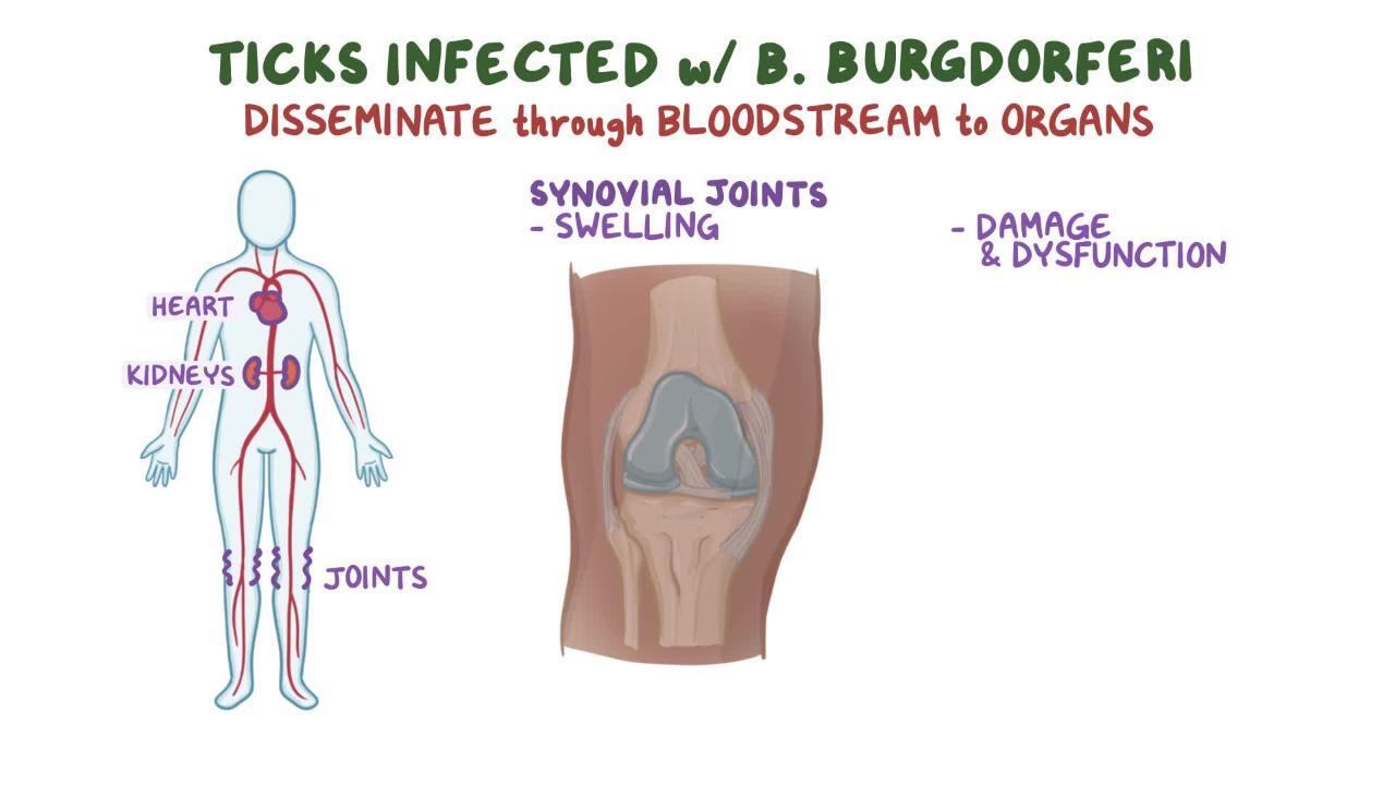
Numerous clinical syndromes have been attributed to Lyme borreliosis in domestic animals, including limb and joint disease and kidney, neurological, and cardiac abnormalities. However, the clinical signs in different animal species vary considerably. Certain clinical signs of Lyme borreliosis are well documented in horses and dogs and, although similar, are less common in cats.
In dogs, the most common clinical signs include the following:
intermittent, shifting leg lameness
fever
anorexia
lethargy
lymphadenopathy, with or without swollen, painful joints
Additionally, a renal syndrome, Lyme nephropathy, has been attributed to B burgdorferi infection in dogs; however, a causal relationship has yet to be established. This syndrome is characterized by severe protein-losing nephropathy, uremia, and hyperphosphatemia, often accompanied by peripheral edema and thromboembolic disease. It is thought to be related to immune-complex deposition in the glomerulus, and dogs typically present with acute or chronic kidney injury, with or without secondary cardiac or neurological signs (eg, blindness, hypertension, heart murmurs, thromboembolic events) (3). Disease typically presents well after the bite of the tick, between 2 and 5 weeks after transmission of the organism.
Clinical signs typically include fever, lameness, lethargy, and lymphadenopathy. Proteinuric kidney disease, a chronic manifestation, can be mistaken for an acute illness because it results in secondary, serious signs such as ascites, hypertension, or thromboembolic events. The incidence of this chronic form of Lyme borreliosis is extremely low.
In horses, well-documented clinical signs of Lyme borreliosis include neuroborreliosis, uveitis, nuchal bursitis, and cutaneous pseudolymphoma. Horses with neuroborreliosis can exhibit any of the following clinical signs:
atrophy of spinal muscles
dysphagia
laryngeal dysfunction
facial paresis
spinal cord ataxia and paresis
behavioral changes
hyperesthesia
Horses with neuroborreliosis can also exhibit clinical signs common to other neurological disorders (eg, meningitis, cranial nerve dysfunction). Other clinical signs in horses, such as intermittent, shifting lameness, have not been conclusively linked to Lyme borreliosis.
Most seropositive dogs and horses do not have any clinical signs. This stands in contrast to cases in humans, in whom infection with B burgdorferi causes true acute illness in an estimated 90% of people. Soon after infection via a tick bite, most people develop a characteristic bull's-eye rash (erythema migrans) at the site of infection, and some experience influenza-like symptoms. Common clinical signs and symptoms of Lyme borreliosis in humans include arthritis with severe joint pain and swelling, facial paralysis, dizziness, heart palpitations, nerve pain, and severe headaches.
Pearls & Pitfalls
|
Diagnosis of Lyme Borreliosis
History of tick bite
Clinical signs, laboratory data, response to therapy, and exclusion of other diseases
Serological testing
Cytological and histological evaluation
A diagnosis of Lyme borreliosis is generally reached through the exclusion of other diseases, in conjunction with a history of exposure to ticks, clinical signs, supporting laboratory data, and response to therapy. Because clinical signs are nonspecific, other diseases can mimic the signs of Lyme borreliosis. Moreover, most infected animals demonstrate no clinical signs, so a positive test result for B burgdorferi has a low positive predictive value for disease. In other words, a positive test result neither demonstrates that current clinical signs are caused by infection with B burgdorferi nor suggests that the animal is likely to develop clinical illness in the future. Thus, a positive test result in a clinically ill animal might be an incidental finding.
A number of infectious agents can be transmitted by ticks (eg, Ehrlichia, Anaplasma, Babesia, Rickettsia), some of which can cause clinical signs similar to those of Lyme borreliosis. Thus, infection with these agents, or mixed infections with B burgdorferi, should be considered. Anaplasma phagocytophilum, for example, is also transmitted by Ixodes ticks and can cause clinical signs resembling those of Lyme borreliosis in dogs and horses. In addition to excluding infectious causes, orthopedic disorders (eg, trauma, immune-mediated diseases) should be excluded as well. In some areas the seroprevalence of coinfections is as high as 10–20% (6).
Results of laboratory testing are generally normal, except for results pertaining directly to the affected system (eg, soft tissue swelling in limbs, neutrophil accumulation in synovial fluids of affected joints). Frequently used tests and analyses used to diagnose Lyme borreliosis include the following:
CBC
serum chemistry
antinuclear antibody test
rheumatoid factor test
direct Coombs test
C-reactive protein test
urine protein:creatinine ratio test
SDS-polyacrylamide gel electrophoresis,
radiographic examination
arthrocentesis with fluid analysis
Serological testing for antibodies specific for B burgdorferi sensu lato is an adjunct to clinical diagnosis. Antibodies can be detected with ELISA (which tests for C6 peptide) and with multiplex fluorescence assays (3). The C6 peptide is only expressed by B burgdorferi after natural antibody or exposure. A positive C6 result will not be reported from cross-reactivity with vaccinal antibodies. A positive test result indicates exposure. The use of anti-C6 antibody is being investigated as a diagnostic test in human medicine.Because of possible cross-reactions with other spirochetal infections, the serological testing methods whole cell ELISA, immunofluorescent antibody assays, and Western blot are no longer recommended.
Pearls & Pitfalls
|
Antibodies against C6 peptide, VlsE (variable major protein-like sequence, expressed), and OspF (outer surface protein F) indicate natural exposure, because these antigens are not contained in any vaccines. Although rare, false-negative serological results can occur in the case of an acute infection (< 1 month), infection of an immune-privileged site (eg, eye), or an abnormal host response (ie, immunodeficiency). In particular, horses with neuroborreliosis are often found to be seronegative.
Isolation of B burgdorferi sensu lato by culture or detection of specific DNA by PCR assay from joints, skin tissue samples, or other affected tissues can also help in diagnosis. Direct detection of the organism, however, is difficult and time-consuming (up to 6 weeks for culture) and yields negative results in most cases. In horses, PCR assays can be performed on CSF but are not very sensitive (7). Blood samples are generally negative because the organism resides in tissue and not in the circulation. Characteristic inflammatory changes in histological or cytological samples of affected tissues can also be used to support a diagnosis.
Treatment of Lyme Borreliosis
Antimicrobials
Pain management
Supportive therapy as needed
Immunosuppressive therapy when indicated
Antimicrobial therapy is indicated in all animals with clinical signs attributed to Lyme borreliosis. Treatment of healthy seropositive animals is problematic because of the inability to monitor response to treatment and the potential for adverse drug effects. Antimicrobials in the tetracycline and beta-lactam families have been shown to be effective in treating Lyme borreliosis in dogs and horses. A long course of antimicrobials (4 weeks) is indicated. Chronic infection in dogs and horses is not well documented.
Persistence of clinical signs should prompt consideration of reinfection, another disease process, or the occurrence of immune-complex disease; in the case of immune-complex disease, immunosuppressive therapy may be indicated.
Directing specific treatments toward the affected organ system and any clinicopathological abnormalities that are present (eg, fever, pain, proteinuria) is also important.
The 2018 American College of Veterinary Internal Medicine consensus update on Lyme borreliosis in dogs and cats is the recommended source for dosages of drugs used for medical management. In dogs, doxycycline (10 mg/kg, PO, every 12–24 hours for 1 month) is considered first-line treatment, and amoxicillin (20 mg/kg, PO, every 8 hours) can be used in doxycycline-sensitive or growing dogs as an alternative (3). Although it is rare for cats testing positive for Lyme disease to demonstrate clinical signs of illness, they can be treated with either doxycycline or amoxicillin in the same dosage as dogs. In cats, doxycycline should be administered as a suspension or given with food. If cats have difficulty swallowing or vomit/regurgitate, they should be assessed via endoscope for ulceration or stricture (1).
A rapid response (1–3 days) to treatment in both dogs and cats occurs in most cases; however, incomplete or transient resolution of clinical signs occurs in many affected animals. Doxycycline is preferred over penicillins because mixed infections with other tickborne pathogens that are more susceptible to tetracyclines are often found in animals with clinical signs.
Pearls & Pitfalls
|
Treatment for neuropathic pain might be indicated, in which case gabapentin is the recommended therapy (dogs 10–20 mg/kg, PO, every 8 hours [8]; cats 8 mg/kg, PO, every 6 hours [9], as needed). In dogs with kidney dysfunction that is suspected to be related to Borrelia infection, supportive therapy is indicated, and immunosuppressive agents such as mycophenolate (5 mg/kg, PO, every 12 hours; increase to 10 mg/kg, PO, every 12 hours if no GI upset [3]) might be required for severe, rapidly progressing cases.
In horses, a number of tetracycline (administered PO and parenterally) and beta-lactam (administered parenterally) antimicrobials have been successful in treating Lyme borreliosis. Treatment is more challenging in horses because of the poor bioavailability of antimicrobials and because horses are often diagnosed long after they have been infected. Four-week courses of doxycycline (10 mg/kg, PO, every 12 hours) or minocycline (4 mg/kg, PO, every 12 hours) have reportedly been successful in treating Lyme borreliosis in horses (10).
Although beta-lactam antimicrobials are less practical in horses because of parenteral administration and carry a risk of toxicity, they are preferable for treatment of neuroborreliosis. In these cases, parenteral high-dose penicillin (44,000 U/kg, IV, every 4–6 hours) or cefotaxime (25–50 mg/kg, IV, every 6–8 hours) can be useful (10). Certain antimicrobials (eg, ceftriaxone) have resulted in a high incidence of adverse effects in horses and should not be used (10).
Quantitative C6 antibody assays are sometimes performed before and after treatment to monitor response to therapy; however, titers do not always decrease after treatment, and some animals remain seropositive. Successful treatment is focused on the resolution of clinical signs rather than serological data. An increased titer after treatment could represent a recrudesence or reinfection.
Control and Prevention of Lyme Borreliosis
Tick avoidance using perimeter control (creating a barrier between well-groomed areas in lawns and low brush, high grass, and leaves) plays an important role in the control of Lyme borreliosis. A number of effective repellents and acaricides in collars, sprays, and spot-ons are available for use in dogs. Tick checks and prompt removal of ticks can decrease the likelihood of transmission, because at least 24 hoursis required for transmission of spirochetes from tick to host. However, Ixodes ticks (nymphs and adults) are so small and difficult to see in thick fur that tick checks are less effective than repellents and acaricides. A multimodal approach to tick control is required for prevention of Lyme borreliosis.
Ticks must be removed carefully (see ). If the tick mouthparts are damaged and left in the animal, transmission of spirochetes can still occur, even after the tick has been removed.


Tick remover tool: Slide the tool under the tick at the point where it makes contact with skin (Step 1), lift the hook lightly, and turn the tool 2–3 times in the same direction to release the tick OR gently pull back on the bar, using leverage to extract the tick (Step 2).
Fine point tweezers: With tweezers, grasp the head or neck (not the body) of the tick as close to the skin as possible (Step 1), and gently pull straight upward with steady traction to remove the tick from the skin (Step 2). Do not use a twisting motion with tweezers.
Courtesy of Dr. Nadine A. Vogt.
A number of bacterin and recombinant vaccines are licensed for use in dogs. The duration of immunity is 1 year (3). Dogs that have been exposed to ticks should be tested serologically for established infection before vaccination. Postinfection vaccination has little to no therapeutic effect on established infections.
Zoonotic Risk of Lyme Borreliosis
Lyme borreliosis is a vector-borne zoonotic disease. Ticks transmit spirochetes from wildlife reservoirs to humans. Companion and farm animals are not a reservoir; however, they can harbor unattached infected ticks that can subsequently attach to a person and transmit spirochetes.
Key Points
Lyme borreliosis is a bacterial, Ixodes tick–transmitted disease affecting companion and farm animals (dogs, horses, cats) that can harbor unattached infected ticks that transmit disease to humans.
Dogs with Lyme borreliosis typically develop fever and intermittent, shifting leg lameness, whereas horses develop neurological signs, nuchal bursitis, or uveitis.
Diagnosis is based on history, clinical signs, laboratory data, response to therapy, and exclusion of other diseases.
Clinically ill animals should be treated with a long course (4 weeks) of antimicrobials (eg, doxycycline) and immunosuppressive therapy if an immune-mediated reaction occurs.
Preventive measures to decrease the risk of Lyme borreliosis include perimeter control over ticks, tick avoidance, prompt tick removal, and use of acaricides and vaccines.
For More Information
Field guide to ticks. University of Rhode Island.
Lyme disease. CDC.
Also see pet owner content regarding Lyme borreliosis in dogs, cats, and horses.
References
Hoyt K, Chandrashekar R, Beall M, Leutenegger C, Lappin MR. Evidence for clinical anaplasmosis and borreliosis in cats in Maine. Top Companion Anim Med. 2018;33(2):40-44. doi:10.1053/j.tcam.2018.05.002
Comstedt P, Jakobsson T, Bergström S. Global ecology and epidemiology of Borrelia garinii spirochetes. Infect Ecol Epidemiol. 2011;1(1)1. doi:10.3402/iee.v1i0.9545
Littman MP, Gerber B, Goldstein RE, Labato MA, Lappin MR, Moore GE. ACVIM consensus update on Lyme borreliosis in dogs and cats. J Vet Intern Med. 2018;32(3):887-903. doi:10.1111/jvim.15085
Duffy DC, Campbell SR. Ambient air temperature as a predictor of activity of adult Ixodes scapularis (Acari: Ixodidae). J Med Entomol. 1994;31(1):178-180. doi:10.1093/jmedent/31.1.178
Gustafson JM, Burgess EC, Wachal MD, Steinberg H. Intrauterine transmission of Borrelia burgdorferi in dogs. Am J Vet Res. 1993;54(6):882-890. https://lyme.org/wp-content/uploads/2024/05/1993-Gustafson-Burgess-Wachal.pdf
Reif KE. 2026 annual pet parasite forecasts. Companion Animal Parasite Council. March 23, 2026. Accessed April 24, 2026. https://capcvet.org/articles/2026-annual-pet-parasite-forecasts/
Johnstone LK, Engiles JB, Aceto H, et al. Retrospective evaluation of horses diagnosed with neuroborreliosis on postmortem examination: 16 cases (2004-2015). J Vet Intern Med. 206;30(4):1305-1312. doi10.1111/jvim.14369
KuKanich B, Cohen RL. Pharmacokinetics of oral gabapentin in greyhound dogs, Vet J. 2011;187(1):133-135. doi:10.1016/j.tvjl.2009.09.022
Siao KT, Pypendop BH, Ilkiw JE. Pharmacokinetics of gabapentin in cats. Am J Vet Res. 2010;71(7):817-821. doi:10.2460/ajvr.71.7.817
Divers TJ, Gardner RB, Madigan JE, et al. Borrelia burgdorferi infection,and lyme disease in North American horses: a consensus statement. J Vet Intern Med. 2018;32(2):617-632. doi:10.1111/jvim.15042