Infectious and inflammatory diseases of the spinal column and spinal cord include bacterial, rickettsial, viral, fungal, protozoal, and parasitic infections and idiopathic inflammatory disease. Many of these diseases can also affect the brain (see Meningitis, Encephalitis, and Encephalomyelitis).
Bacterial Diseases of the Spinal Column and Cord
Diskospondylitis is inflammation of the intervertebral disk and adjacent vertebral bodies. Vertebral osteomyelitis is inflammation of the vertebra without concurrent disk infection. Vertebral physitis is infection centered on the vertebral physis, with no initial involvement of the disk space. These diseases are usually caused by hematogenous transmission of bacterial or, less commonly, fungal infection. Immunosuppression might play a role in some infections.
Diskospondylitis is most common in dogs, especially larger breeds. Vertebral osteomyelitis of the lumbar vertebrae can develop in dogs secondary to migration of plant awns. Vertebral physitis generally affects the lumbar vertebrae of dogs < 2 years old. Vertebral infection is rare in cats and usually due to direct transmission of infection from an adjacent wound. Diskospondylitis and vertebral osteomyelitis have also been reported in horses, ruminants, and pigs, especially neonates (1, 2, 3). Infection can occur at any disk space, and multiple lesions can occur. (See .)

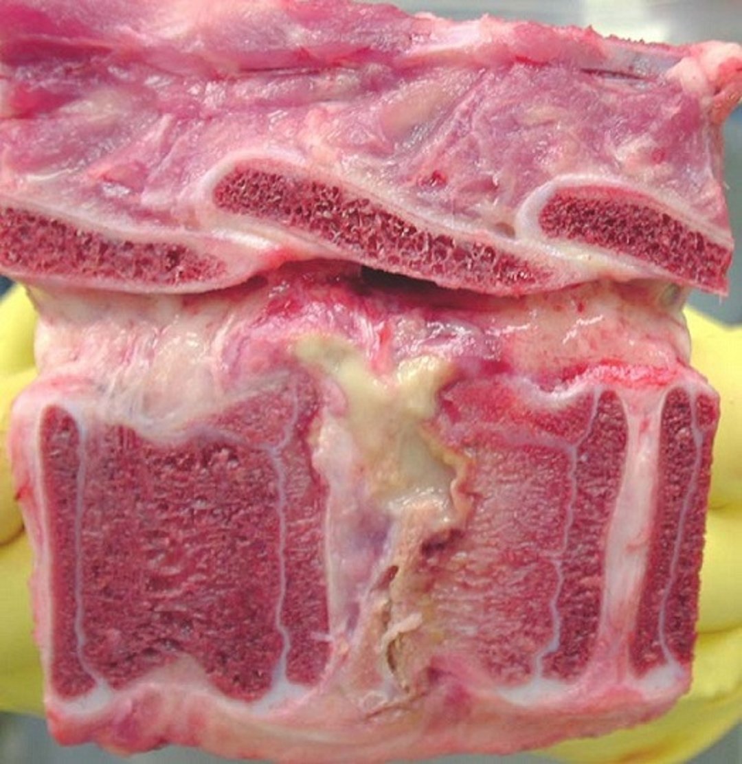
Gross pathology photograph of a spinal vertebral body abscess in cross-section. The pus visible in the affected disk space extends into the epidural space. The adjacent vertebral end plate shows bony lysis.
Courtesy of Dr. Sameeh M. Abutarbush.
In canine diskospondylitis, the most commonly isolated organisms are Staphylococcus spp. Other organisms include Brucella canis, Streptococcus spp, Escherichia coli, Proteus spp, Corynebacterium diphtheroides, Nocardia spp, and Aspergillus spp.
Spinal pain is the most consistent clinical finding. Systemic clinical signs, such as fever, depression, and weight loss, are less common. Neurological deficits might develop because of spinal cord compression caused by proliferative tissue or, rarely, transmission of infection to the spinal cord or pathological fracture.
Early radiographic findings for diskospondylitis consist of destruction of the adjacent vertebral end plates and collapse of the disk space (see ). More advanced lesions also have variable extents of osteophyte formation. Microbial cultures often identify the causative organism; samples for culture include blood, urine, or aspirate from the infected disk space. Affected dogs should be tested for brucellosis (Brucella canis).

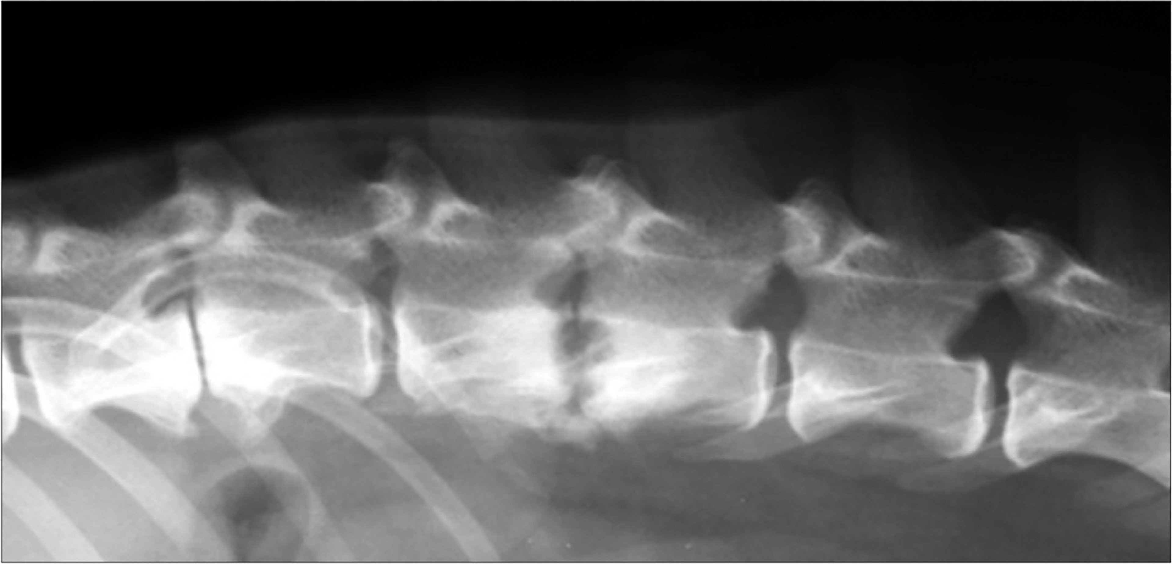
Lateral radiographic view of a dog with lysis of the vertebral end plates at L2 through L3, consistent with diskospondylitis.
Courtesy of Dr. William Thomas.
Although clinical signs of diskospondylitis usually resolve within 5 days of treatment with an appropriate antimicrobial, treatment should be continued for at least 12–16 weeks. Administration of amoxicillin potentiated with clavulanic acid for presumed Staphylococcus spp infection is a good choice if cultures yield no growth (4).
Pearls & Pitfalls
|
Rickettsial Diseases of the Spinal Column and Cord
Neurological abnormalities, including clinical signs of spinal cord dysfunction, occur in up to one-third of dogs with Rocky Mountain spotted fever or ehrlichiosis (5). Rickettsial infection should be suspected in dogs when neurological signs are accompanied by systemic signs, such as fever, lymphadenopathy, and petechial hemorrhage.
Rocky Mountain spotted fever (Rickettsia rickettsii) is most common in the spring and summer and initially causes fever, lethargy, and decreased appetite. Neurological signs can include seizures, ataxia, spinal pain, or vestibular dysfunction. Common laboratory findings are thrombocytopenia, leukocytosis, and neutrophilic pleocytosis and mildly increased protein on CSF analysis. Diagnosis is based on a 4-fold increase in serum antibody concentration.
Dogs with ehrlichiosis (Ehrlichia canis) often have thrombocytopenia, anemia, leukopenia, hyperglobulinemia, and mononuclear pleocytosis and marked increase in protein on CSF analysis. Paresis, spinal pain, vestibular dysfunction, and seizures are potential clinical signs. A single serum antibody titer is usually sufficient for diagnosis of E canis.
Treatment of rickettsial myelitis consists of administration of doxycycline (5 mg/kg, PO, every 12 hours for 14–21 days). Other antibiotics, including enrofloxacin, have been shown to clear rickettsial infection in dogs. Chloramphenicol, used historically to treat rickettsial infection, is no longer recommended due to potentially fatal idiosyncratic aplastic anemia seen in humans.
The prognosis for affected animals is good with early treatment; however, neurological deficits occasionally progress despite treatment (6).
Viral Diseases of the Spinal Column and Cord
Canine distemper encephalomyelitis, caused by a paramyxovirus, remains one of the most common CNS disorders in dogs worldwide.
Onset of neurological deficits can be acute or slowly progressive, reflecting the location of the lesions within the CNS. The brainstem and spinal cord are the regions most commonly affected in mature dogs. Clinical neurological signs are usually not preceded by, nor coincident with, systemic illness in young dogs.
Definitive antemortem diagnosis of canine distemper encephalomyelitis is difficult. There can be active or inactive chorioretinitis on fundoscopy. A lymphocytic pleocytosis with increased protein concentration is the most common finding on CSF analysis. Reverse transcription-PCR assay on urine or CSF is useful in diagnosis.
No specific treatment exists, and the prognosis is poor for severely affected dogs.
Vaccination is usually successful in preventing the systemic form of distemper; however, previously vaccinated dogs can be affected by the neurological form.
Caprine arthritis and encephalomyelitis is caused by a lentivirus that can also cause pneumonitis and arthritis; CNS disease is most common in goats 2–4 months old; however, older animals can also be affected. Acute onset of slightly asymmetrical spastic paraparesis can progress to tetraplegia with exaggerated reflexes. Mononuclear pleocytosis and increased protein in the CSF are present in approximately 90% of the cases (7).
Serological testing can help detect infection with caprine arthritis and encephalomyelitis; however, false-negative results do occur. Histologically, severe nonsuppurative inflammation with demyelination or necrosis is present, most prominently in the white matter of the spinal cord.
No treatment exists, and recovery is unlikely.
A related lentivirus is a rare cause of chronic encephalomyelitis in sheep (maedi). Affected sheep are usually > 2 years old and develop insidious onset of progressive ataxia, paraparesis, or tetraparesis.
Equine infectious anemia occasionally produces encephalomyelitis in horses. Neurological deficits are usually referable to spinal cord disease and include ataxia and weakness in the hindlimbs. The protein concentration of the CSF and the number of lymphocytes are often increased.
Diagnosis is by positive results of agar gel immunodiffusion testing.
No treatment exists, and affected horses are usually euthanized to prevent transmission of the disease.
Equine herpesvirus 1 (EHV-1) encephalomyelopathy is a neurological disorder that affects horses worldwide. Equine herpesvirus 1 infects vascular endothelial cells, particularly those within the CNS, and causes immune-mediated vasculitis with secondary infarction and hemorrhage throughout the brain and spinal cord. EHV-1 has also been associated with meningoencephalitis in alpacas and llamas. In horses, neurological signs can be evident as the primary disease or follow rhinopneumonitis or abortion. Animals of any age can be affected.
Neurological deficits in EHV-1 have an abrupt onset, vary from mild hindlimb ataxia to paraplegia, and usually do not progress after 24 hours. Urine dribbling, fecal retention, and sensory deficits in the perineum and tail are common. The CSF is often xanthochromic with increased protein content and normal numbers of cells.
Diagnosis is based on clinical findings and an increase in antibody concentration in paired serum samples, isolation of virus from nasal or pharyngeal secretions, or PCR assay.
There is no evidence that pharmacologic treatments are effective once clinical signs have developed. Moderate evidence suggests that administration of valacyclovir to horses prior to EHV-1 exposure can reduce the risk of developing EHM, and this approach may be effective as preexposure prophylaxis during an outbreak. Anti-inflammatory agents such as dexamethasone (0.05–0.07 mg/kg, IM or IV, every 24 hours for 5 days ) and NSAIDs can help (8).
Supportive care is important to prevent complications such as urine retention, cystitis, and decubitus. Mildly affected horses often recover with supportive care. Even recumbent horses can eventually recover with meticulous nursing care.
Vaccination does not protect from the neurological form of this disease.
Feline infectious peritonitis (FIP) is a disease of domestic cats caused by a dysregulated immune response to infection with feline coronavirus (FCov). Involvement of the CNS is common. Pyogranulomatous lesions may involve the neural parenchyma, choroid plexuses, ependyma, and leptomeninges.
Clinical signs of spinal cord involvement include spinal hyperesthesia and paraparesis or tetraparesis. Involvement of other organs, especially the eyes, is common.
Hyperglobulinemia is a common but nonspecific clinicopathological finding. Serum antibody tests have limited sensitivity and specificity (ie, antibodies associated with FCoV are not specific for FIP). Mixed (neutrophilic and mononuclear) pleocytosis with increased protein concentration is the most common finding on CSF analysis.
Remdesivir (or its active metabolite GS-441524) has demonstrated efficacy in the treatment of FIP. Treatment protocols for FIP are evolving and differ for neurological and nonneurological manifestations. One protocol for cats with neurological signs is GS-441524 at 12 mg/kg, every12 hours (9).
Rustrela virus (RusV) has been identified as the etiological agent of feline staggering disease, a nonsuppurative encephalomyelitis in cats in several regions worldwide (10). Borna disease virus 1 (BoDV-1), which causes neurological disorders in various mammals, including humans, can infect cats and was historically implicated but is not currently considered the cause of feline staggering disease.
Initially, clinical signs include ataxia with increased muscle tone in the limbs and tail, protracted claws, lumbar pain, and behavior changes such as vocalizations and abnormally affectionate behavior. Usually, paraplegia and death follow; however, some cats recover spontaneously.
No effective treatment exists (11).
Feline leukemia virus–associated myelopathy occurs in some cats infected with feline leukemia virus (FeLV) for ≥ 2 years. Ataxia and weakness of the pelvic limbs progresses to paraplegia within 1 year. Other clinical signs include diffuse spinal pain and abnormal behavior. Diagnosis is based on clinical signs, serological testing, and exclusion of other causes, such as spinal lymphoma and myelitis due to toxoplasmosis or fungal infection. No treatment exists; affected cats are typically eventually euthanized because of disability. Pathological findings consist of white matter degeneration, swollen axons, and dilatation of myelin sheaths in the spinal cord and brainstem. Feline leukemia virus antigen is present in the nervous system, indicating that the lesions are due to viral infection.
Teschovirus encephalomyelitis, also called Teschen disease, Talfan disease, and porcine polioencephalomyelitis, is caused by a neurotropic teschovirus previously classified as an enterovirus. Peracute or subacute onset of hindlimb ataxia and paresis occurs, with hyporeflexia, depression, seizures, and death. Older pigs might survive, but the mortality rate is high in young pigs.
Porcine hemagglutinating encephalomyelitis virus is a coronavirus that causes both vomiting and wasting disease and encephalomyelitis. It is most common in piglets < 3 weeks old, and there is some overlap of these syndromes. The CNS disease starts with several days of vomiting, which is followed by hyperesthesia, muscle tremors, ataxia, paresis, opisthotonos, coma, and death. Histopathologically, there is diffuse nonsuppurative encephalomyelitis, primarily involving gray matter. Diagnosis is based on necropsy or an increase in antibody titer in paired sera. No treatment exists.
Rabies is caused by a neurotropic rhabdovirus that reaches the CNS via peripheral nerves. It produces multifocal, nonsuppurative polioencephalomyelitis in all domestic mammals. Clinical signs of spinal cord involvement include ataxia and progressive paralysis, usually with absent reflexes. Affected animals typically, but not invariably, die with progressive neurological signs within 2–7 days of illness.
Fungal Diseases of the Spinal Column and Cord
Cryptococcus neoformans is the most common fungus to involve the CNS in animals. Infection is most common in dogs and cats and occurs occasionally in horses. Other fungal organisms can invade the CNS, including Blastomyces dermatitidis, Histoplasma capsulatum, Coccidioides immitis, Aspergillus spp, and phaeohyphomycoses. Affected animals often have involvement of other organs, such as the lungs, eyes, skin, or bones.
Clinical signs of spinal cord involvement include paresis or paralysis and spinal hyperesthesia. Diagnosis is based on serological testing, culture, or identifying the organism in CSF or extraneural tissue.
Fluconazole is often effective for cryptococcosis and coccidioidomycosis. Itraconazole or amphotericin B is recommended for histoplasmosis and blastomycosis; however, the prognosis is guarded to poor.
Also see Fungal Infections in Animals.
Protozoal Diseases of the Spinal Column and Cord
Equine protozoal myeloencephalitis is a common disease of horses that produces nonsuppurative, often necrotizing, meningoencephalomyelitis. Horses are likely an aberrant host for the causative organism, usually Sarcocystis neurona; less commonly, however, other protozoa cause the disease. Neurological signs are extremely variable and often asymmetrical, reflecting involvement anywhere in the CNS. Ataxia and paresis are common. Other potential clinical signs include obscure lameness, focal muscle atrophy, and cranial nerve dysfunction. Most affected horses improve with treatment (see the chapter on equine protozoal myeloencephalitis); however, permanent neurological deficits are possible and relapse is not rare.
Neosporosis is caused by Neospora caninum, a protozoon that can cause a nonsuppurative encephalomyelitis, most commonly in dogs. Infection in young puppies typically causes ascending paralysis with rigid contraction of the muscles of one or both pelvic limbs. Other organs, including muscle, liver, and lungs, can be affected. Diagnosis is based on detection of antibodies against the organism by immunohistochemical analysis or PCR assay. Early treatment with clindamycin or sulfadiazine and pyrimethamine might be effective; however, the prognosis is poor.
Toxoplasmosis is caused by Toxoplasma gondiiand can occasionally cause nonsuppurative encephalomyelitis in puppies, kittens, and piglets. Diagnosis is based on identifying the organism in tissue or a 4-fold increase in IgG antibody in paired sera. In cats, a high concentration of IgM antibody in serum or CSF supports the diagnosis. Administration of clindamycin or sulfadiazine and pyrimethamine is recommended for treatment.
Parasitic Diseases of the Spinal Column and Cord
Verminous myelitis is inflammation of the spinal cord caused by parasite migration. Organisms that cause verminous myelitis include the following:
Parelaphostrongylus tenuis in sheep, goats, and llamas
Hypoderma bovis in cattle
Strongylus vulgaris, Halicephalobus deletrix, and Setaria spp in horses
Stephanurus dentatus in pigs
Cuterebra spp in cats
Baylisascaris procyonis in dogs
Clinical signs of spinal cord involvement are usually acute, often asymmetric, and can be progressive.
Antemortem diagnosis is difficult. Increased numbers of eosinophils in the CSF are suggestive; however, CSF findings are variable.
Treatment with fenbendazole, thiabendazole, or ivermectin is recommended; however, the prognosis is guarded.
Also see Central Nervous System Diseases Caused by Helminths and Arthropods.
Idiopathic Inflammatory Diseases of the Spinal Column and Cord
Meningoencephalomyelitis of unknown origin (MUO) is a group of inflammatory diseases of the CNS in dogs. Subtypes include granulomatous meningoencephalitis (GME), necrotizing meningoencephalitis (NME), and necrotizing leukoencephalitis (NLE). The cause is unknown; however, an autoimmune response is suspected (ie, immune-mediated meningoencephalomyelitis).
MUO has a worldwide distribution. Adult dogs of any breed can be affected; however, young to middle-aged small- and toy-breed dogs are predisposed.
Clinical signs of MUO are variable and might indicate focal or multifocal brain or spinal cord dysfunction. Spinal pain and varying degrees of paresis are the most common clinical signs of spinal cord involvement. Clinical signs can be acute or slowly progress over several months.
Spinal radiographs are normal; however, MRI might show focal or multifocal T2 hyperintense lesions with varying degrees of contrast enhancement. CSF usually has increased protein and pleocytosis, with either mononuclear cells or neutrophils predominating.
Dogs often improve with administration of immunosuppressive doses of corticosteroids and other immunomodulating drugs such as cytarabine, cyclosporine, and procarbazine; however, relapse is possible, and many dogs eventually become refractory to treatment.
Steroid-Responsive Meningitis-Arteritis in Dogs
Steroid‐responsive meningitis‐arteritis (SRMA) is an immune‐mediated disease characterized by inflammation of the meninges and associated arteries in dogs.
Affected dogs are typically 6–18 months old. Any breed of dog can be affected; however, Beagles, Boxers, and Bernese Mountain Dogs are predisposed.
The acute form of SRMA is characterized by spinal pain, especially cervical pain, and fever. Affected dogs often stand with a hunched posture, are reluctant to move, and exhibit generalized pain or hyperesthesia. Left untreated, SRMA can progress to cause spinal cord dysfunction characterized by ataxia and paresis.
Diagnosis is based on signalment, clinical signs, and CSF analysis. In the acute phase, the CSF shows marked neutrophilic pleocytosis with increased protein and negative results of bacterial culture. In the chronic phase, there is a mild to moderate mononuclear pleocytosis with increased protein.
Treatment consists of immunosuppressive doses of glucocorticoids with a gradual taper over 4–6 months. The prognosis is good; however, relapse is possible if treatment is stopped too soon.
For More Information
Flegel T. Overview of non-infectious inflammatory CNS disease in dogs. WSAVA Congress Proceedings. 2016.
References
Muggli E, Schmid T, Hagen R, Schmid B, Nuss K. Diagnosis and treatment of lumbosacral discospondylitis in a calf. BMC Vet Res. 2011;7:53. doi:10.1186/1746-6148-7-53
Hillyer MH, Innes JF, Patteson MW, Barr AR. Discospondylitis in an adult horse. Vet Rec. 1996;139(21):519-521. doi:10.1136/vr.139.21.519
Doige CE. Discospondylitis in swine. Can J Comp Med. 1980;44(2):121-128. https://pmc.ncbi.nlm.nih.gov/articles/instance/1320047/pdf/compmed00022-0017.pdf
Vallios VL, Sourla E, Low D, Volk HA, Charalambous M. Treatment of discospondylitis in dogs: a systematic review. Vet Rec. 2026;199(4):e1-e17. doi:10.1002/vetr.70334
Greene CE, Burgdorfer W, Cavagnolo R, Philip RN, Peacock MG. Rocky Mountain spotted fever in dogs and its differentiation from canine ehrlichiosis. J Am Vet Med Assoc. 1985;186(5):465-472. doi:10.2460/javma.1985.186.05.465
Thomas WB. Inflammatory diseases of the central nervous system in dogs. Clin Tech Small Anim Pract. 1998;13(3):167-178. doi:10.1016/S1096-2867(98)80038-8
Norman S, Smith MC. Caprine arthritis-encephalitis: review of the neurologic form in 30 cases. J Am Vet Med Assoc. 1983;182(12):1342-1345. doi:10.2460/javma.1983.182.12.1342
Pusterla N, Hussey GS, Goehring LS. Equine herpesvirus-1 myeloencephalopathy. Vet Clin North Am Equine Pract, 2022;38(2):339-362. doi:10.1016/j.cveq.2022.05.006
Taylor SS, Coggins S, Barker EN, et al. Retrospective study and outcome of 307 cats with feline infectious peritonitis treated with legally sourced veterinary compounded preparations of remdesivir and GS-441524 (2020-2022). J Feline Med Surg. 2023;25(9). doi:10.1177/1098612X231194460
Matiasek K, Pfaff F, Weissenböck H, et al. Mystery of fatal "staggering disease" unravelled: novel rustrela virus causes severe meningoencephalomyelitis in domestic cats. Nat Commun. 2023;14(1):624. doi:10.1038/s41467-023-36204-w
Wensman JJ, Jäderlund KH, Holst BS, Berg M. Borna disease virus infection in cats. Vet J. 2014;201(2):142-149. doi:10.1016/j.tvjl.2013.12.012