Also see Management of Reproduction: Sheep.
Although many of the toxins that cause abortion in cows also cause abortion in ewes, some plant-associated reproductive toxins are particularly important in sheep. For example, consumption of goitrogenic kale during pregnancy can impair reproductive efficiency and cause congenital goiter, weak or nonviable lambs, and perinatal losses (1, 2, 3).
Toxins produced by subterranean clover (Trifolium subterraneum) are a common cause of early embryonic death and abortion in sheep.
The major infectious agents that cause abortions in sheep are the bacteria Campylobacter spp, Chlamydia abortus, Coxiella burnetii, Listeriamonocytogenes, Brucella spp, and Salmonella spp; the protozoa Toxoplasma spp; border disease virus; and Cache Valley virus.
Bluetongue as a Cause of Abortion in Sheep
Bluetongue virus (BTV) is an arbovirus transmitted by biting midges (Culicoides spp). Infected sheep show fever, swollen tongue, oral ulcers, nasal discharge, lameness, and, rarely, a blue-tinged tongue due to cyanosis. The infection also causes abortion, fetal mummification, stillbirth, and congenital brain malformation in lambs.
Nearly 30 different BTV serotypes affect sheep. BTV-3 caused a major outbreak of bluetongue in sheep in the Netherlands in 2023, which subsequently spread to other European countries, including Belgium, Germany, and the UK. Sheep experienced severe clinical disease and high mortality rates during the outbreak (4, 5). BTV-8 is known for high mortality rates and transplacental transmission (6). Infection during early to midgestation causes the highest risk of embryonic death and abortion.
Diagnosis: ELISA is used to detect antibodies against BTV, and PCR assay is used to detect the virus. Whole blood, spleen, or fetal tissue should be collected in cases of abortion.
Prevention: Bluetongue is controlled in sheep via management to decrease exposure to biting midges and by vaccination. Inactivated and modified live vaccines are available and widely used; however, the availability of each vaccine varies by country. Modified live vaccines are used predominantly in regions with a long history of bluetongue, such as the US and Africa, but their use is controversial.
The modified live vaccine should not be given to pregnant ewes. It should also not be given when Culicoides spp are active, because these midges can transmit the vaccine virus to unvaccinated animals, including pregnant ewes.
Pearls & Pitfalls
|
Border Disease as a Cause of Abortion in Sheep
Border disease occurs worldwide and, in sheep, is an important cause of embryonic and fetal deaths, weak lambs, and congenital abnormalities. Border disease is caused by a pestivirus closely related to bovine viral diarrhea virus (BVDV) and classical swine fever (hog cholera) virus.
Sheep susceptible to border disease become infected after the introduction of persistently infected animals. Venereal transmission from shedding rams is possible. Abortion can occur at any stage of gestation.
Dams show no clinical signs of border disease, except for, occasionally, mild fever and leukopenia. Affected lambs born alive usually are undersized, and they often have congenital tremors and an abnormally hairy coat (leading to the name hairy shaker lambs; see ).


Aborted lamb with marked hypertrichosis (“hairy shaker”) and characteristic arthrogryposis (“camel limbs”), consistent with congenital infection by border disease virus.
Courtesy of Dr. Vincenzo Di Marco Lo Presti.
Diagnosis: Diagnosis of border disease is based on the identification of border disease virus in the placenta or fetal tissues (kidneys, lungs, spleen, thyroid glands, abomasum; see ) via fluorescent antibody staining, virus isolation, or demonstration of precolostral antibodies.


Aborted fetal lamb organs showing thymic hypoplasia (circled), consistent with in utero infection with border disease virus.
Courtesy of Dr. Vincenzo Di Marco Lo Presti.
Prevention and control: No vaccine is currently available that provides reliable protection against border disease virus. Vaccines developed against BVDV have limited and inconsistent cross-protection and should not be relied on to prevent transplacental infection with border disease virus (7, 8).
Control of border disease is primarily based on identification and removal of persistently infected animals, which are the major source of virus transmission within a flock. Persistently infected lambs should be identified by antigen or virus testing and removed from the breeding population. New animals should be quarantined and tested before introduction, and replacement animals should originate from flocks with a known negative border disease virus status.
Maintaining a closed flock, when practical, decreases the risk of introducing infection. Pregnant ewes should be protected from contact with persistently infected sheep and, where relevant, with cattle that might be persistently infected with pestiviruses. Good biosecurity measures and surveillance are particularly important before and during the breeding season.
Brucellosis as a Cause of Abortion in Sheep
Brucella ovis is a major cause of contagious epididymitis in rams. On a flock basis, brucellosis results in infertility; it also causes late-term abortions, stillbirths, and the birth of weak lambs.
Brucella melitensis is rare in the US but causes abortion in areas where it is found. Brucella abortus occasionally causes abortion in sheep.
Brucella abortions occur late in gestation, resulting in placentitis with edema and necrosis of the cotyledons, as well as thickened, leathery intercotyledonary areas. Many fetuses aborted as a result of B ovis infection are alive at the beginning of expulsion, but some are mummified or autolyzed. Most fetuses aborted because of B melitensis are autolytic.
Diagnosis: Diagnosis of brucellosis is based on culture of the placenta, fetal gastric contents, and the dam’s vaginal discharge.
Prevention: A vaccine against B melitensis is available in some countries.
Brucellosis is zoonotic, affecting humans worldwide.
Cache Valley Virus as a Cause of Abortion in Sheep
Cache Valley virus is a mosquito-transmitted virus that causes infertility, abortions, stillbirths, and multiple congenital abnormalities in sheep. The virus is endemic in most parts of the US, Canada, and Mexico. Often, epizootics caused by Cache Valley virus affect sheep over a wide geographical area that can include several states.
The most noticeable effects of Cache Valley virus infection in sheep are stillbirths and the birth of live lambs with congenital abnormalities that affect the CNS and the musculoskeletal system. Hydranencephaly, hydrocephalus, cerebral and cerebellar hypoplasia, arthrogryposis, scoliosis, torticollis, and hypoplasia of skeletal muscles are common.
Infection by Cache Valley virus before 32 days of pregnancy in sheep results in early embryonic death. Infection between 32 and 37 days of pregnancy results in musculoskeletal and CNS lesions. Infection between 37 and 48 days results in primarily musculoskeletal lesions.
Diagnosis:Infected ewes clear Cache Valley virus before fetuses are aborted. Therefore, diagnosis relies on testing of fetal samples (eg, fluid around the heart or lungs, thoracic fluid) for antibodies and on PCR assay.
Vaccines against Cache Valley virus are not available.
Campylobacter spp Infection as a Cause of Abortion in Sheep
Infection of sheep with Campylobacter fetus fetus or with Campylobacter jejuni results in abortions in late pregnancy or stillbirths. The route of infection is oral. Ewes can develop metritis after expelling the fetus. Placentitis occurs with hemorrhagic necrotic cotyledons and edematous or leathery intercotyledonary areas.
A fetus aborted because of Campylobacter is usually autolyzed and might have characteristic orange-yellow, 1- to 2-cm necrotic foci in the liver (9). Fetuses can have accumulated serosanguineous fluid in the thoracic and peritoneal cavities.
Diagnosis: Diagnosis of Campylobacter infection is based on finding the bacteria in dark-field or fluorescent antibody preparations or on isolating the bacteria from placental smears, uterine discharge, or fetal abomasal contents, liver, and lungs.
Prevention:Tetracyclines can help prevent ewes exposed to Campylobacter from aborting. However, many isolates could be resistant to tetracycline. Campylobacter infection tends to be cyclical, with epizootics occurring every 4–5 years; therefore, vaccination programs, which help prevent outbreaks, should be consistently practiced.
Identification of the Campylobacter species involved is important, because in some areas C jejuni is as common as C fetus, and some vaccines do not include C jejuni. Strict hygiene is necessary to stop an outbreak.
C jejuni is zoonotic and is a common cause of enteritis in humans.
Chlamydiosis (Enzootic Abortion of Ewes)
The bacterium Chlamydia abortus is an obligate intracellular organism and the world's leading cause of enzootic abortion of ewes (EAE), which is characterized by late-term abortions (see ), stillbirths, and weak lambs. Chlamydia pecorum is the cause of chlamydial arthritis and chlamydial conjunctivitis of sheep.
Pearls & Pitfalls
|


Aborted lamb with hepatomegaly and accumulation of serosanguineous fluid in the abdomen, consistent with Chlamydia abortus infection.
Courtesy of Dr. Vincenzo Di Marco Lo Presti.
EAE occurs worldwide, except in Australia and New Zealand, and it is most important in intensively managed sheep.
Ovine abortions due to C abortus occur during the last 2–3 weeks of gestation regardless of when infection occurs, and the fetuses are fresh, with minimal autolysis. Gross lesions in the fetus, although rare, can include ascites, lymphadenopathy, and liver congestion. There is placentitis with necrotic, reddish-brown cotyledons, and thickened brown intercotyledonary areas are covered by exudate (see ).

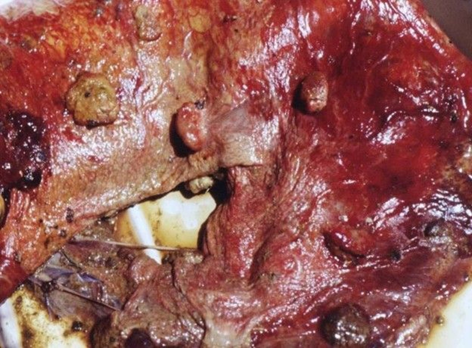
Necropsy photograph from a sheep, showing multifocal necrotizing placentitis characterized by thickened intercotyledonary areas (leathery appearance) and raised necrotic foci (“plaques”), consistent with Chlamydia abortus infection.
Courtesy of Dr. Vincenzo Di Marco Lo Presti.
The chlamydial life cycle is characterized by the alternation of two morphologically distinct forms: an infectious, nonreplicative elementary body, and a noninfectious, replicative reticulate body. The elementary bodies, after attachment to and uptake by host cells, differentiate into metabolically active reticulate bodies, which replicate intracellularly. The reticulate bodies then reorganize into infectious elementary bodies, which are released from the cell and initiate the infection of additional cells (10).
Chlamydial elementary bodies can be found by examination of Gimenez- or Giemsa-stained smears of the placenta or vaginal discharge; however, it can be difficult to differentiate them from Coxiella burnetii, which also occasionally causes abortion in sheep.
Diagnosis: Definitive diagnosis of C abortus infection is based on identification of the bacterium by ELISA, fluorescent antibody staining, PCR assay, or isolation.
Ewes infected with C abortus seldom abort more than once; however, they remain persistently infected and shed C abortus from their reproductive tract for 2–3 days before and after ovulation. Rams can be infected and transmit the organism venereally.
Prevention: Control of C abortus infection consists of isolating all affected ewes and lambs and treating in-contact ewes with long-acting oxytetracycline or oral tetracycline.
C abortus bacterins are available and decrease ovine abortions. In parts of Europe, a modified live vaccine is available.
C abortus is zoonotic; infectious elementary bodies are shed in the environment and inhaled by humans. Pregnant women can develop life-threatening infections, resulting in septic abortion and stillbirth.
Coxiella burnetii Infection as a Cause of Abortion in Sheep
The bacterium Coxiella burnetii, the agent of Q fever, is an important cause of abortion and reproductive loss in sheep and other ruminants, with major zoonotic implications.
C burnetii shows strong tropism for placental trophoblasts, reaching extremely high loads in the placenta and birth fluids. Infected ruminants shed the bacteria in the placenta, fetal fluids, vaginal discharge, milk, feces, and urine, contaminating the environment and facilitating aerosol spread.
Diagnosis: The recommended method for diagnosing C burnetii infection in sheep is PCR assay on the placenta and fetus, combined with histological examination and serological testing.
Prevention and control: Vaccination with inactivated phase I C burnetii vaccines in previously infected sheep flocks boosts humoral immunity in sheep; however, it does not reliably prevent shedding at the next lambing season.
Control of C burnetii–caused abortion and zoonotic risk should rely on strict handling and disposal of aborted fetuses and placentas, limiting lambing-area access, and pasteurization of milk products from small ruminants.
Listeriosis as a Cause of Abortion in Sheep
Abortion caused by the bacterium Listeria monocytogenes in ewes usually occurs in late gestation. Aborting ewes show variable clinical signs of listeriosis, including fever, depression, and anorexia; and some succumb to septicemia.
In cases of ovine abortion due to listeriosis, there is some necrosis of cotyledons and the intercotyledonary areas, and the fetus is usually autolyzed. Mummification is possible. The fetal liver (and possibly lung) might have necrotic foci 0.5–1 mm in diameter.
Diagnosis: Diagnosis of listeriosis is based on culture.
Treatment, prevention, and control: Ewes with listeriosis should be isolated and treated promptly with appropriate systemic antimicrobials. Early treatment improves the likelihood of recovery; however, treatment of ewes that have already aborted is unlikely to prevent fetal loss. Penicillin or ampicillin, combined with oxytetracycline, is commonly used, and antimicrobial selection is guided by susceptibility testing when possible.
Prevention and control of listeriosis should focus primarily on maintaining good feed hygiene and removing sources of environmental contamination. Spoiled or poorly fermented silage should not be fed, and silage associated with an outbreak should be removed from the ration. Feed should be protected from fecal contamination, and old, wet, or decomposing feed should be discarded.
Aborted fetuses, placentas, and contaminated bedding should be removed promptly and disposed of appropriately, and affected ewes should be segregated from the remainder of the flock. Maintaining good hygiene in lambing areas and preventing the contamination of feed and water with feces or reproductive discharges are important.
Salmonellosis as a Cause of Abortion in Sheep
Five serotypes of Salmonella enterica—Abortusovis, Arizonae, Brandenburg, Dublin, and Typhimurium—are known to have caused abortions in sheep. Salmonella Abortusovis is endemic in England and Europe but has not been reported in the US. Salmonella Brandenburg has been reported in Europe and New Zealand. The other serotypes occur worldwide.
Most ewes with salmonellosis are sick and febrile before aborting. There are no specific placental lesions, and the fetus is autolyzed (see ).


Sheep fetus aborted as a result of infection by Salmonella enterica serovar Abortusovis. The fetus is fresh but premature.
Courtesy of Dr. Giuseppe Cuttone.
Diagnosis: Diagnosis of salmonellosis is based on culture of the placenta, fetus, or uterine discharge.
Treatment, prevention, and control: Treatment of ewes salmonellosis should be based on antimicrobial susceptibility testing when possible, because antimicrobial resistance among Salmonella isolates is common. Antimicrobial therapy can decrease mortality rates and, in some outbreaks, decrease subsequent abortion losses; however, its efficacy in controlling abortion outbreaks is inconsistent. Supportive care should be provided to systemically affected ewes (11, 12, 13).
Ewes that abort as a result of salmonellosis should be isolated promptly, and aborted fetuses, placentas, and contaminated bedding should be removed and appropriately disposed of. Lambing areas should be thoroughly cleaned and disinfected, and contamination of feed and water with feces or reproductive discharges should be minimized. Replacement animals should be obtained from flocks with a known negative disease status and quarantined before introduction.
Because subclinical carriers (particularly of S Abortusovis) can contribute to transmission, flock surveillance is important. In endemic areas, vaccination before breeding can decrease rates of abortion associated with S Abortusovis; however, vaccine protection is serotype and strain dependent, and vaccination does not replace biosecurity measures. Salmonella spp are zoonotic, and appropriate precautions should be taken when handling affected sheep, fetuses, placentas, and contaminated materials.
Salmonellosis is zoonotic.
Toxoplasmosis as a Cause of Abortion in Sheep
The protozoon Toxoplasma gondii is a major cause of abortion in small ruminants throughout the world. Ingestion of sporulated coccidian oocytes early in gestation results in fetal resorption or mummification (see ).


Mummified aborted sheep fetus and dry, leathery placenta resulting from Toxoplasma gondii infection.
Courtesy of Dr. Vincenzo Di Marco Lo Presti.
If ewes contract toxoplasmosis late in gestation, abortions or perinatal deaths occur. Ewes do not usually appear sick. In an outbreak, there is usually a wide range in gestational age of aborted fetuses.
Diagnosis: In most cases of toxoplasmosis in sheep, there are no gross lesions; occasionally, however, there are distinct, small, white foci (1–3 mm in diameter) in some cotyledons (see ). The fetal brain often shows focal areas of nonsuppurative inflammation on histological examination. Fetal serological tests (indirect hemagglutination inhibition, latex agglutination, or fluorescent antibody staining) can also be used.


Necropsy photographs of sheep, showing severe placentitis, typical of Toxoplasma gondii infection in small ruminants. A. Multifocal, punctate, caseonecrotic, and mineralized foci evident within the cotyledonary tissue. B. Multifocal, markedly calcified foci at the cotyledonary level.
Courtesy of Dr. Vincenzo Di Marco Lo Presti.
Prevention: Once infected with T gondii, ewes are immune to toxoplasmosis, so running unbred ewes with aborting ewes can help the unbred animals develop immunity. Transplacental transmission of T gondii is possible. Preventing contamination of feed by cat feces that might be infected with toxoplasmosis can help decrease exposure to T gondii.
Toxoplasmosis is a zoonotic disease.
Other Causes of Abortion in Sheep
Akabane virus causes abortion and congenital anomalies in sheep. Akabane virus infection is a differential diagnosis for Cache Valley virus infection.
Neospora caninum has been reported to cause occasional abortions in sheep (14), with the lesions resembling those of Toxoplasma gondii.
Fungal placentitis also occurs; however, it is not as common in sheep as in cattle or horses.
Other pathogens associated with sheep abortion include Schmallenberg virus, Rift Valley fever virus, Nairobi sheep disease virus, Wesselsbron disease virus, Francisella tularensis, and Leptospira spp.
Parachlamydia acanthamoebae is an emerging cause of abortion in sheep and goats. It was detected in necrotizing placentitis when other causes were excluded (15). P acanthamoebae is a Chlamydia-related organism considered to be an emerging agent of pneumonia in humans.
For More Information
de la Concha-Bermejillo A, Romano J. Pregnancy loss in small ruminants. Clin Theriogenol. 2021;13(3):194-205.
References
Wright E, Sinclair DP. The goitrogenic effect of thousand-headed kale on adult sheep and rabbits. N Z J Agric Res. 1958;1(4):477-485. doi:10.1080/00288233.1958.10431532
Willliams HLL, Hill R, Alderman G. The effects of feeding kale to breeding ewes. Br Vet J. 1965;121:2-14. doi:10.1016/S0007-1935(17)41402-3
Knowles SO, Grace ND. Serum total iodine concentrations in pasture-fed pregnant ewes and newborn lambs challenged by iodine supplementation and goitrogenic kale. J Anim Sci. 2015;93(1):425-432. doi:10.2527/jas.2014-7854
van den Brink KMJA, Santman-Berends IMGA, Harkema L, et al. Bluetongue virus serotype 3 in ruminants in the Netherlands: clinical signs, seroprevalence and pathological findings. Vet Rec. 2024;195(4):e4533. doi:10.1002/vetr.4533
Holwerda M, Santman-Berends IMGA, Harders F, et al. Emergence of bluetongue virus serotype 3, the Netherlands, September 2023. Emerg Infect Dis. 2024;30(8):1552-1561. doi:10.3201/eid3008.231331
Thabet S, Lajnef R. Potential mechanisms underlying bluetongue virus emergence and spread. Front Virol. 4:1448192. doi:10.3389/fviro.2024.1448192
EFSA AHAW Panel (EFSA Panel on Animal Health and Welfare), More S, Bøtner A, et al. Scientific opinion on the assessment of listing and categorisation of animal diseases within the framework of the Animal Health Law (Regulation (EU) No 2016/429): border disease. EFSA Journal 2017;15(10):e04993. doi:10.2903/j.efsa.2017.4993
Righi C, Petrini S, Pierini I, Giammarioli M, De Mia GM. Global distribution and genetic heterogeneity of border disease virus. Viruses. 2021;13(6):950. doi:10.3390/v13060950
Wolf-Jäckel GA, Boye M, Angen Ø, Müller M, Jensen TK. Fluorescence in situ hybridization in species-specific diagnosis of ovine Campylobacter abortions. J Vet Diagn Invest. 2020;32(3):413-419. doi:10.1177/1040638720915678
Cossé MM, Hayward RD, Subtil A. One face of Chlamydia trachomatis: the infectious elementary body. Curr Top Microbiol Immunol. 2018;412:35-58. doi:10.1007/82_2016_12
Amagliani G, La Guardia ME, Dominici S, Brandi G, Omiccioli E. Salmonella abortusovis: an epidemiologically relevant pathogen. Curr Microbiol. 2021;79(1):3. doi:10.1007/s00284-021-02689-1
Pardon P, Sanchis R, Marly J, et al. Experimental ovine salmonellosis (Salmonella Abortusovis): pathogenesis and vaccination. Res Microbiol. 1990;141(7-8):945-953. doi:10.1016/0923-2508(90)90134-c
Cagiola M, Severi G, Forti K, et al. Abortion due to Salmonella enterica serovar Abortusovis (S. Abortusovis) in ewes is associated to a lack of production of IFN-gamma and can be prevented by immunization with inactivated S. Abortusovis vaccine. Vet Microbiol. 2007;121(3-4):330-337. doi:10.1016/j.vetmic.2006.12.018
Hässig M, Sager H, Reitt K, et al. Neospora caninum in sheep: a herd case report. Vet Parasitol. 2003;117(3):213-220. doi:10.1016/j.vetpar.2003.07.029
Ruhl S, Goy G, Casson N, et al. Parachlamydia acanthamoebae infection and abortion in small ruminants. Emerg Infect Dis. 2008;14(12):1966-1968. doi:10.3201/eid1412.080582