


Brucella melitensis infection in certain breeds of sheep causes clinical signs of disease similar to those in goats. However, B ovis produces a disease unique to sheep, in which epididymitis and orchitis impair fertility—the key economic effect. Occasionally, placentitis and abortion occur, and there may be perinatal mortality. The disease was first described in New Zealand and Australia and has since been reported in many sheep-raising regions of the world. B ovis infection among sheep in the US is rare.
There is no evidence that the disease is present in any other animal species. Rare natural and experimental infections in farmed red deer stags have been reported in New Zealand.
Rams as young as 8 weeks have been infected experimentally via various nonvenereal routes. The disease can be transmitted among rams by direct contact (mounting behavior). Active infection in ewes is unusual but has developed after breeding with naturally infected rams. Transmission from ewes to uninfected rams is possible but not as common as direct contact from young rams mounting each other. Contaminated pastures do not appear to be important in spread of the disease. Infection frequently persists in rams, and a high percentage shed B ovis intermittently for several years.
Primary manifestations of brucellosis in sheep are lesions of the epididymis, tunica, and testis in rams; placentitis and abortion in ewes; and occasionally perinatal death in lambs. Lesions may develop rapidly. In rams, the first detectable abnormality may be a marked deterioration in semen quality associated with the presence of inflammatory cells and organisms. An acute systemic phase is rarely seen in naturally occurring infections.
After regression of the acute phase—which may be so mild as to go unobserved—lesions may be palpated in the epididymis and scrotal tunics. Epididymal enlargement may be unilateral or bilateral (see photo). The tail of the epididymis is involved more frequently than the head or body, and the most prominent lesion is spermatoceles of variable size containing partially inspissated spermatic fluid. The tunics frequently become thickened and fibrous, and extensive adhesions develop between them. The testes may show fibrous atrophy; these lesions are usually permanent. In a few cases, palpable lesions are transient, whereas in others, organisms may be present in semen for long periods without clinically detectable lesions.

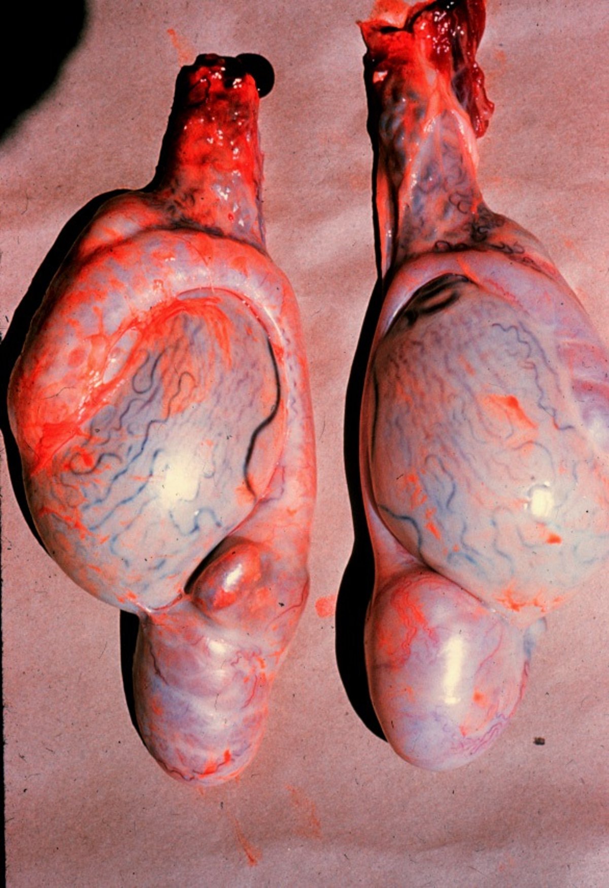
Lesions in the tail of the epididymis of a Dorset ram caused by B ovis infection.
Courtesy of Dr. John Larsen.
Not all infected rams develop palpable abnormalities of scrotal tissues and not all cases of epididymitis are due to brucellosis. Therefore, rams in an infected flock must be repeatedly examined via scrotal palpation and serologic testing.
Options for eradication of ovine brucellosis from heavily infected flocks include:
Culling of all rams and the purchase of fresh rams from a flock known to be free of infection. This is expensive in the short term but can be far less costly in the long term, especially with heavily infected flocks.
An eradication program consisting of regular scrotal palpation and serologic testing.
In eradication programs the initial testing interval should be 2–4 weeks, depending upon the number of rams on the farm and proportion infected. Isolating rams in discrete mobs, such as by age or breeding groups, can slow the spread of infection between tests. Confidence in eradication requires two consecutive negative tests at least 30–90 days after the last infected ram is removed. Replacement rams should then be sourced from a known, "accredited-free" flock, or else palpated and tested and held in isolation before being mixed with the main ram mob until confirmed as negative for infection.
Rams shedding organisms, but having no lesions, can be identified by means of microbial culture of semen samples, although repeated examinations may be necessary to identify intermittent shedders. Microscopic examination of stained semen smears may also be helpful; fluorescent antibody examination is a highly specific diagnostic aid. Serologic tests used for eradication of disease and certification of animals include indirect ELISA, complement fixation, hemagglutination inhibition, indirect agglutination, and gel diffusion.
Incidence and spread of the disease may be reduced by regular examination of rams before the breeding season and culling of those with obvious genital abnormalities. Because susceptibility in rams increases markedly with age, it is advantageous to keep a young ram flock and isolate noninfected rams from older, possibly infected rams.
Immunization of weaner rams with attenuated (Rev. 1) B melitensis has been recommended in some countries. Because infection in ewes apparently originates almost exclusively from service by infected rams, economic effects of lamb losses due to infection of ewes may be controlled by restricting vaccination to rams. There is no recommended vaccination in the US.
Chlortetracycline and streptomycin administered concurrently have effected bacteriologic cures. However, treatment is not economically feasible except in especially valuable rams, and even if infection is eliminated, fertility may remain impaired.


